Neuroendocrine tumor of parapharyngeal space
Marlinda Adham, Novra Widayanti
Department of Ear, Nose and Throat, Head and Neck Surgery Faculty of Medicine University of Indonesia
Cipto Mangunkusumo Hospital Jakarta
ABSTRACT
Background: Parapharyngeal space tumors account for some 0.5% of tumors of the head and neck, most of them benign. The most common benign neoplasms are salivary gland neoplasm, paragangliomas and followed by neurogenic tumors. The importance of these tumors lies mainly in two aspects, the difficulty of early diagnosis, due to the lack of symptoms in the initial stages and, on the other hand, the extreme risk of complications in
performing surgery in the parapharyngeal region. Purpose: We present this case to enlighten general
practitioners and also otorhinolaryngologist about diagnosis and management of parapharyngeal tumor. Case:
One clinical case of neuroendocrine tumor in parapharyngeal space on a 37 years old man. Management: The patient underwent diagnosis procedure and extirpation of the tumor mass. Conclusion: Parapharyngeal tumor is one of head and neck tumors that has good prognosis, especially if diagnosed early and adequately treated.
Keywords: neuroendocrine tumor,parapharyngeal space, benign tumor.
ABSTRAK
Latar belakang: Tumor parafaring meliputi sekitar 0,5% dari seluruh tumor kepala dan leher, sebagian besar jinak. Tumor jinak yang paling sering adalah tumor kelenjar liur, paraganglioma dan tumor neurogenik. Tumor parafaring ini penting disebabkan sulit untuk melakukan diagnosis dini karena sedikitnya gejala pada tahap awal dan kemungkinan komplikasi yang dapat terjadi pada saat dilakukan tindakan bedah di daerah parafaring. Tujuan: Kasus ini diajukan agar para dokter umum dan spesialis Telinga Hidung Tenggorok dapat mengenali diagnosis dan penatalaksanaan tumor parafaring. Kasus: Dilaporkan satu kasus tumor neuroendokrin parafaring pada laki-laki usia 37 tahun. Penatalaksanaan: Pada pasien ini dilakukan prosedur untuk mendiagnosis tumor dan dilakukan ekstirpasi massa tumor. Kesimpulan: Tumor parafaring merupakan salah satu dari tumor kepala dan leher yang mempunyai prognosis baik terutama bila didiagnosis secara dini dan diterapi secara adekuat.
Kata kunci: Tumor neuroendokrin, spatium parafaring, tumor jinak.
Alamat korespondensi: Marlinda Adham, Department of ENT-HN Faculty of Medicine University of Indonesia- Cipto Mangunkusumo Hospital. Jl. Diponegoro 71, Jakarta. e-mail: [email protected]
INTRODUCTION
Tumors of the parapharyngeal space
(PPS) are rare, most of them being diagnosed
in adults, but occasionally found in
children. PPS tumors often presented as an
asymptomatic growth and may be detected
in a routine medical checkup or as an
accidental finding when scanning for
another reason. There are many difficulties
in diagnosis of parapharyngeal mass because
of late manifestation of the symptoms and the
image of the mass on CT Scan or MRI.1,2
PPS tumors are infrequent, accounting
and neck. Most of these tumors (70-80%) are
benign and approximately 50% originate
from the deep lobe of the parotid or from the
minor salivary glands, particularly the
pleomorphic adenoma.3,4
The symptoms are manifested when the
tumor becomes larger than 2.5–3 cm and
related to the prestyloid–poststyloid
locali-zation.3,5
Tumors of the PPS include primary
neoplasms, direct extension from adjacent
regions, and metastatic disease.1,2,5
The diagnosis is done by physical
examination, radiological imaging with CT
or MRI with contrast and pathological
examination which could be performed by
fine needle aspiration cytology (FNAC).3,6
Choosing the appropriate approach
before surgery is very important when
dealing PPS. Transcervical approach allows
direct access to PPS. Infratemporal fossa
skull base surgery approach provides a very
wide operative field, which is preferable for
the resection of large PPS.7,8
We present this case to enlighten ENT
surgeons about diagnosis and management of
parapharyngeal tumor. Diagnosis performed
with nasopharyngeal CT and Magnetic
Resonance Angiography (MRA) that showed
big mass that had vascularisation from left
external carotid artery and the management
done by transcervical approach with vessel
ligation.
CASE REPORT
A 37 years old male came to ENT
Oncology Division in Cipto Mangunkusumo
Hospital (CMH) with chief complaint a
lump in the left neck since two years. There
were difficulty in swallowing, nasal voice and
snoring but no pain. Physical examination
showed assymetric pharyngeal arch, uvula
was pushed to the right side, left lateral
pharyngeal wall was bulged to medial and
lump in the left neck with the size 5x5x2 cm,
hard, fixated and painless. From
nasoendos-copic evaluation there was no mass on
naso-pharynx but fossa Rossenmuller, torus tubarius
and Eustachian tube were protruded.
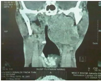
Naso-pharyngeal computed tomography (CT) scan
showed left parapharyngeal mass that
heterogenly enhanced after given contrast,
pushed vascular structures to lateral, no
vascular infiltration, no left neck
lympha-denopathy. It was suggested to do neck CT
angiography.
Figure 2. Sagittal CT scan of parapharyngeal mass.
Parapharyngeal biopsy result showed
moderate-severe dysplasia with chronic
inflammation. FNAB result from the left neck
lymphnode showed a metastasis from poorly
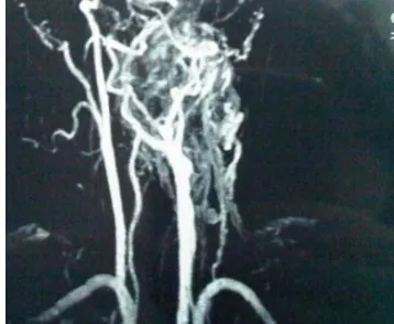
differentiated carcinoma. MRI evaluation
showed a big mass that got vascularisation
from left external carotid artery.
Figure 3. MRA of parapharyngeal mass showed the feeding artery from left external carotid artery.
Histopathology result from
paraphary-ngeal biopsy was carcinoma that difficult to
determine the type and origin, could be
from neuroendocrine or vascular and
suggest-ed for immunohistochemistry examination
and the final result was neuroendocrine
mass of small cell carcinoma. Thyroid USG
result was left lymphadenopathy colli in
accord with malignancy with normal thyroid.
Extirpation of left parapharyngeal
neuro-endocrine tumor was done by transcervical
approach and preceded by external carotid
artery ligation. After the procedure, from
flexible laryngoscopy evaluation there was
left vocal cord paralysis. Thorax CT scan
was taken 15th month after surgery and
there was right lung consolidation on 6th
segment with diferential diagnosis of lung
tuberculosis or metastasis and hepatomegaly.
Echo cardiography result was normal. The
result of cytology taken by bronchoscopy
showed no malignancy sign. The patient
was planned to be given chemotherapy.
DISCUSSION
A patient complained of a lump in the
left neck, difficulty in swallowing and snoring.
These were in accordance with literature that
the most common symptoms of
parapha-ryngeal tumors were neck mass, dysphagia,
hoarseness, vocal cord paralysis,
orophary-ngeal mass and hearing loss. The symptoms
are manifested when the tumor size 2.5–3
cm and related to the prestyloid–poststyloid
localization. Pain in the neck accompanied
with lock-jaw and/or paralysis of any of the
pairs of cranial nerves suggested malignancy
in origin.3,5,9
From physical evaluation there were
asymetric pharyngeal arch, uvula was pushed
to the right side, left lateral pharyngeal
wall bulged to medial as there was a big
Tumors of the PPS may include primary
neoplasms, direct extension from adjacent
regions or metastatic disease. The presence
of a mass in the parotid is often associated
with mass in the oral cavity. The presence
of pain or neuropathy should direct the
clinician to suspect a primary or metastatic
cancer but lesions of the posterior PPS may
also present with a neuropathy. A tumor
originating in the cervical sympathetic
chain may produce Horner's syndrome. 2,5
Malignant tumors can invade the PPS
from the nasopharynx, oropharynx, mandible,
maxilla, oral cavity, or parotid gland and
could extend intracranially through the
jugular foramen, or other foramina of the
skull base; they may also erode bone or
invade or expand to involve other proximal
spaces such as the retropharyngeal space or
infratemporal fossa. Neoplastic pathology
of the parapharyngeal space can mainly be
divided into salivary, neurogenic and vascular
tumors.1,2,10
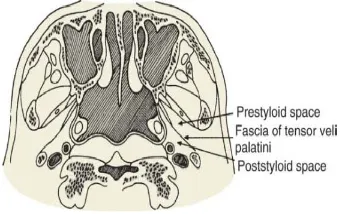
Figure 4. The fascia of the tensor veli palatini muscle divides the parapharyngeal space into a prestyloid and a poststyloid compartment.2
From the anatomical point of view the
prestyloid PPS contains the deep lobe of the
parotid gland, adipose tissues, small blood
vessels, lymphatics, and minor nerves. The
poststyloid PPS contains the carotid sheath
and is also traversed by cranial nerves IX,
X, and XII. The cervical sympathetic chain
lies posterior to the carotid artery.This tumor
should be extirpated but we should consider
the possible complication of surgery like
bleeding and paralysis of the fascial nerve.1,2
The diagnosis is done by physical
exa-mination, radiological imaging like CT or
MRI with contrast and pathological
exami-nation which could be performed by fine
needle aspiration cytology (FNAC).
Although MRI is better than CT, contrast
CT is the modality of choice. MRI provides
useful information on tumor localization
and extent, and distinguishes tumors of the
deep lobe, neurogenic lesions, intravagal
paraganglioma or carotid body tumors and
their relations with the internal carotid
artery and adjacent structures. Angiography is
recommended if paragangliomas or the
invol-vement of the carotid artery is suspected.2,6
On CT scan, parapharyngeal mass that
pushed vascular structures to lateral was
noted. The mass was seen in post styloid
region and might be a primary tumor.
FNAC can be an easy, rapid, and
effec-tive method of diagnosing these lesions.
Diagnostic difficulty persists due to their
similar mode of presentations and at times
morphological overlap. Hence, some cases
can only be confirmed by histopathological
examination and some by
FNA via a transoral approach for visible
parapharyngeal space lesions is an option
with an accuracy of 78–86%, but a
false-negative rate as high as 19% secondary to
inadequate stabilization of the lesion. The
value of FNA includes its low invasiveness,
the use of small needles in an area where
several vascular structures are present.7
Open biopsy is not advised, due to the
risk of bleeding, opening of the capsule
and, accordingly, relapse and seeding to
neighbouring tissues. The result from
parapha-ryngeal biopsy was neuroendocrine or vascular,
supported by immuno-histochemistry.9
Papadogeorgakis et al11 consider the
following five points to be the main
para-meters when deciding on the best PPS
surgical approaches. First, the proximity
and the projection of the tumor to the
oropharyngeal wall or the neck. Second, the
size of the tumor (the bigger the size, the
wider the access required). Third, the
suspicion of malignancy, because if a
malignant PPS tumor must be removed, it is
required wide access in order to obtain clear
margins. Fourth, the vascularity of the tumor,
because in PPS tumors of high vascularity,
the intraoperative risks of massive blood loss
make wide access important for effective
surgical handling. Fifth, the relation of the
tumor to the neck neurovascular bundle is of
great importance.
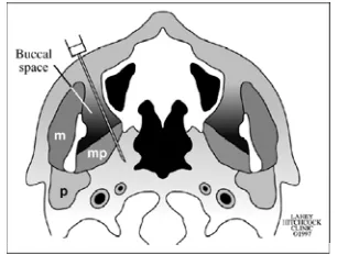
Figure 5. Schematic diagram shows the buccal space and biopsy needle. .m= masticator muscle; mp = medial pterygoid muscle; p= parotid gland. 12
To maximize the safety of this
procedure, wide access to the PPS is
recom-mended. The surgical approach must be as
minimal as possible, but wide enough to
ensure the complete removal of the tumor
and the safety of the vessels and nerves of
the neck. During the operation the surgeon
must be ready to modify the plan, if
unexpected difficulties in the complete and
safe removal of the tumor arise.13
In our case neuroendocrine tumor
extirpation was performed with carotid
artery ligation prior the surgery to prevent
massive bleeding. Neurogenic tumor was
only about 13% from parapharyngeal space
tumor. From the literatures, there were three
grades of neuroendocrine tumor: low
grade, intermediate grade and high grade
tumor. The neuroendocrine tumors can be
found in lung, thymus and pancreas. Result
from bronchoscopy-cytology there was no
Transcervical approaches was indicated
for removal of tumors originating from minor
salivary gland, schwannommas and
para-gangliomas in the poststyloid space, and
presumably benign tumor. Transcervical
approach allow directly acces to PPS.
Removal of the tumor is often preceded by
vessel ligation. On follow up the patient
doesn’t have any complaint, no hoarseness
nor difficulty in swallowing.14
Successful treatment of disseminated
neuroendocine tumors requires a
multi-modal approach; radical tumor surgery may
be curative but is rarely possible. Therapy
with radionuclides may be used for tumors
exhibiting uptake to a diagnostic scan, either
after surgery to eradicate microscopic residual
disease or later if conventional treatment or
biotherapy fails. Maintenance of the quality
of life should be a priority, particularly
because patients with disseminated disease
may experience prolonged survival.
REFFERENCES
1. Bradley PJ, Bradley PT, Olsen KD. Update on
the management of parapharyngeal tumors. Curr Opin Otolaryngol Head Neck Surg 2011;19:92-8.
2. Myers EN, Johnson JT. Management of tumors
of the parapharyngeal space. In: Myers EN, editors. Operative otolaryngology. Head and Neck Surgery. 2nd ed. Philadelphia: Saunders, 2008. p. 657-70
3. Ferro MF, Sanroman JF, Lopez AC, Gutierrez JS, Sanchez AL. Surgical treatment of benign parapharyngeal space tumors. Presentation of two clinical cases and revision of the literature. Med Oral Patol Oral Cir Bucal 2008; 13(1):E61-4.
4. Riskalla A, Arora A, Vaz F, O’Flynn P. Novel
use of ultrasound-guided endo-cavitary probe to evaluate an impalpable parapharyngeal mass. J Laryngol Otol 2010; 124:328-9
5. Dimitrijevic MV, Jesic SD, Mikic AA, Arsovic
NA, Tomanovic NR. Parapharyngeal space tumors: 61 cases reviews. Int J Oral Maxillofac Surg 2010; 39: 983–9
6. Mondal P, Basu N, Gupta SS. Fine needle
aspiration cytology of parapharyngeal tumors. J Cytol 2009; 26:102-4
7. Kanzaki S, Nameki H. Standardized method of
selecting surgical approaches to benign parapharyngeal space tumors, based on pre-operative images. J Laryngol Otol 2008; 122:628-34
8. Bozza F, Vigili MG, Ruscito P, Marzetti A,
Marzetti F. Surgical management of para-pharyngeal space tumors: result of 10-year follow–up. Acta Otolaryngol Ital 2009; 29:10-15
9. Gangopadhyay M, Bandopadhyay A, Sinha A.
Clinicoptahologic study of parapharyngeal tumors. J Cytol 2012; 29:26-9
10. Klimstra DS, Modlin IR, Coppola D, Lioyd
RV, Suster S. The pathologic classification of neuroendocrine tumors. A review of nomenclature, grading and staging systems. Pancreas 2010; 39:707-12
11. Papadogeorgakis N, Petsinis V, Goutzanis L.
Parapharyngeal space tumors: surgical approaches in a series of 13 cases. Int J Oral Maxillofac Surg 2010; 39:243-50
12. Tu AS, Geyer CA, Mancall AC, Baker RA.
The buccal space: A doorway for percutaneus CT-guided biopsy of the parapharyngeal region. AJNR Am J Neuroradiol 1998; 19:728-31
13. Shirakura S, Tsunoda A, Akita K. Parapharyngeal space tumors : Anatomical and image analysis findings. Auris Nasus Larynx 2010; 37:621-5
14. Kaltsas GA, Besser GM, Grossman AB. The
diagnosis and management of advanced neuroendocrine tumors. Endocrine Review 2004; 25(S):488-511