The Indonesian Journal of Gastroenterology, Hepatology and Digestive Endoscopy 86
Correlation of Carcinoma Percentage (CP) in Colorectal
Adenocarcinoma with Several Clinical Pathological
Aspects in Anatomical Pathology Department
Faculty of Medicine Universitas Indonesia/Cipto
Mangunkusumo Hospital Jakarta in Year 2012-2013
Elisabeth Indria Sari, Ening Krisnuhoni
Department of Anatomic Pathology, Faculty of Medicine, Universitas Indonesia/ Dr. Cipto Mangunkusumo General National Hospital, Jakarta
Corresponding author:
Ening Krisnuhoni. Division of Gastroentero-hepatology, Department of Anatomical Pathology, Dr. Cipto Mangunkusumo General National Hospital. Jl Diponegoro No.71 Jakarta Indonesia. Phone: +62-21-31930373; Facsimile: +62-21-3912477. E-mail: ening.krisnuhoni@yahoo.com.
ABSTRACT
Background: Colorectal carcinoma is malignant tumour of the large intestinal epithelial, where more than 90% of colorectal carcinoma is adenocarcinoma. Desmoplastic stroma in primary tumour holds an important role in tumour development process. Mesker et al described carcinoma percentage (CP) evaluation in routine preparations with Haematoxylin-eosin (HE) staining as one of the prognostic factors.
Method: This retrospective study was performed by collecting forms and slides of colorectal carcinoma resection cases from the archives of Anatomical Pathology Department, Faculty of Medicine, Universitas Indonesia/Cipto Mangunkusumo Hospital in 2 years period (2012-2013). Collection of clinical data and re-evaluation of HE slides by 2 people which were the writer and supervisor were performed to determine CP value by observing the comparison between tumour percentage and desmoplastic stroma.
Results: We included 92 cases of colorectal adenocarcinoma within those 2 years period (2012-2013). Comparison of total cases between male and female was 3:2, age > 40 years old were the most commonly found; in regard to location in the colon, most were found in the left side compared to the right side of the colon with the ratio of 3:7. Tumour histological grading with good differentiation were the most commonly found and most pT value being found were pT3; most lymph node involvement was N1. The most CP value attained in every parameter was CP-Low.
Conclusion: Carcinoma percentage (CP) which was divided as CP-High and CP-Low could be used as a prognostic factor. In this study, we found more cases of CP-Low compared to CP-High, either based on sex, age, location, degree of differentiation, pT value, or even lymph node involvement. CP-Low which showed worse prognosis could also be used as a marker of patients who were at higher risk of colorectal carcinoma.
Keywords: colorectal carcinoma, carcinoma percentage (CP), CP-Low, CP-High
ABSTRAK
Volume 18, Number 2, August 2017 87
(CP) dari sediaan rutin dengan pulasan Haematoxylin-eosin (HE) sebagai salah satu faktor prognostik. Metode: Telaah retrospektif ini dilakukan dengan mengumpulkan formulir dan slaid kasus reseksi karsinoma kolorektal (CRC) dari arsip di Departemen Patologi Anatomik FKUI-RSCM selama kurun waktu 2 tahun (2012-2013). Dilakukan pencatatan data klinis dan pembacaan ulang slaid HE oleh 2 orang yaitu penulis dan pembimbing untuk menentukan nilai CP dengan melihat perbandingan antara presentase tumor dan stroma desmoplastik.
Hasil: Terseleksi didapatkan 92 kasus adenokarsinoma kolorektal selama kurun waktu 2 tahun (periode 2012-2013). Perbandingan jumlah kasus antara jenis kelamin laki-laki dan perempuan adalah 3:2, usia > 40 tahun terbanyak ditemukan, untuk lokasi kolon terbanyak pada sisi kiri dibanding kolon sisi kanan dengan perbandingan 3:7. Grading histologik tumor dengan diferensiasi baik yang paling banyak ditemukan dan untuk nilai pT terbanyak ditemukan adalah pT3, keterlibatan kelenjar getah bening (KGB) paling banyak pada N1. Nilai CP yang terbanyak ditemukan untuk tiap-tiap parameter adalah CP-Low.
Simpulan: Carcinoma percentage (CP) yang terbagi atas CP-High dan CP-Low dapat digunakan sebagai faktor prognostik. Pada penelitian ini didapati lebih tingginya kasus CP-Low dibandingkan CP-High, baik berdasarkan jenis kelamin, usia, lokasi, derajat diferensiasi, nilai pT maupun keterlibatan KGB. CP-Low yang menunjukkan prognosis yang lebih buruk dapat pula dipakai sebagai marker untuk pasien-pasien yang lebih beresiko terhadap karsinoma kolorektal.
Kata kunci: karsinoma kolorektal, carcinoma percentage (CP), CP-Low, CP-High
INTRODUCTION
Colorectal carcinoma (CRC) is malignant tumour of epithelial cells deriving from epithelial cells of the colon which invade mucosal to submucosal muscle layer; more than 90% of CRC is an adenocarcinoma.1,2,3 CRC is one of the most common causes of morbidity and mortality worldwide with an approximate of 9% from all cancer cases.3,5 Incidence varies based on age, sex, and tumour location. Interactions of genetics, environmental factor, life style, and socio-economical condition play role in tumour development.1,3,4,5 CRC is the fourth most frequent cancer in male after lung, prostate, and gastric cancer and is the third most frequent cancer after breast and cervical cancer with the ratio of similar total cases between male and female.1 Incidence of CRC increases with increasing of age and more than 90% are sporadic CRC which happen at the age of > 40 years old.1,3,5 Based on Indonesian National Registry of Cancer (Badan Registrasi Kanker Indonesia) in 2011, malignancy in colorectal reached up to 9.66%.6
CRC is located in the right side (caecum, ascending colon, hepatic flexure, transverse colon) and left side (spleen flexure, descending colon, sigmoid colon, recto-sigmoid).2,7 Molecularly, the difference of location is associated with microsatellite instability (MSI) pathway which is differentiated into microsatellite high (MSI-H) and microsatellite low (MSI-L). This molecular identification of CRC can be used in detection as well as prognosis determination.8 Pathological examination of the resected specimen
of CRC play important role in providing information regarding prognosis and therapy.9 Cuthbert Dukes in 1929-1935 introduced a staging system including Tumour-Node-Metastasis (TNM) classification which is currently being used.1,10 Periodically, American Joint Committee on Cancer (AJCC) and International Union for Cancer Control (UICC) renew the tumour-node-metastasis (pTNM) staging system. This system explains that T is the extension of primary tumour, N is the metastasis to regional lymph nodes, and M is distant metastasis. Lymph node involvement is an important aspect of staging system in CTC, where metastasis to regional lymph node is one of the factors in prognosis determination.4,7,10 Clinical stages according to AJCC consist of stage I-IV based on TNM classification.1 Histological tumour grading can also be used as other factor to determine CRC prognosis. WHO divides tumour grading based on glandular formation into well, moderately, poorly differentiated, or undifferentiated colon adenocarcinoma.1,9
The Indonesian Journal of Gastroenterology, Hepatology and Digestive Endoscopy 88
later this CP value is divided into High and CP-Low. CP determination is aimed to evaluate survival [five years survival rates for overall survival (OS) and disease free survival (DFS)] between CP-High versus CP-Low as one of the parametric candidate to determine prognosis in routine diagnosis and in addition to pTNM classification.7,11,12 This retrospective study referred to the study which has been performed by Mesker et al in colorectal adenocarcinoma patients to determine carcinoma-percentage (CP) in primary tumour which is evaluated based on routine/daily preparation with HE staining.7 Patients with high stromal percentage or CP-Low between primary tumours have poor prognosis, while those with CP-High have better prognosis.7,12 The objective of this study is to determine carcinoma-percentage (CP) value to differentiate patients with good or bad prognosis in addition to pTNM classification and several other parameters. Sample of this study was obtained from patients’ data with diagnosis of stage I-III colorectal adenocarcinoma in Anatomical Pathology Department in 2012-2013.
METHOD
This retrospective study is a descriptive study with cross-sectional design. Demographic characteristics, clinical, and histopathological data were presented in tabular and graph format. Data were obtained from archives of Anatomical Pathology Department, Faculty of Medicine, Universitas Indonesia/Cipto Mangunkusumo Hospital. Cases searching were performed using topographical and morphological codes based on International Classification of Disease-10 (ICD-10) with topographical code of C.18 and C.20 and morphological code of M8140/3. Inclusion criteria were all resection cases with diagnosis of stage I-III colorectal adenocarcinoma within 2 years period (2012 and 2013). Exclusion criteria were diagnosis other than adenocarcinoma and distant metastasis. Determination of diagnosis of tumour type referred to histological type AJCC year 2010.
All forms and paraffin block slides were collected. Re-evaluation and re-inscription of all forms completion were performed, including age, location, sex, histopathological diagnosis, pTNM, and degree of differentiated by writer. Re-interpretation of paraffin block slides was performed by the writer; if any difficulty in determining CP value in this study was found, slide evaluation was performed together with supervisor. Data analysis was performed using SPSS
for windows (version 17.0) by using Fisher’s exact test and Kolmogorov Smirnov test to identify the presence of significant difference between each parameters being used with CP value.
CP identification was performed by microscopic examination of HE stained slides preparation with the thickness of 5µm and was conducted to all area of primary tumour. Magnification being used were 2.5 or 5x and invasive area with stromal involvement was chosen. Later with 10x magnification, percentage from CP area was determined. Chosen CP area was the most invasive area from desmoplastic stroma which was infiltrated by smallest tumour nest. Generally, CP area was determined from one of the most representative side of primary tumour. The next stage was percentage determination between carcinoma and desmoplastic stroma.7
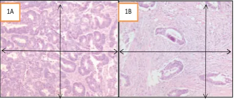
Tumour cell in scoring determination had to be present in all sides of viewing field (upper, lower, left, and right) (Figure 1A and 1B); if during evaluation, there was mucin component or necrotic area in the viewing field, those components need to be kept aside as it could cause over-or underscoring. Tumour percentage was estimated in tenth viewing field (10%, 20%, 30%, etc.). The lowest percentage was used to determine CP value. Cut-off CP value being used was 50%. CP value was further divided into CP-High (> 50%) and CP-Low (< 50%). For example, CP 70% is mentioned as CP-High which describe carcinoma proportion of 70% and 30% proportion of desmoplastic stroma.7,11,13
Figure 1. Slide with HE staining showing the least invasive part from primary tumour. Tumour cell should be present in all sides in the field of view (upper, lower, right, left).1A. CP-High (80%) and 1B. CP-Low
(20%).12
RESULTS
Based on the archive data in Anatomical Pathology Department FMUI/CMGNH, there was a total of 140 colon resection cases in 2 years period (2012 and 2013). In this study, we included stage I-III colorectal adenocarcinoma with morphology code M8140/3 to determine carcinoma-percentage (CP). After re-inscription was performed, we found 24 cases with diagnosis of mucinous adenocarcinoma, 7 cases of Signet Ring Cell carcinoma, 5 cases of lymphoma, 5 cases of mixed adenoneuroendocrine carcinoma (MANEC), and 7 cases of colorectal adenocarcinoma with metastasis; thus, we included 92 cases of colon resection to this retrospective study (Figure 2).
Figure 2. Number of colorectal adenocarcinoma cases in Anatomical Pathology Department FMUI/CMGNH with a total of 92 cases in 2012 and 2013
1B 1A
Figure 1. Slide with HE staining showing the least invasive part
from primary tumour. Tumour cell should be present in all sides
in the field of view (upper, lower, right, left).1A. CP-High (80%) and 1B. CP-Low (20%).12
RESULTS
Volume 18, Number 2, August 2017 89
After re-inscription was performed, we found 24 cases with diagnosis of mucinous adenocarcinoma, 7 cases of Signet Ring Cell carcinoma, 5 cases of lymphoma, 5 cases of mixed adenoneuroendocrine carcinoma (MANEC), and 7 cases of colorectal adenocarcinoma with metastasis; thus, we included 92 cases of colon resection to this retrospective study (Figure 2).
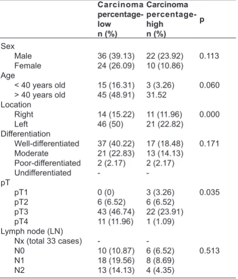
N value (metastasis to regional lymph node) which was not included in this study was 33 cases (35.88%). From all 59 cases (64.12%) of N value which could be studied, mostly were N1 with CP-Lowvalue in 18 cases (19.56%) (Table 1).
Myofibroblast was a component from desmoplastic stroma which has important role in tumour development process, invasion process, metastasis process or even as a prognostic indicator in primary tumour.14,15 Myofibroblast which is close to the nest of tumour cells may express α-smooth muscle actin (α-SMA)-positive myofibroblast though secretion of metalloproteinase (MMP), cytokines, chemokines, and neoplastic cells, which can produce lytic enzymes to degrade basal membrane and membrane around the tumour gland. One of the important mediators is transforming growth factor (TGF-β).7,11,14
TGF-β in many carcinoma has excessive expression and is known as pro-oncogenic and play important role in epithelial-to-mesenchymal transition (EMT) process. During EMT process, stromal cells lose binding and polarity causing tumour cells be able to infiltrate surrounding tissue and metastasis to distant organ. In molecular level, losing expression from E-cadherin may be a main marker of EMT. E-Cadherin expression may be inhibited by Cyclin B1 which has role in cell cycle regulation, cell growth, differentiation, apoptosis, and metastasis.7,11,14,15,16
Figure 2. Number of colorectal adenocarcinoma cases in
Anatomical Pathology Department, Faculty of Medicine,
Universitas Indonesia/Cipto Mangunkusumo Hospital with a total of 92 cases in 2012 and 2013
Study with 92 cases of colorectal adenocarcinoma found the highest distribution in 61-70 years old age group with 24 cases (Figure 3).
Figure 3. Colorectal adenocarcinoma in year 2012-2013 in
Anatomical Pathology, Faculty of Medicine, Universitas
Indonesia/Cipto Mangunkusumo Hospital based on age
Most cases in this study were found in male with 58 cases (63.05%), while female with 34 cases (36.95%) (Table 1). Most results of CP value for male and female were CP-Low, 36 cases in male (39.13%) and 24 cases in female (26.09%). Patients whose aged were > 40 years old or even < 40 years old had highest value which was CP-Low, where age > 40 years old in 45 cases (48.91%) and age < 40 years old in 15 cases (16.3%). Tumour were mostly found in the left side of the colon in 67 cases (72.82%) with highest CP value which was CP-Low in 46 cases (50%) (Table 1).
In this retrospective study, the most common degree of differentiation was well-differentiated in 54 cases (58.7%) with highest CP value of CP-Low in 37 cases (40.22%). The highest pT value in this study was pT3 with a total of 65 cases (70.65%) with highest value of CP-Low with a total of 43 cases (46.74%).
Table 1. Study results of carcinoma-percentage (CP-High) and (CP-Low) in colorectal adenocarcinoma in year 2012-2013
in Anatomical Pathology Department, Faculty of Medicine,
The Indonesian Journal of Gastroenterology, Hepatology and Digestive Endoscopy 90
Based on this study, there was several theories; first theory stated a hypothesis that higher stromal percentage or desmoplastic response has bad prognosis. The second theory stated that presence of fibrosis desmoplastic may decrease the ability of immune response to tumour by encaging the malignant tumour cells and preventing the destruction.13
Increased stromal cells which can be detected although just a small part from overall primary tumour may suggest bad prognosis; possibility which may occur is that the main part of tumour mass has the ability to directly facilitate the surrounding environment on invasion and metastasis.7,11,12,13 CRC patients with CP-Low with higher percentage of desmoplastic stroma compared to tumour mass, may produce a factor to promote tumour growth.11,13
Mesker et al stated that carcinoma percentage (CP) could be implemented as prognostic factor to evaluate five-year survival rate (OS/DFS). OS and DFS in CP-Low has lower value compared to CP-High or it could be explained that CP-Low (< 50%) showed worse prognosis, while CP-High(> 50%.) has better prognosis.7,11,12 Similar methods was also used by Courrech Stall et al to determine CP in oesophageal adenocarcinoma and CP evaluation in breast tumour by Moorman et al.11,12,14
In this retrospective study, the number of colorectal carcinoma tend to be relatively more common in male compared to female. 58 cases (63.05%) were found in male patients with 36 cases (39.13%) of CP-Low and 22 cases (23.92%) of CP-High. 34 cases (36.95%) were found in female patients also has highest value of CP which were CP-Low in 24 cases (26.09%). Results of statistical test for CP value found no significant difference between male and female (p = 0.113). This was in line with the study performed by Courrech Stall et alwho evaluated CP in oesophageal adenocarcinoma cases where there was no significant difference. Meanwhile, in the study conducted by Mesker et alin CRC, there was higher value of CP-High in male, while in this retrospective study, CP-Low value was higher.7,11
Literature described that incidence of CRC increased with increasing of age, where 90% of cases occurred in age > 40 years old. CRC patients with young age (< 40 years old) may be influenced by genetic factors; however, there is still no explanation on the association of age with CP value.1,3 In this study, more number of CRC cases were found in age > 40 years old compared to those aged < 40 years old. There were 74 subjects (80.43%) whose age was more than 40 years old, with CP-Low in 45 cases
(48.91%) and CP-High in 29 cases (31.52%). Results of statistical analysis for CP values showed that there was no significant difference between age < 40 and > 40 years old (p = 0.060).
Molecularly, 10-15% of sporadic CRC has MSI pathway which is one of the molecular marker for detection as well as prognostic determination. MSI pathway with high level of MSI (MSI-H) has better prognosis compared to those with low level of MSI (MSI-L).8 Based on its anatomical location, there was difference which was associated with carcinogenesis process through mismatch repair defective ratio in the right side of the colon and mismatch repair competent ratio in the left side. As much as 30% CRC which is located in the right side is mismatch repair defective and has MSI-H, while CRC in the left side is only 2%. Molecular appearance of MSI-H in the right side of the colon shows lower genetic mutation in TP53, K-ras, and c-MYC expression, while MSI-L which is located in the left side has higher frequency of genetic mutation in TP53, K-ras and c-MYC expression.5,17 The results of this study found more cases in the left side of the colon in 67 cases (72.82%) with CP-Low in 46 cases (50%) and CP-High in 21 cases (22.82%). Results of statistical analysis in this retrospective study showed significant difference between anatomical location and CP value (p = 0.000). This was in accordance with the study performed by Mesker et al who stated that there was significant difference between right side and left side of the colon, where there was lower value of OS/ DFS in the left side.7
CRC histological grading using WHO grading system is one of the prognostic determinant factors which is evaluated based on gland formation. It is categorised as well-differentiated if glandular structure is > 95%, moderately differentiated if glandular structure is between 50-95%, and poorly differentiated if it has solid pattern with glandular structure of < 50%.1,9. In this study, well-differentiated degree with CP-Low was found in 37 cases (40.22%), and CP-High in 17 cases (18.48%). Results of statistical analysis revealed no significant difference between differentiation degree with CP value (p = 0.171). In the study performed in oesophagus and CRC found high results of CP-Low in well-differentiated cases, but no statistical analysis was performed.7,11 In this study, we also obtained high CP-Low in well-differentiated cases.
Volume 18, Number 2, August 2017 91
E-Cadherin expression. Increased level of Cyclin B1 might decrease the expression of E-Cadherin; this explained that high level of cyclin B1 was related to poor overall survival due to the increased capacity of migration and invasion of tumour cells.16 In this study, there were highest rate of pT3 in 43 cases (46.74%) of CP-Low and 22 cases (23.91%) of CP-High. Results of statistical analysis for CP values exhibited the presence of significant difference between pT value and CP value (p = 0.035). Results of studies by Mesker et aland Courrech Stall et al to patients with CP-Low showed that pT value was less significant because it was said that tumour has its own mechanism to allow metastasis.7,11
Other important factor as a prognostic determinant in CRC is the number of lymph node involvement. Presence of positive lymph nodes and the number of involvement may influence prognosis. According to International Union Against Cancer and AJCC, minimum there should be 12 lymph nodes from colon resection tissue.7,9,18 Number of lymph node being obtained depends on many other factors, including the capability of surgeon, tumour location, length of resected specimen, and capability of anatomical pathologist in searching for small lymph nodes, particularly in the mesentery fat. If the number of lymph nodes were less than 12, additional technique such as fat clearance may be necessary.9
Figure 4A. Well-differentiated colorectal adenocarcinoma CP-Low< 50% (HE, 2,5X), Figure 4B. (HE,10X)
Figure 4C. Well-differentiated colorectal adenocarcinoma, CP-High> 50% (2,5X), Figure 4D. (HE,10X)
Figure 4E. Poorly-differentiated colorectal adenocarcinoma, CP-high> 50% (2,5X), Figure 4F.(HE,10X)
4B 4A
4C 4D
4E 4F
Figure 4A. Well-differentiated colorectal adenocarcinoma CP-Low< 50% (HE, 2,5X), Figure
4B. (HE,10X)
Figure 4C. Well-differentiated colorectal adenocarcinoma, CP-High > 50% (2,5X), Figure 4D. (HE,10X)
Figure 4E. Poorly-differentiated colorectal adenocarcinoma, CP-high> 50% (2,5X), Figure
The Indonesian Journal of Gastroenterology, Hepatology and Digestive Endoscopy 92
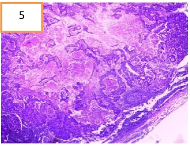
There are several studies on the important relationship between tumour cells and stromal cells in tumour development process as has been described. Zidar et al reported the correlation between α-SMA-positive myofibroblast and lymphatic-microvessel density(LMVD). Component from LMVD which is podoplanin (PDPN) is a specific marker of lymphatic endothelial cells. Myofibroblast in desmoplastic stroma can express PDPN to detect lymphatic involvement in colon cancer. This study explained that myofibroblast in peritumoural area hold essential role in lymphangiogenesis, and myofibroblast proliferation degree in peritumoural area can be used to predict the presence of metastasis to lymph nodes; besides of having association with lymphatic invasion and bad prognosis (Figure 5).19,20
CONCLUSION
Desmoplastic stroma plays role in the development process of tumour, invasion, metastasis, and as a prognostic factor in CRC patients. Mesker et al described carcinoma percentage (CP) as another prognosis determination factor which could be used in addition to the available pTNM classification. We included 92 cases of colorectal adenocarcinoma in Anatomical Pathology Department, Faculty of Medicine, Universitas Indonesia/Cipto Mangunkusumo Hospital in 2 years period (2012-2013). We found more results of CP-Low compared to CP-high in several parameters being used (sex, age, location, histological grading, pT value, and lymph node involvement).
Based on the results of statistical analysis, we found there was no significant difference between sex, age, degree of differentiation, and lymph node involvement with CP value, while for location and pT value there was significant difference. This CP value which can be a marker to determine patients with higher risk or even as a prognostic factor could be evaluated from routine/daily preparation with HE staining; therefore, meticulousness was highly required in determining deepest invasion in every case of colon resection, to obtain more accurate CP value.
ACKNOWLEDGEMENT
I would like to thank dr. Ening Krisnuhoni, SpPA(K) who has provided me with the protected time to give knowledge and supervision to complete this retrospective study amidst her demanding and busy schedule. I also thank Ibu Darmi and Ibu Upi in Archieve Division, as well as Bapak Udin in histopathological laboratory, for all of their support. I am also grateful to my fellow anatomical pathology residents for the support, suggestions, and corrections. I hope this study will be useful in evolving our knowledge in the field of anatomical pathology.
REFERENCES
1. Hamilton SR, Bosman FT, Boffeta P, Ilyas M, Morreau H,
Nakamura SI, et al. Carcinoma of the colon and rectum. In: Bosman FT, Carneiro F, Hruban R, Theise ND, eds. World Health Organization classification of tumors: pathology and genetics of tumours of the digestive system. Lyon: IARC Press, 2010.p.131-46.
2. Nawa T, Kato J, Kawamoto H, Okada H, Yamamoto H, Kohno H, et al. Differences between right-and left-sided colon cancer in patient characteristics, cancer morphology and histology. J Gastroenterology and hepatology 2008;23:418-23.
Figure 5. Metastasis to lymph node. (HE,2,5X)
A total of 33 cases (35.88%) of lymph nodes in this study could not be evaluated, due to the
number of lymph nodes were less than 12 or even not included or not found in the colon resection
tissue specimen. There were 59 cases (64.12%) of lymph nodes which could be evaluated, for CP
value from lymph node were mostly were in N1 with highest value of CP-Low in 18 cases
(19.56%). The results of statistical test in this study showed no significant difference of lymph node
involvement and CP value (p = 0.513). This was in accordance with the study performed by
Courrech Staal et al who stated that there was no significant difference. Meanwhile, in the study by
Mesker et al, there was highest value of CP-High in N0 without performing statistical analysis.
Adequate lymph node examination for CRC patients without metastasis was very much
needed and important. Inadequate lymph nodes being assessed possess serious risk which may cause
a patient with positive lymph node to not have the opportunity of the correct treatment; besides, not
resecting the positive lymph nodes may cause the high recurrence rate in CRC patients.
18CONCLUSION
Desmoplastic stroma plays role in the development process of tumour, invasion, metastasis,
and as a prognostic factor in CRC patients. Mesker et al described carcinoma percentage (CP) as
another prognosis determination factor which could be used in addition to the available pTNM
classification. We included 92 cases of colorectal adenocarcinoma in Anatomical Pathology
Department FMUI/CMGH in 2 years period (2012-2013). We found more results of CP-Low
compared to CP-high in several parameters being used (sex, age, location, histological grading, pT
5
Figure 5. Metastasis to lymph node. (HE,2,5X)
A total of 33 cases (35.88%) of lymph nodes in this study could not be evaluated, due to the number of lymph nodes were less than 12 or even not included or not found in the colon resection tissue specimen. There were 59 cases (64.12%) of lymph nodes which could be evaluated, for CP value from lymph node were mostly were in N1 with highest value of CP-Low in 18 cases (19.56%). The results of statistical test in this study showed no significant difference of lymph node involvement and CP value (p = 0.513). This was in accordance with the study performed by Courrech Staal et al who stated that there was no significant difference. Meanwhile, in the study by Mesker et al, there was highest value of CP-High in N0 without performing statistical analysis.
Volume 18, Number 2, August 2017 93
3. Haggar F, Boushey R. Colorectal cancer epidemiology: incidence, mortality, survival, and risk factors. Colorectal Cancer Epidemiol 2009;22:191-5.
4. Brozek W, Kriwanek S, Bonner E, Peterlik M, Cross H. Mutual associations between malignancy, age, gender, and subsite incidence of colorectal cancer. Anti cancer Res 2009;29:3721-5.
5. Al-Sohaily S, Biankin A, Leong R, Corish MK, Warusavitarne J. Molecular pathways in colorectal cancer. J Gastroenterol Hepatol 2012;29:1423-31.
6. Badan Registrasi Kanker Perhimpunan Dokter Spesialis Patologi Indonesia. Kanker di Indonesia tahun 2011, Jakarta (Indonesia): Data histopatologik 2011.
7. Mesker WE, Junggeburt JM, Szuhai K, De HP, Morreau H, Tanke HJ, et al. The carcinoma-stromal ratio of colon carcinoma is an independent factor for survival compared to lymph node status and tumor stage. Cell Oncol 2007;5:387-98. 8. Pawlik TM, Raut CP, Rodriguez-bigas MA. Colorectal
carcinogenesis: MSI-H versus MSI-L. Dis Markers 2004;20:199-206.
9. Marzouk O, Schofield J. Review of histopathological and
molecular prognostic features in colorectal cancer. Cancers 2011;3:2767-810.
10. Edge SB, Compton CC. The American joint Committee on Cancer: the 7th Edition of the AJCC Cancer Staging Manual and Future of TNM. Ann surg Oncol 2010;17:1471-4. 11. Staal EF, Wouters MW, Sandick JW, Tankenberg MM, Smith
VT, Junggeburt JM, et al. The Stromal part of adenocarcinomas of the oesophagus: does it conceal targets for therapy? Eur J Cancer 2010;4:720-8.
12. Hujibers A, Tollenaar RA, Pelt GW, Zesstraten EC, Dutton S, McConkey CC, et al. The proportion of tumor-stroma as strong prognosticator for stage II and III colon cancer patients: validation in VICTOR trial. Ann Oncol 2012;10:1-7. 13. Stall EF, Smit VT, Velthuysen ML, Naaykens JM, Wouters
MW, Mesker WE, et al. Reproducibility and validation of tumour stroma ratio scoring on oesophageal adenocarcinoma biopsies. Eur J Cancer 2010;5:118-32.
14. Unlu M, Cetinayak HO, Onder D, Ecevit C, Akman F, Ikiz AO, et al. The Prognostic value of tumor-stroma proportion in laryngeal squamous cell carcinoma. T J Path 2013;29:27-35. 15. Conti J, Thomas G. The role of tumour stroma in colorectal
cancer invasion and metastasis. Cancers 2011;3:2160-8. 16. Fang Y, Liang X, Jiang W, Li J, Xu J, Cai X. Cyclin B1
suppresses colorectal cancer invasion and metastasis by regulating E- Cadherin. PLOS One 2015;10:1-16.
17. Glebov OK, Rodriguez LM, Nakahara K, Jenkins J, Cliatt J, Humbyrd CJ, et al. Distinguishing right from left colon by the pattern of gene expression. Am Assoc Cancer Res J 2003;12:755-62.
18. Baxter NN, Virnig DJ, Rothenberger DA, Morris AM, Jessurun J, Virnig BA. Lymph node evaluation in colorectal cancer patients : a population-based study. J Natl Cancer Inst 2005;97:219-25.
19. Kitano H, Kageyama S, Hewitt SM, Hayashi R, Doki Y, Ozaki Y, et al. Podoplanin expression in cancerous stroma induces lymphangiogenesis and predicts lymphatic spread and patient survival. Arch Pathol Lab Med 2010;134:1520-6.