478
Biologi, Sains, Lingkungan, dan Pembelajarannya dalam Upaya Peningkatan Daya Saing Bangsa KEPADATAN TULANG METACARPAL SIMPANSE USIA 0 SAMPAI 44 TAHUNTetri Widiyani1, Bambang Suryobroto2, Yuzuru Hamada3 1
Jurusan Biologi FMIPA Universitas Sebelas Maret, Jl. Ir. Sutami 36 A Surakarta 57126
2
Departemen Biologi FMIPA IPB, Gedung Fapet Lt. 5 Kampus IPB Dramaga Bogor 16681 3
Primate Research Institute, Kyoto University Japan email: [email protected]
ABSTRAK
Sifat fisik tulang merupakan indikator yang baik untuk studi pertumbuhan dan penuaan. Tulang adalah jaringan dinamis karena adanya proses modeling dan remodeling. Tulang berubah tidak hanya pada ukuran dan bentuknya, tetapi juga kepadatannya yang disebabkan karena perubahan kandungan mineralnya. Osteoporosis atau kekeroposan tulang merupakan salah satu tanda umum penuaaan manusia. Osteoporosis di kalangan anthropoid masih belum diketahui. Dalam penelitian ini kami melakukan pengukuran kepadatan korteks tulang metacarpal simpanse (Pan troglodytes) usia 0 sampai 44 tahun berdasarkan radiografi. Pengukuran dilakukan dengan metode mikro-densitometri pada 68 simpanse betina dan 49 simpanse jantan. Kami menemukan bahwa kepadatan tulang meningkat pesat sampai usia sekitar 10 tahun. Pada simpanse jantan kepadatan tulangnya terus meningkat sampai usia 44 tahun, sedangkan pada simpanse betina kepadatan tulangnya menurun mulai usia 20 tahun. Penurunan kepadatan tulang simpanse betina dapat disebabkan karena kalsium tulang digunakan pada masa kehamilan dan menyusui. Namun demikian, simpanse betina diketahui tidak mengalami menopause. Jadi tidak seperti wanita, kejadian osteoporosis pada simpanse betina bukanlah akibat dari menopause. Kemungkinan hal ini berkaitan dengan berkurangnya kadar estrogen pada simpanse lanjut usia.
Kata Kunci: osteoporosis, kepadatan tulang, metakarpal, simpanse ABSTRACT
The physical properties of bone are good indicators of growth and aging. Bone is a dynamic tissue as its modeling and remodeling change not only the size and shape but also its density due to changes in mineral content. The incidences of bone loss in aging (osteoporosis) among anthropoids major interests for aging study. We assessed the age change of the density of second metacarpal bone in chimpanzees (Pan troglodytes) which was measured, by micro-densitometry on radiographs. We found that chimpanzee exhibited an initial increase in metacarpal density with age. It increased rapidly until around 10 years of age. It continued to grow slowly for several years and acquired maturation at around age 16 years. Sexual difference which was greater in males than in females, has been apparent since younger stage. In adulthood, male has a longer plateau than female. During elderly, the bone density of many females continued to decline since age 20 years but males continued its plateau phase or even slight increasing instead. The decline of bone mass in female chimpanzee elderly could not be explained as the result of menopause as female chimpanzees have no clear menopausal stage. It may be caused by the decline in fertility which occurs in parallel with decline in overall aging. This aging effect on fertility may include the diminishing estrogen.
Keywords: osteoporosis, bone density, metacarpal, chimpanzee.
INTRODUCTION
The physical properties of bone are good indicators of growth and aging. Bone characterizes unique life history of human, that is, extended lifespan, presence of adolescent growth and long postmenopausal life, and in turn it relates to quality of life in elderly. As the storage of calcium, bone is a dynamic tissue that always changes itself by apposition and absorption of bone materials throughout life. Bone grows in length and width and yet maintains its general shape. Nevertheless, bone grows in more complicated way than simple elongation or enlargement. It involves modeling and remodeling which change not only the size and shape but also its density due to changes in mineral content. During growth, bone modeling results in diameter enlargement and cortical thickening while remodeling results in bone mineral content accrual to decrease diameter and cortical thickness (Buckwalter et al., 1995; Robling et al., 2006).
Bone mineral content has been determined by many methods including assessment using radiographs. The radiograph of human metacarpal bones is commonly used to provide a general index of skeletal maturity (Ives and Brickley, 2004). The metacarpal is assessed easily using standard anteroposterior radiographs of the hand with low and safe doses of radiation (Tothill, 1989; Ives and Brickley, 2004; Barker
et al., 2005). Their anatomical areas are considered as fair representatives of long tubular bones. They give
precise information on status of bone resorption and apposition. A method to measure bone density based on radiography is photodensitometry (radiographic absorptiometry). It has been used extensively to estimate bone mass in the total skeleton and anatomical sites of interest (Wahner et al., 1984). The principle of photodensitometry is quantitative measurement of optical density of an object captured on light-sensitive medium due to exposure to light. The greater the density of the object is, the lesser the amount of light is permitted. Under X-ray, the mineralized mass of bone is of greater density than surrounding tissues so it blocks the exposure of the light-sensitive film. Therefore the amount of the light reaching the film after
Seminar Nasional IX Pendidikan Biologi FKIP UNS
479
passing through the bone is a measure of the BMC of the whole bone (Seeman, 2001; Haidekker et al., 2004; Symmons, 2004).
Age-changes in human bone density have been well-documented. It increases with age during infant and juvenile stages until late adolescence and reaches a plateau during adulthood. An adolescent acceleration of increase (spurt) in bone density was found by Martin et al. (1997), Bass et al. (1999), and Braillon (2003). There are sexual differences in adult bone density which are almost always greater in males than in females. Bone loss (osteoporosis) is most apparent during elderly. Women experience a greater decline in bone mass during aging than men do. It is well-known that menopause had a major effect to the evidence of bone loss in many older women (Schneider et al., 1997; Osei-Hyiaman et al., 1998; Ahlborg et al., 2003). With demographic shifting into older cohort in population pyramid, bone porosity raises medical concern.
Bone loss is a well established characteristic of human aging. It is found in all human populations. Thus, it is often considered to be a universal aspect of human ontogeny, although there are important sex and population differences in the rates at which bone loss occurs. Comparative investigation on age change of bone density in non-human primates is therefore necessary for the evolutionary consideration of growth (ontogeny) and aging and to find suitable model to study bone loss. The majority of reports of age-related bone density changes in nonhuman primates were made on macaques (Macaca spp.); DeRousseau, 1985; Pope et al., 1989; Jayo et al., 1994; Champ et al., 1996; Colman et al., 1999a; b; Hotckiss, 1999; Cerroni et al., 2000; Black et al., 2001). Few reports dealt with baboons (Papio spp.); Kammerer et al., 1995; Havill et al., 2003; Carlson and Pickering, 2004) and green monkeys (Chlorocebus sp.); Hiyaoka et al., 1996). Although chimpanzee (Pan troglodytes and Pan paniscus) is the closest species to human within extant non-human primates (Perelman et al., 2011), studies on the changes in bone density across the lifespan have not been well described, excepted some reports on the bone density states in adulthood (Sumner et al., 1989; Kikuchi, 2003; Kikuchi et al., 2003) and in elderly (Gunji et al., 2003). The paucity of chimpanzee studies was due to the fact that the population of chimpanzee accessible for the study is small, as they are large animals with long lifespan and growth periods.
The objective of the present study is to delineate the cross-sectional and longitudinal age-related changes in the MCI and cortical density at the mid-shaft of the second metacarpal of chimpanzee aged 0 to 43.6 years using radiographs. Our study comprised a large sample of individuals to cover the long lifespan so that present study provides a new coherent data.
MATERIALS AND METHODS
A total of 167 radiographs of chimpanzee's proximodistal left hands, collected by Primate Research Institute of Kyoto University, were used for the study. They were taken from 46 females and 35 males aged 0 to 43.62 years old. The chimpanzees belong to the Primate Research Institute of Kyoto University, Japan Monkey Center and the Kumamoto Primates Research Park of the Sanwa Kagaku Kenkyusho Co. Ltd. in Japan. We measured bone density of the second metacarpal cortex from the radiographs. The radiographs were transferred to the digital data before analysis. Bitmap images were captured from the radiographs using a Fujifilm FinePix F100fd digital camera. We applied an image analysis system, Scion Image Release Alpha 4.0.3.2 software for Windows (Scion Corporation, 2005) to measure linear dimensions and gray scale value of the pixels. Bone density of second metacarpal cortex was assessed from radiographs by applying photodensitometry. It is expressed as the thickness of an aluminum equivalent (mmAl) showing corresponding X-ray absorption (Seeman, 2001; Haidekker et al., 2004; Symmons, 2004).
All data were presented as an age-change curve. We drew age-change curves for cross-sectional data by applying generalized additive models for location, scale and shape (GAMLSS) (Rigby and Stasinopoulos, 2005) with 9 levels of percentiles (3%, 5%, 10%, 25%, 50%, 75%, 90%, 95%, and 97%). Age-changes in cortical density were analyzed in the different life stages by the application of ANOVA model and followed by the use of Tukey HSD (honest significant difference) post hoc test for multiple comparisons. All statistical procedures were performed using the R software version 2.9.1 (R Development Core Team, 2009).
480
Biologi, Sains, Lingkungan, dan Pembelajarannya dalam Upaya Peningkatan Daya Saing Bangsa RESULTWe defined the bone age stage into five phases for tracing the change in bone density. It is a combination of life stages reported by Hamada et al. (1996) for captives and Sumner et al. (1989) for wild chimpanzees. Hamada et al. (1996) defined the growth period of captive chimpanzee into four phases from the viewpoint of somatic growth. They are infancy (from birth to 1 year of age), juvenile (from 1 to 6 years of age in females or from 1 to 7 years of age in males), adolescence (from 6 to 8 years of age in females or from 7 to 9 year of age in males), and the late growth phase (from 8 to 13 years of age in females or from 9 to 15 years of age in males). Sumner et al. (1989) classified wild chimpanzee into five life stages: infancy (from birth to 5 years of age), childhood (from 5 to 7 years of age), adolescence (from 8 to15 years of age), maturity (from 16 to 33 years of age), and old age (>33 years of age). Phases of Hamada et al. covered age 0 to 15 years only while our data extended into >40 years of age. We therefore incorporated it to the
Sumner’s et al. phases although life stages of the wild chimpanzee were rather different to that of captive. Growth stage of wild chimpanzee is one to four years longer in each stage.
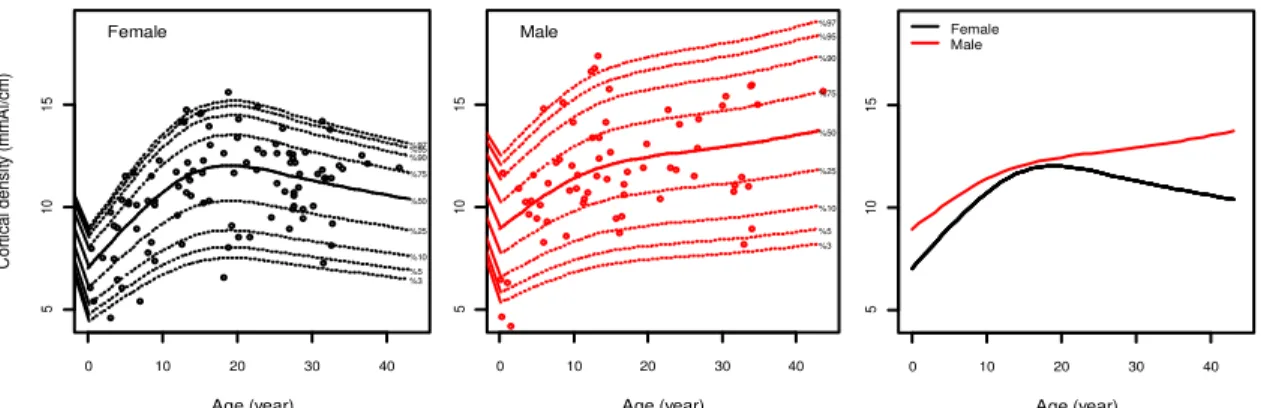
We examined mineral density age-change of the second metacarpal cortex. A comparison between female and male is shown in Figure 1, in which data on cortical density was plotted against age. It can be seen there is a significant sex difference in cortical density (p<0.05, ANOVA), that female had a lower cortical density. Cortical density increased steadily for first 9 years of life, from about 7 mmAl/cm into 10.5 mmAl/cm in female and from about 9 mmAl/cm into 11.5 mmAl/cm in male. Increment of the female cortical density therefore was higher during these early stages than that of male. However, because of wide variation among individuals, there were not significant increases (p>0.05, Tukey HSD) from infancy to juvenile and from adolescence to adult in both sexes. At about age 13 years, female attained full maturation of bone density, while male did approximately at the age of 12 years. The density at maturation does not differ between sexes, ca. 12mmAl/cm. Female had a short plateau and decreased soon since 21 years of age through elderly, although the decrease was statistically insignificant (p>0.05, Tukey HSD). In contrast, male had a longer plateau stage during adulthood. Cortical density consistently grew with low rates instead through elderly phase in males, even though their increments were also insignificant (p>0.05, Tukey HSD). The variation among individuals was very wide in both sexes e.g., 19 years in females, the density ranged from 7 to 15 mmAl (about 2 times), and in males, ranges from 8.5 to 17 mmAl (about 2 times).
DISCUSSION
The size, shape and density of bone are good indicators of both growth and aging, not only intraspecific but also interspecific assessment, e.g., skeletal maturation. According to the calcium demands, modeling and remodeling processes occur in bone in humans, which are shared by non-human primates, but not by such experimental animals as rats. Representing the growth and aging processes, the state of bone would be beneficial to study the evolution of unique life-history of humans, which is characterized by the long life-span, long growth period with adolescent growth spurt (acceleration), and long postmenopausal life. Within extant nonhuman primates the chimpanzee (Pan troglodytes) is the closest species to humans (Perelman et al., 2011). Knowledge of growth and aging of bone, especially the bone mineral density, in
Figure 1 Curves of age-change of the cortical density in the second metacarpal in female (left) and male (center) chimpanzees from the cross-sectional study. Solid line for 50th percentile and dotted-lines for 3rd, 5th, 10th, 25th, 75th, 90th, 95th, and 97th
percentiles (bottom to top). Combined of both sexes curves (right) were derived from 50th percentile.
Age (year) 0 10 20 30 40 5 10 15 %97 %95 %90 %75 %50 %25 %10 %5 %3 Male Age (year) C o rt ica l d e n si ty (m m A l/cm ) 0 10 20 30 40 5 10 15 %97 %95 %90 %75 %50 %25 %10 %5 %3 Female Age (year) 0 10 20 30 40 5 10 15 Female Male
Seminar Nasional IX Pendidikan Biologi FKIP UNS
481
chimpanzee at present is scarce because its measurement is difficult and the population accessible for study is small, as they are large animals with long lifespan and growth periods.
We delineated a general pattern of age change in cortical density of the second metacarpal bone in chimpanzees. Cortical density estimates absolute bone mass in the cortical sites (Wahner et al., 1984). Based on cross-sectional analysis, chimpanzee exhibited an initial increase in bone density with age. It increased for a longer period, until around 10 years of age. Sexual difference in bone density, which was greater in males than in females, has been apparent since the early ages. It is earlier than the onset of sexual difference on body length and weight, both of which were reported approximately at the age of 8 years (Leigh, 1992; Hamada and Udono, 2002). Bone density continued to increase slowly for several years and acquired maturation at around age 16 years. Maturation age in bone density was much later than reproductive maturity which is observed at the age of 7 years in females (menarche) and 8 years in males (development of testicular size; Hamada and Udono, 2002). Bone density decreases in females from about 20 years of age, though not in males. We found, thus, that the pattern of change in density of cortical bone
of chimpanzee’s second metacarpal was quite similar to the human growth pattern. Nevertheless there were differences due to difference in age of each stage which reflects the life span distinction between chimpanzee and human (Bogin and Smith, 1996; Wallis, 1997, Thompson et al., 2007).
Bone aging in humans is characterized by gradual decrease in adulthood and rapid loss in postmenopausal women, which is another point of interest of age change pattern of bone density in chimpanzees. In adulthood through elderly, male chimpanzees show a longer plateau than female chimpanzee, that is, bone density is maintained. Some female chimpanzees experienced bone loss since approximately age 20 years, as it was reported by Sumner et al. (1989) and Gunji et al. (2003). However, in both of female and male age-related BMC changes during adult through elderly were statistically insignificant. Individual variation is rather wide, especially in cortical density. Despite these, the sequential decline of bone mass in female may be said to be genuine.
Some evidence supports the aging of bone in female chimpanzees, for instance Gunji et al. (2003) reported extraordinary low bone density in wild elderly (estimated about 40 years at death) female chimpanzee. The low bone mineral content in adult female might be caused by the demands of pregnancy and lactation (Sumner et al. 1989). Female chimpanzees have high fertility rate during age 15 years until 30 years (Thompson et al., 2007). Several studies in human reported that maternal bone acts as a calcium storage depot for the fetal and neonatal mineral nutrient (Drinkwater and Chesnut, 1991; Sowers et al., 1993; More et al., 2001). Adult bone loss due to pregnancy and lactation was also reported in rhesus macaques (deRousseau, 1985), green monkeys (Hiyaoka et al., 1996) and cynomolgus macaques (Lees et al., 1998). These phenomena might also be true for chimpanzee. So we may conclude that female chimpanzees experience sequential decline in bone mass as it is observed in human.
For our study, estrogen deficiency cannot be dismissed out of hand as a factor causing the loss of bone density since diminished ovarian estrogen levels has been observed in old captive chimpanzees, after the age of 40 years (Gould et al., 1981; Videan et al., 2006). In human, a normal level of estrogen inhibits bone resorption by directly inducing apoptosis of the bone-resorbing osteoclasts (Kameda et al., 1997; Frost, 1999; Riggs et al., 2002). Nevertheless the low bone density in elderly female chimpanzee could not be explained by the postmenopausal hormonal changes as it had been thought to occur in human. Menopause is not a typical characteristic of chimpanzee life histories (Thompson et al., 2007; Lacreuse et al., 2008). According to the study on wild chimpanzee, females continue to reproduce in their life-span although the inter-birth interval is rather long in elderly. On the other hand their survival rate is constantly decreasing from birth. Therefore their fertility decline occurs in parallel with declines in overall aging (Thompson et al., 2007). This aging effect on fertility decline may include the diminishing of estrogen which might be responsible for the decline of bone mass.
CONCLUSION
Chimpanzee exhibited an increase of bone density until adulthood. It increased until around 10 years of age. Sexual difference, which was greater in males than in females, has been apparent since the early ages. Bone density continued to increase more slowly for several years and acquired maturation at around age 16 years. In adulthood, male has a longer plateau phase than female. Some female chimpanzees
482
Biologi, Sains, Lingkungan, dan Pembelajarannya dalam Upaya Peningkatan Daya Saing Bangsaexperienced bone loss approximately since age 20 years. During aging, the bone density of many females
declined but male’s continued its plateau phase or increase instead. Despite there was a statistical insignificant in age-related changes in aging, the decline of bone mass in female may be said to be genuine.
REFERENCES
Ahlborg, H.G., Johnell, O., Turner, C.H., Rannevik, G., & Karlsson, M.K. (2003). Bone loss and bone size after menopause. N Engl J Med, 349 (4), 327334.
Bass, S., Delmas, P.D., Pearce, G., Hendrich, E., Tabensky, A., & Seeman, A. (1999). The differing tempo of growth in bone size, mass, and density in girls is region-specific. J Clin Invest 104 (6), 795–804.
Barker, D.S., Schultz, C., Krishnan, J., & Hearn, T.C. (2005). Bilateral symmetry of the human metacarpal: Implications for sample size calculations. Clin Biomech, 20, 846–852.
Black, A., Tilmont, E.M., Handy, A.M., Scott, W.W., Shapses, S.A., Ingram, D.K., Roth, G.S., & Lane, M.A. (2001). A nonhuman primate model of age-related bone loss: a longitudinal study in male and premenopausal female rhesus monkeys. Bone 28(3), 295–302.
Braillon, P.M. (2003). Annual changes in bone mineral content and body composition during growth. Horm Res 60, 284– 290.
Bogin, B. & Smith, B.H. (1996). Evolution of the human life cycle. Am J Hum Biol 8, 703-716.
Buckwalter, J.A., Glimcher, M.J., Cooper, R.R., & Recker, R. (1995). Bone biology. J Bone Joint Surg Am 77-A(8), 12761289.
Carlson, K.J. & Pickering, T.R. (2004). Shape-adjusted bone mineral density measurements in baboons: other factors explain primate skeletal element representation at Swartkrans. J Archaeol Sci 31, 577–583.
Cerroni, A.M., Tomlinson, G.A., Turnquist, J.E., & Grynpas, M.D. (2000). Bone mineral density, osteopenia, and osteoporosis in the rhesus macaques of Cayo Santiago. Am J Phys Anthropol 113, 389–410.
Champ J.E., Binkley, N., Havighurst, T., Colman, R.J., Kemnitz, J.W., & Roecker, E.B. (1996). The effect of advancing age on bone mineral content of female rhesus monkeys. Bone 19(5): 485492.
Colman, R.J., Kemnitz, J.W., Lane, M.A., Abbott, D.H., & Binkley, N. (1999a). Skeletal effects of aging and menopausal status in female rhesus macaques. J Clin Endocrinol Metab 84(11), 41444148.
Colman, R.J., Lane, M.A., Binkley, N., Wegner, F.H., & Kemnitz, J.W. (199b). Skeletal effects of aging in male rhesus monkeys. Bone 24(1), 17–23.
deRousseau, C.J. (1985). Aging in the musculoskeletal system of rhesus monkeys: III. Bone loss. Am J Phys Anthropol
68, 157167.
Drinkwater, B.L. & Chesnut, C.H. (1991). Bone density changes during pregnancy and lactation in active women: a longitudinal study. Bone Miner 14(2), 153160.
Frost, H.M. (1999). Perspective on the estrogen–bone relationship and postmenopausal bone loss: a new model. J Bone Miner Res 14(9), 1473–1477.
Gould, K.G., Flint, M., & Graham, C.E. (1981). Chimpanzee reproductive senescence: a possible model for evolution of the menopause. Maturitas 3, 157166.
Gunji, H., Hosaka, K., Huffman, M.A., Kawanaka, K., Matsumoto-Oda, A., Hamada, Y., & Nishida, T. (2003). Extraordinarily low bone mineral density in an old female chimpanzee (Pan troglodytes schweinfurthii) from the Mahale Mountains National Park. Primates 44, 145–149.
Haidekker, M.A., Stevens, H.Y., & Frangos, J.A. (2004). Computerized methods for X-ray-based small bone densitometry. Comp Meth Prog Biomed 73, 3542.
Hamada, Y. & Udono, T. (2002). Longitudinal analysis of length growth in the chimpanzee (Pan troglodytes). Am J Phys Anthropol 118, 268–284.
Hamada, Y., Udono, T., Teramoto, M., & Sugawara, T. (1996). The growth pattern of chimpanzees: somatic growth and reproductive maturation in Pan troglodytes. Primates 37, 279295.
Havill, L.M., Mahaney, M.C., Czerwinski, S.A., Carey, K.D., Rice, K., & Rogers, J. (2003). Bone mineral density reference standards in adult baboons (Papio hamadryas) by sex and age. Bone 33(6), 877888.
Hiyaoka, A., Yoshida, T., Cho, F., & Yoshikawa, Y. (1996). Changes in bone mineral density of lumbar vertebrae after parturition in African green monkeys (Cercopithecus aethiops). Ex Anim 45(3), 257259.
Hotchkiss, C.E. (1999). Use of peripheral quantitative computed tomography for densitometry of tha femoral neck and spine in cynomolgus monkeys (Macaca fascicularis). Bone 24(2), 104107.
Ives, R. & Brickley, M.B. (2004). A procedural guide to metacarpal radiogrammetry in archaeology. Int. J. Osteoarchaeol
14, 7–17.
Jayo, M.J., Jerome, C.P., Lees, C.J., Rankin, S.E., & Weaver, D.S. (1994). Bone mass in female cynomolgus macaques: a cross-sectional and longitudinal study by age. Calcif Tissue Int 54, 231236.
Kameda, T., Man, H., Yuasa, T., Mori, Y., Miyazawa, K., Shiokawa, M., Nakamaru, Y., Hiroi, E., Hiura, K., Kameda, A., Yang, N.N., Hakeda, Y., & Kumegawa, M. (1997). Estrogen inhibits bone resorption by directly inducing apoptosis of the bone-resorbing osteoclasts. J Exp Med 186(4), 489–495.
Kammerer, C.M., Sparks, M.L, & Rogers, J. (1995). Effects of age, sex, and heredity on measures of bone mass in baboons (Papio hamadryas). J Med Primatol 24: 236 –242.
Kikuchi Y. 2003. Age change of bone mineral density in distal radius of chimpanzee and Japanese macaques. Anthropol Sci 111(2), 165186.
Seminar Nasional IX Pendidikan Biologi FKIP UNS
483
Kikuchi, Y., Udono, T., & Hamada, Y. (2003). Bone mineral density in chimpanzees, humans, and Japanese macaques.
Primates 44, 151–155.
Kimura, K. (1990). Age and sex difference in bone density of the second metacarpal in its midshaft cross-section. Ann Hum Biol 17(5), 399406.
Lacreuse, A., Chennareddi, L., Gould, K.G., Hawkes, K., Wijayawardana, S.R., Chen, J., & Herndon, J.G. (2008). Menstrual cycles continue into advanced old age in the common chimpanzee (Pan troglodytes). Biol Reprod 79, 407–412.
Lees, C.J., Jerome, C.P., Register, T.C., & Carlson, C.S. (1998). Changes in bone mass and bone biomarkers of cynomolgus monkeys during pregnancy and lactation. J Clin Endocrinol Metab 83, 4298–4302.
Leigh, S.R. (1992). Patterns of variation in the ontogeny of primate body size dimorphism. J Hum Evol 23, 2750. Martin, A.D., Bailey, D.A., McKay, H.A., & Whiting, S.(1997).Bone mineral and calcium accretion during puberty. Am J
Clin Nutr 66, 611615.
More, C., Bettembuk, P., Bhattoa, H.P., & Balog, A. (2001). The effects of pregnancy and lactation on bone mineral density. Osteoporos Int 12, 732–737.
Osei-Hyiaman, D., Satoshi, T., Ueji, M., Hideto, T., & Kano, K. (1998). Timing of menopause, reproductive years, and bone mineral density a cross-sectional study of postmenopausal Japanese women. Am J Epidemiol 148(11), 10551061.
Perelman, P., Johnson, W.E., Roos, C., Seuánez, H.N., Horvath, J.E., Moreir, M.A.M., Kessing, B., Pontius, J., Roelke, M., Rumpler, Y., Schneider, M.P.C., Silva, A., O’Brien, S.J., & Pecon-Slattery, J. (2011). A molecular phylogeny of living primates. PLoS Genet 7(3), e1001342.
Pope, N.S., Gould, K.G., Anderson, D.C., & Mann, D.R. (1989). Effects of age and sex on bone density in the rhesus monkey. Bone 10, 109112.
R Development Core Team. (2009). R: A language and environment for statistical computing. R Foundation for Statistical Computing, Vienna, ISBN 3-900051-07-0, http://www.R-project.org/.
Rigby, R.A. & Stasinopoulos, D.M. 2005. Generalized additive models for location, scale and shape. Appl Statist 54(3), 507554.
Riggs, B.L., Khosla, S., & Melto, L.J. (2002). Sex steroids and the construction and conservation of the adult skeleton.
Endocrine Rev 23(3), 279–302.
Robling, A.G., Castillo, A.B., & Turner, C.H. (2006). Biomechanical and molecular regulation of bone remodeling. Annu Rev Biomed Eng 8, 455–498.
Scion Corporation. (2005). Scion image release beta 4.0.3. http://www.scioncorp.com.
Seeman, E. (2001). Sexual dimorphism in skeletal size, density, and strength. J Clin Endocrinol Metab 86(10), 4576– 4584.
Schneider, D.L., Barrett-Connor, E.L., & Morton, D.J. (1997). Timing of postmenopausal estrogen for optimal bone mineral density: The Rancho Bernardo Study. JAMA 277(7), 543547.
Symmons, R. (2004). Digital photodensitometry: a reliable and accessible method for measuring bone density. J Archaeol Sci 31, 711–719.
Sowers, M., Corton, G., Shapiro, B., Jannausch, M.L., Crutchfield, M., Smith, M.L., Randolph, J.F., & Hollis, B. (1993). Changes in bone density with lactation. JAMA 269(24), 31303135.
Sumner, D.R., Morbeck, M.E., & Loblick, J.J. (1989). Apparent age-related bone loss among adult female Gombe chimpanzees. Am J Phys Anthropol 79, 225–234.
Thompson, M.E., Jones, J.H., Pusey, A.P., Brewer-Marsden, S., Goodall, J., Marsden, D., Matsuzawa, T., Nishida, T., Reynolds, V., Sugiyama, Y., & Wrangham, R.W. (2007). Aging and fertility patterns in wild chimpanzees provide insights into the evolution of menopause. Curr Biol 17(24), 2150–2156.
Tothill, P. (1989). Methods of bone mineral measurement. Phys Med Biol 34, 543572.
Videan, E.N., Fritz, J., Heward, C.B., & Murphy, J. (2006). the effects of aging on hormone and reproductive cycles in female chimpanzees (Pan troglodytes). Comp Med 56(4), 291299.
Wahner, H.W., Dunn, W.L., & Riggs, B.L. (1984). Assessment of bone mineral. Part 1. J Nucl Med, 11341141.
Wallis, J. (1997). A survey of reproductive parameters in the free-ranging chimpanzees of Gombe National Park. J Repr Fertil 109, 297307.