83
DETERMINING THE ABNORMALITY OF BULL SPERM TAIL MORPHOLOGY
USING SUPPORT VECTOR
a
Stevanus Hardiristanto, bI Ketut Eddy Purnama, cAdhi Dharma Wibawa, dMira Candra Kirana, eBudi Santoso, fMunawir, gSlamet Hartono, hI Nyoman Tirta Ariana, iDian
Ratnawati, jLukman Affandhy a,b,c,d,e,f
Department of Multimedia and Network Engineering, Faculty of Industrial Technology, Institute of Technology Sepuluh Nopember, Surabaya, Indonesia
g
Balai Pembibitan Ternak Unggul Sapi Bali, Ministry of Agriculture, Republik of Indonesia h
Faculty of Animal Science, University of Udayana, Bali, Indonesia i,j
Loka Penelitian Sapi Potong Grati, Ministry of Agriculture, Republik of Indonesia E-mail: [email protected]
Abstrak
Penilaian atas ketidaknormalan spermatozoa bisa dilakukan dari sisi motilitas maupun morfologi (kepala dan ekor). Penelitian ini mengevalusi ketidaknormalan spermatozoa dari sisi morfologi bagian ekor spermatozoa sapi. Data berupa 50 citra mikroskopis spermatozoa yang diperoleh dari Loka Penelitian Sapi Potong Grati, Pasuruan digunakan dalam penelitian ini. Prosedur yang ditetapkan terdiri atas beberapa tahap. Tahap pertama adalah melakukan segmentasi spermatozoa untuk memisahkan spermatozoa dari latar belakang dan memisahkan bagian ekor spermatozoa dari bagian yang lain. Selanjutnya dari hasil segmentasi dicari garis tengah ekor (skeleton) menggunakan metode medial axis transform. Berdasarkan garis tengah yang dihasilkan, dilakukan prosedur ekstraksi fitur menggunakan metode polynomial curve fitting. Kemudian, metode Support Vector Machine (SVM) digunakan untuk menentukan ketidaknormalan bentuk ekor spermatozoa. Untuk pembelajaran digunakan 25 data spermatozoa normal dan 10 data spermatozoa tidak normal. Testing kemudian dilakukan atas 15 data spermatozoa tersisa. Ketelitian SVM dalam menentukan ketidaknormalan bentuk ekor spermatozoa mencapai 73.33%. Dengan demikian ketidaknormalan bentuk ekor spermatozoa dapat ditentukan dengan menggunakan SVM.
Kata kunci: Ekor Sperma sapi, Morphology, Polynomial Curve Fitting, SVM.
Abstract
Determinining the abnormality of spermatozoa can be done by inspecting its motility or morphology (head or tail). This study examined 50 data of sperm microscopic images. The semen was obtained from Loka Penelitian Sapi Potong Grati, Pasuruan. A sequence of procedure consist of several steps were then carried out. The first step was to obtain sperm tails by segmenting the sperms from its background and removing the heads and the necks parts. The skeletons of the tails were then obtained using a method of medial axis transform. The features of the tails were then extracted using polynomial curve fitting. Then, Support Vector Machine (SVM) was used as a classifier. In the training phase, 25 normal sperm and 10 abnormal sperm were utilized. Afterward, the remaining 15 data were used in the testing phase. The accuracy of SVM was 73.33%. Hence, the abnormality of spermatozoa based on the shape of sperm tail can be determined using SVM.
INTRODUCTION
Analysis of sperm shape has been done on either on human or animal [1-3]. However, assesing the abnormality of bull sperm especially based on its morphological condition is still crucial issue since morphological condition related to the quality of bull sperm especially for artificial insemination program for both beef and dairy industry [4,5]. The quality of sperm could be determined by several criterions, such as the size and the form (morphology) of the head, the length and the form of the tail and its movement behaviour (motility) [6,7]. Sperm or also called Spermatozoa are cells of the male reproductive system. Spermatozoa are highly specialized cells and dense that no longer have cleavage or growth, derived from the gonosit into spermatogonia, primary and secondary spermatocytes, subsequently changed to be spermatids and finally turn into spermatozoa. Spermatozoa consists of two functionally important parts, the head and tail [8]. In the normal adult male, spermatogenesis continues throughout life, although the quality and quantity decline with age.
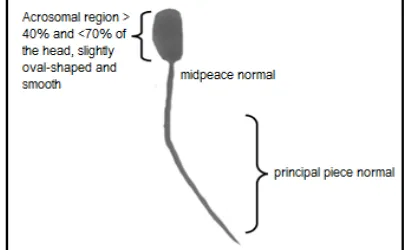
The head of spermatozoa is oval with approximately 5 microns in length, 3 microns in diameter, 2 microns in thickness and mainly formed by the nucleus. Spermatozoa tail section consists of neck, middle, principal, and the end piece. The tail length is about 55 microns and 1 micron thickness. The length of principal piece 45 microns, 0.5 microns thick and the tip of 4-5 microns long, 0.3 microns thick. The tail could not be observed using light microscope but electron microscope should be used [9]. Sperm that has abnormal shape loss its ability to fertilize ovum in the fallopian tubes.
Figure 1 shows the normal bull sperm morphology that has a normal head, midpeace and tail. The figure was derived from one of the images used in this study.
Previous studies in evaluating bull sperm morphology have been done by some researchers in the past using two main methods [10], the first is bright-field (BF) microscopy method and differential interference phase contrast (DIC) microscopy method. It was reported that there was no difference in percentage of normal sperm that
can be seen between each method, however DIC was more effective invisualizing main defect. Moreover, BF is considered to make the sperm looks smaller. From the result point of view, DIC is recognized as a better viewing tool in assessment of bull sperm morphology compared to BF method [11].
Figure 1. Normal Sperm Morphology.
Previous studies in evaluating bull sperm morphology have been done by some researchers in the past using two main methods [10], the first is bright-field (BF) microscopy method and differential interference phase contrast (DIC) microscopy method. It was reported that there was no difference in percentage of normal sperm that can be seen between each method, however DIC was more effective invisualizing main defect. Moreover, BF is considered to make the sperm looks smaller. From the result point of view, DIC is recognized as a better viewing tool in assessment of bull sperm morphology compared to BF method [11].
In another research, the identification and classification of spermatozoa has been performed by experts on different sperm samples such as human, horses, pigs and bull spermatozoa [12]. Ahmad proposed in their
study that the identification of human sperm’
tail can be done through detecting shapes using Structural Similarity Index (SSIM) and Local Entropy. The advantage of the this method was being able to detect the sperm tail at low contrast but the disadvantage was it required a long execution time [13].
MATERIAL AND METHODS
Data Acquisition
CASA (Computer Assisted Semen Analisys) as one of the key player device in measuring
morphological condition of sperm
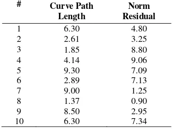
morphologically. Casa provides many advantages to the researchers but also presents some drawbacks such as high device installation cost and need to be stationed at a fix position like in laboratory. Developing a low cost device and mobile could be a breakthrough in solving some limitations of CASA such as for gathering and analysing data from many beef or dairy industries in remote areas. In this study we implemented our own tool for observing and analysing bull sperm tail based on its morphological condition. The tool was implemented using a microscope equiped with CCD camera (Sony IMX035 CMOS, model: FL3-U3-13S2C-CS) connected to a computer via usb. Using this tool images of sperm samples prepared on slides were taken. The sperm samples were prepared by droping sperm from straw onto slides, colored using eosin-negrosin agents to distinguish the dead from the live, smear carefully and evenly over the slides. The sperm in straw packaging was obtained from Loka Penelitian Sapi Potong, Grati, Pasuruan, East Java, under the auspices of the Ministry of Agriculture, Republic of Indonesia. Fifty images were obtained in this study. Four out the 50 were shown in Figure 2 as an example.
Figure 2. Example of bull sperm images (a) and (b) normal spermatozoa , (c) and (d) abnormal spermatozoa.
Support Vector Machine
Support Vector Machine (SVM) is a famous method for classification since it can find an exact classification boundary using a
so-called support vectors, hyperplanes and kernels. Numerous applications of SVM have been performed by several researchers such as in remote sensing application [14] and medical application [15]. In this research we apply one type of SVM, linear SVM [16], to classify the sperm abnormality based on the
shape of sperm’s tail.
Work flow
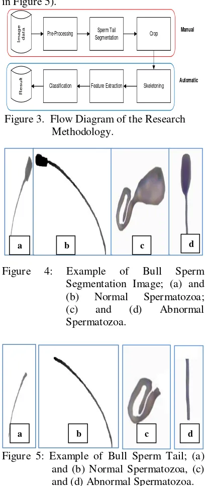
The fifty images obtained were processed. The process followed the steps as depicted in Figure 3. Manual part of Figure 3 were to separate sperm from the background (as in example of Figure 4) and to crop the tail (as in Figure 5).
Automatic
R
esul
t
Ima
ge
da
ta
Pre-Processing SegmentationSperm Tail Crop
Skeletoning Feature Extraction
Classification
Manual
Figure 3. Flow Diagram of the Research Methodology.
Figure 4: Example of Bull Sperm Segmentation Image; (a) and (b) Normal Spermatozoa;
(c) and (d) Abnormal
Spermatozoa.
Figure 5: Example of Bull Sperm Tail; (a) and (b) Normal Spermatozoa, (c) and (d) Abnormal Spermatozoa.
a b c d
a b c d
Figure 6: Example of Bull Sperm Tail Skeleton Image; (a) and (b) Normal Spermatozoa; (c) and (d) Abnormal Spermatozoa.
The automatic part of Figure 3 starts with using medial axis transform method to get the skeleton of the segmented tails as shown in Figure 6. Features were extracted from the skeletons using polynomial curve fitting. The skeleton were fit using a polynomial of corresponding pixel of the obtained polynomial, known as norm of residual, were calculated using Equation (2).
curve length constitutes the features extracted from the data in this study.The next step were classification of the features obtained above using support vector machine. SVM implementation in this study uses linier function. For example: {x1,...,xn}
is dataset and yiЄ{+1, -1} is class label from
xi data. The two classes are separated by pair
of field barrier. Field barrier of class 1 is position to the centre of coordinate. Whereas margin value is distance between field barrier of class 1 and class 2 that is shown in Equation (5).
RESULT AND DISCUSSION
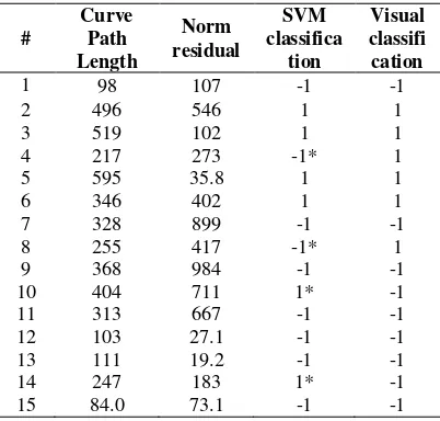
For the purpose of training of the linear SVM classifier, 35 data were selected. Twenty five out of the 35 known to be normal data (shown in Table 1) while 10 out of the 35 known to be abnormal data (shown in Tabel 2).
Table 1. Twenty five Training Normal Sperm Data (in pixel x 102).
Table 2. Ten training Abnormal sperm data (in pixel x 102). were then classified using the support vector to be normal (+1) or abnormal (-1). The result of SVM classification is shown in column 4 of Table 3. Visual classification is also shown in column 5 for comparison.
Table 3. Comparison of SVM and Visual classified incorrectly. Those are samples number 4, 8, 10, and 14. This gave accuracy of 73.33 %.
CONCLUSSION
This classification result were confirmed by researchers at Loka Penelitian Sapi Potong Grati, Pasuruan.
In conclusion, applying SVM for determining the abnormalities of bull sperm based on tail morphology in this study has shown good results. Simplicity of the classification process both training data and testing data with SVM provided some advantages in terms of design and speed.
However, evaluation and improvement of the method need to be carried out classes in the classification process rather than only two classes, normal and abnormal
REFERENCES
[1] L. Jun, L. Clemen, Z. Lu, and S. Yu,
“Quantitative Analysis of Locomative Behavior of Human Sperm Head and Tail,” IEEE Transaction on Medical Engineering, vol. 60, no. 2, pp. 390-396, 2013.
[2] M. Hidalgo, I. Rodriguez, J. Dorado,
and C. Soler, “Morphometric
classification of Spanish thoroughbred stallion sperm heads,” Animal Reproduction Science, vol. 103, no. 3, pp. 374–378, 2008.
[3] M. F. Skowronek, J. Alciaturi, G. Casanova, A. Capurro, J.M. Montes, and R. Sapiro, “Value of Quantitative Ultramorphological Sperm Analysis in Infertile Men,” Reproductive Biology, vol. 10, no. 2, pp. 125- 139, 2010.
[4] A. R. P. Yowell, “The Use of Computer Assited Semen Analysis to Predict Fertility in Holstein Bulls,” Master Thesis, Colorado State University, Fort Colins, 2011.
[5] G. C. Ostermeier, R. Sartor-Bergfelt, J. L. Susko-Parrish, and J. J. Parrish, “Bull
Fertility and Sperm Nuclear Shape,” AgBiotechNet, vol. 2, pp. 1- 6, 2000.
[6] C. Alvarez, J. A. Castilla, J.P. Ramirez, F. Vergara, A. Yoldi, A. Fernandez, and J. J. Gaforio, “External Quality Control Program for Semen Analysis: Spanish Experience,” Journal of Assisted Reproduction and Genetics, vol. 22, pp. 379 – 387, 2005.
[7] P. J. Chenoweth, “Genetic Sperm Defects,” Theriogenology, vol. 64, pp. 457–468, 2005.
[8] B. Hafez, Reproduction in Farm Animal, 7th edition. Baltimore, USA: Lippincot William and Walkins, 2000.
[9] Y. Wildan, Biologi Modern Histologi. Bandung: Tarsito, 2011.
[10] G. E. Freneau, P. J. Chenoweth, R. Ellis, and G. Rupp, ”Sperm Morphology of Beef Bulls Evaluated by Two Different
Methods,” Animal Reproduction
Science, vol. 118, pp. 176–181. 2010.
Moments-based Classification of the
Acrosome Integrity of Boar
Spermatozoa Images,” Computer
Methods and Program in Biomedicine, vol. 108, no. , pp. 873-881, 2012.
[12] A. Bijar, “Sperm’s Tail Identification and Discrimination in Cicroscopic Image of Stained Human Semen Smear,” in Proceedings of 7th International Symposium on image and Signal Processing and Analysis (ISPA), 2011.
[13] Enrique, B. Michael, P. Nicolai, S. Lidia, “Automatic Classification of the Acrosome Status of Boar Spermatozoa using Digital Image Processing and LVQ”, Computer in Biology and Medicine, vol. 38, pp. 461-468, 2008.
[14] T. Rao, N. Rajasekar, and T. V. Rajinikanth, “Classification of Remote
sensed Data Using Linear Kernel Based Support Vector Machines,” in Proceedings of 2013 International Conference on Control Communication and Computing (ICCC), 2013.
[15] M. A. Alolfe, W. A. Mohamed, A. B. M. Youssef, A. S. Mohamed, and Y. M. Kadah, “Computer Aided Diagnosis in Digital Mammography using Combined Support Vector Machine and Linear Discriminant Analysis Classification,” in Proceedings of 16th IEEE International Conference on Image Processing (ICIP), 2009.
K. Z. Arreola, J. Fehr, and H.Burkhardt,