Indrawati, Rahmayani, Soeripto, Department of Anatomic Pathology, Faculty of Medicine, Gadjah Mada University.
Susi Hariyati, Department of Pediatric, Sardjito General Hospital, Yogyakarta
Pancreatoblastoma : A case report
Indrawati1, Rahmayani1, Susi Hariyati2, Soeripto1
1Department of Anatomic Pathology, Faculty of Medicine, Gadjah Mada University
2Department of Pediatric, Sardjito General Hospital, Jogjakarta
ABSTRACT
Indrawati, Rahmayani, Susi Haryati, Soeripto - Pancreoblastoma : A case report
A 2.5 year old female child was admitted to Dr. Sardjito Hospital with complaint of enlarging abdomen since 6 months. She was apparently well until 3 months prior to admission when she had anorexia and weight lost. Physical examination revealed a solid intrabdominal mass (15x13 cm) with smooth surface and no tenderness. USG of the abdomen showed a large mass in paraaortal region and pushed the aorta. Operation was done and during exploration the mass was located as high as the stomach level and extended into the pancreas. Gross examination showed the tumor was 11x11x8 cm, encapsulated, nodular, white cut surface, some parts were brown and most of them were fragile. The diagnosis of pancreoblastoma was established by immunohistochemical examination.
The presenting features of pancreoblastoma are generally nonspecific and clinically difficult to distinguish from other intraabdominal tumors such as neuroblastoma, non-Hodgkin lymphoma, Wilms tumor, hepatoblastoma and desmoblastoma. Some clinical tests might suggest these tumors, i.e. multiorgan involvement for non-Hodgkin lymphoma; renal origin, the propensity for venous invasion and for the pulmonary metastasis for Wilms tumor. a–feto protein that is positive in either hepatoblastoma or pancreoblastoma cannot differ both tumors. Another intrabdominal tumor that should be considered is desmoblastoma that positive vimentin stain. The positive CAM5.2. as well as cytokeratin and the negative vimentin in immunohistochemical examination confirmed the diagnosis of pancreoblastoma.
Keywords : pancreoblastoma – child – intraabdominal tumors – immunohistochemical examination
ABSTRAK
Indrawati, Rahmayani, Susi Haryati, Soeripto - Pancreoblastoma : Laporan kasus
Seorang anak perempuan berumur 2.5 tahun dirawat di Rumah Sakit Dr. Sardjito dengan keluhan perut semakin membesar seajk 6 bulan. Sebelumnya anak tampak sehat sampai 3 bulan sebelum masuk rumah sakit, waktu itu dia mulai anoreksia dan berat badannya menurun. Pemeriksaan fisik menunjukkan adanya massa intraabdominal padat (15x13 cm) dengan permukaan rata dan tidak nyeri tekan. Pemeriksaan USG abdomen menemukan satu massa yang besar, di regio para-aorta dan mendesak aorta. Tindakan bedah dilakukan, dan pada waktuj eksplorasi ternyata massa itu terletak setinggi lambung dan meluas ke dalam pancreas. Pemeriksaan luar menunjukkan tumor berukuran 11x11x8 cm, berkapsul, nodular, permukaan potongan berwarna putih, beberapa bagian coklat dan kebanyakan rapuh. Diagnosis pancreoplastoma ditegakkan dengan pemeriksaan immunohistokimiawi.
INTRODUCTION
Pancreatoblastoma is a rare pancreatic tumor with distinct acinar and squamoid cell differentiation that generally affected infants and young children. This tumor accounts for approximately 0.5 % of pancreatic non endocrine tumors and just over 50 cases have been reported in the literature till 1997.1
Pancreatoblastoma has been first reported in the surgery literature by Becker in 1957. The earliest histopathologic descriptionwas published by Frable et al. in 1971. Although the tumorwas initially referred to as “infantile carcinoma of the pancreas,”its histologic resemblance to fetal pancreatic tissue at approximately 7 weeks gestation led Horie et al., in 1977, to propose that the original name be replaced with pancreato-blastoma. Pancreatoblastoma has since become the accepted term and is used interchangeably with pancreaticoblastoma.2,3
Pancreatoblastoma usually affects patients between the age of 1 and 8 years, but it has been reported in neonates and in the elderly.4,5 The congenital form is associated with Beckwith-Wiedemann syndrome and has been described as being cystic. The tumor is slightly more common in male patients, and half of all cases reported in the literature occurred in Asians.5,6
The etiology of pancreatoblastoma is unknown and the head of the gland is affected in about 50% of cases7 The clinical presentation of pancreatoblastoma was variable. Typically, pancreatoblastoma is slow-growing, clinically occult,and quite large at the time of diagnosis.8 Symptoms, when present, are usually related to the mass effect and includeearly satiety, vomiting, constipation, and pain. Cushing syndrome and the syndrome of inappropriate antidiuretic hormone secretion have been reported.These endocrine syndromes are postulated to be the result of adrenocorticotrophic hormone secretion by the tumor.9 Laboratory values may reveal elevated serum levels of a-fetoprotein, a-antitrypsin, or
lactate dehydrogenase .10 Local spread, including omental and peritoneal disease and vascular invasion, has been described. The liver is the most common site for metastasis. Other sites include the lung, bone and posterior mediastinum. 11
Therapy of pancreatoblastoma consists of surgical resection of the localized tumor.The role
is uncertain.12 These tumors tend to have a good prognosis, accounting only for 0.2% of deaths for malignancies in this age13, although poor results and occasional distant metastases (lung, liver or lymph nodes)14 have been reported. Considering the favourable prognosis, pancreatoblastoma should be differentiated from other aggressive tumors in children
A case of pancreatoblastoma is reported and its differential diagnosis will be discussed in this article
CASE
Clinical history and examination
A 2.5 year old child male was admitted to Dr. Sardjito Hospital with chief complaint of an enlarging abdominal mass. Since six months prior to referral, she had a mass in her abdomen which became larger rapidly. She was apparently well until three months prior to referral when she had anorexia and weight lost.
Clinical examination revealed a solid intra-abdominal mass (15 x 13 cm), with smooth surface and no tenderness. USG of the abdomen revealed a large mass which cannot be reached by trans-ducer in para aorta region and pushed the aorta. During exploration, the mass was revealed located as high as the stomach level and extended into the pancreas. There was no abnormality of the kidney and liver.
Gross examination : Tissue 11x11x8 cm, encapsulated, nodular and fragile with white cut surface, some parts were brown and most of them were fragile (FIGURE 1).
Five slides from different areas were made for microscopic examination by routine Hematoxylin-eosin staining and one slide was examined by PAS staining. Three blocks of parrafine embedded tissue were sent to The Department of Pathology, AMC, The Netherlands, for confirmation of the diagnosis and immuno-histochemical examination.
and rosette-like formation. The tumor cells were small, round and oval with vesicular round nuclei and scanty cytoplasm. The other solid area consisted of tumor cells with fusiform and hyperchromatic nuclei. Several areas showed marked necrosis. Infiltration of tumor cells into fibrous capsul tissue and to some vessels were also noted (FIGURE 2-3). Part of tumor cells showed PAS positive staining.
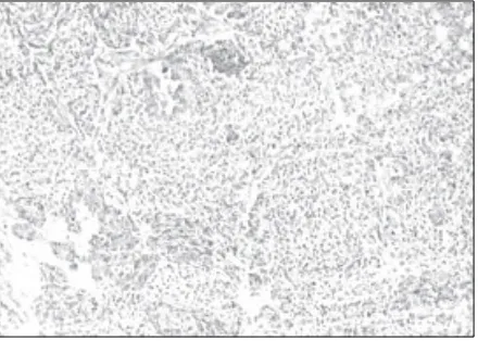
Immunohistochemical examination showed expression of CAM5.2 and in some of the tubular structures showed expression of cytokeratin 7. Vimentin, desmin and neuroendocrine markers were negative (FIGURE 4-5).
CONCLUSSION
Intraabdominal mass : Pancreatoblastoma
FIGURE 2. Microscopic feature of pancreatoblastoma (HE staining, 100X)
FIGURE 1. Macroscopic feature of pancreatoblastoma
FIGURE 3. Microscopic feature of pancreatoblastoma (HE staining, 400X)
FIGURE 4. Positive expression of cytokeratin shows brown cytoplasmic staining
DISCUSSION
The presenting feature of pancreatoblastoma are generally nonspesific. Especially in the pediatric age group, many patients present with an inciden-tally detected abdominal mass. Clinically, distinguishing pancreatoblastoma from other pediatric abdominal masses might be difficult, particularly when the tumor is large and when its origin is uncertain. In such cases, the differential diagnosis in a child includes a consideration of any large intra- or retroperitoneal mass, such as a neuroblastoma, non-Hodgkin lymphoma, or Wilms tumor.2 Neuroblastoma often appears as a large, heterogeneous, calcified abdominal mass. The distinction from pancreatoblastoma can be made clinically on the basis of the predilection for neuroblastoma in very young patients, the characteristic paraneoplastic syndromes and the presence of urine catecholamines. Non-Hodgkin lymphoma may present as a large, abdominal soft-tissue mass or masses. The high incidence of multiorgan involvement at presentation, may help to distinguish non-Hodgkin lymphoma from pancreatoblastoma. A Wilms tumoris another potentially large, heterogeneous, retroperitoneal tumor that occurs in young children. However, its renal origin, its propensity for venous invasion, and its pulmonary metastases help to distinguish it from pancreatoblastoma. 2,15
When the tumor appears to arise from the liver, pancreatoblastoma may resemble other primary pediatric hepatic neoplasms, such as hepatoblastoma.2 Hepatoblastoma is the most common malignant tumor of the liver in children. Nearly 90 % of hepatoblastoma are seen in the first 5 year of life, with 60% presenting in the first 2 years.16 Most patients present with an enlarging abdominal mass. The right lobe is involved three times more commonly than the left, with bilobar involvement seen in 20%-30%, and multicentric involvement in 15%. Less common symptoms are anorexia, weight loss, and pain. Hepatoblastoma is classified by histology as epithelial (56%) or mixed epithelial/mesenchymal (44%).17 Epithelial hepatoblastoma is further broken down to pure fetal (31%), embryonal (19%), macrotrabecular (3%) and small-cell undifferentiated (3%) . The most common mesenchymal elements are osteoid and
been associated with improved prognosis in patients with advanced disease. In completely resected tumors, pure fetal histology confers a better prognosis, while small-cell undifferentiated histologyis associated with a poor prognosis. 17,18,19 The distinction of pancrea-toblastoma from hepapancrea-toblastoma may be difficult as both tumors may occur with an elevated level of a
-fetoprotein. In addition, histopathologhical feature of hepatoblastoma especially mixed epithelial/mesen-chymal type may resemble with pancreatoblastoma in this reported case. However, in contras to pan-creatoblastoma, immunohistochemical examination of vimentin was weakly positive in those hepato-blastomas where mesenchymal tissue was present in the tumor.20,21
by a polyphenotypic profile with expression of epithelial, mesenchymal and neural markers. All tumors stain for epithelial markers, including cytokeratins and epithelial membrane antigen. In addition, although the most useful diagnostic marker is desmin, virtually all desmoplastic small round cell tumors stain for vimentin.29 The positivity of vimentin can differentiate desmoplastic small round cell tumor from pancreatoblastoma.
CONCLUSION
A case of pancreatoblastoma was reported. The positivity of CAM5.2, cytokeratin and negativity of vimentin examined by immuno-histochemical staining can differentiate pancreato-blastoma from hepatopancreato-blastoma and desmoplastic small round cell tumor .
REFERENCE
1. Chun Y, Kim W, Park K, Lee S, Jung S. Pancreatoblastoma. J Pediatr Surg 1997 ; 32 : 1612-65
2. Montemarano H, Lonergan GJ, Dorothy I. Bulas DI, Selby DM. Pancreatoblastoma: Imaging Findings in 10 Patients and Review of the Literature. Radiology 2000; 214:476-82 3. Horie A, Yano Y, Kotoo Y. Morphogenesis of pancreatoblastoma, infantile carcinoma of the pancreas: report of two cases. Cancer 1977; 39: 247-54
4. Levey JM, Banner BF. Adult pancreatoblastoma: a case report and review of the literature. Am J Gastroenterol 1996; 91:1841-44.
5. Klimstra DS, Adair CF, Hefess CS. Pancreatoblastoma: a clinicopathologic study and review of the literature. Am J Surg Pathol 1987; 11:855-65.
6. Drut R, Jones MC. Congenital pancreatoblastoma in Beckwith-Wiedemann syndrome: an emerging association. Pediatr Pathol 1988; 8:331-39.
7. Hamilton SR, Aaltonen LA. Pathology and genetics of Tumors of The Digestive System. WHO Classification of Tumors, IARC 2001 ; 244-45
8. Mergo PJ, Helmberger TK, Buetow PC, et al. Pancreatic neoplasms: MR imaging and pathologic correlation. RadioGraphics 1997; 17:281-301.
9. Passmore SJ, Berry PJ, Oakhill A. Recurrent pancreato-blastoma with inappropriate adrenocorticotropic hormone secretion. Arch Dis Child 1988; 63:1494-96.
10. Willnow U, Willberg B, Schwamborn D. Pancreatoblastoma in children: case report and review of the literature. Eur J Pediatr Surg 1996; 6:369-72.
11. Ohaki Y, Misugi K, Hirose M. Pancreatic carcinoma of childhood: report of an autopsy and review of the literature. Acta Pathol Jpn 1985; 35:1543-54.
12. Dhebri AR, Connor S, Campbell F. Diagnosis, treatment and outcome of pancreatoblastoma. Pancreatology 2004; 4: 441-51
13. Tsukimoto I, watanabe K, Lin J. Pancreatic carcinoma in children in Japan. Cancer 1973 : 53 ; 963-69
14. Buchino JJ, Castello FM, Nagari HS. Pancreatoblastoma: a histochemical and ultrastructural analysis. Cancer 1984; 53:963-69.
15. Friedman AC, Edmonds PR. Rare pancreatic malignancies. Radiol Clin North Am 1989; 27:177-90
16. Stocker J T. Hepatic Tumors in Children in Suchy FJ, Sokol FJ, Balistreri WF. Editors. Liver Disease in Children. 2th ed. Lippincott Williams & Wilkins, 2001 : 927-35
17. Cynthia E. Herzog, Richard J. Andrassy, Farzin Eftekhari Childhood Cancers: Hepatoblastoma The Oncologist ; 5 : 445-53
18. Stocker JT. Hepatoblastoma. Semin Diagn Pathol 1994;11:136-43.
19. Haas JE, Muczynski KA, Krailo M et al. Histo-pathology and prognosis in childhood hepatoblastoma and hepatocarcinoma. Cancer 1989;64:1082-95.
20. O’Brien WJ, Finlay JL, Gilbert-Barness EF. Patterns of antigen expression in hepatoblastoma and hepatocellular carcinoma in childhood. Pediatr hematol Oncol 1989; 6: 361-65
21. Kimura N, Yonekura H, Okamoto H, Nagura H. Expression of human regenerating gene mRNA and its product in normal and neolastic human pancreas. Cancer 1992 ; 70 : 1857-63 22. Bismar TA, Basturk O, Grald WL, Schwarz K, Adsay NV. Desmoplastic small cell tumor in the pancreas. Am J Surg Pathol 2004 ; 28 : 808-12
23. Basade MM, Vege DS, Nair CN. Intra-abdominal desmoplastic small round cell tumor in children : a clinicopathologic study. Pediatr Hematol Oncol 1996; 13 : 95
24. Gerald WL, Miller HK, Battifora H. Intra-abdominal desmoplastic small round cell tumor: Report of 19 cases of a distinctive type of high-grade poly-phenotypic malignancy affecting young individuals. Am J Surg Pathol 1991 ;15: 499–513
25. Weiss SW, Goldblum JR. Enzingers and Weiss Soft Tissue Tumors. 4th ed. Mosby Company, 2001, 1538-45
26. Gerald WL, Ladanyi M, Alava E. Clinical, pathologic, and molecular spectrum of tumors associated with t (11;22) (p13;q12): Desmoplastic small round-cell tumor and its variants. J Clin Oncol 1998 ; 16: 3028–36.
27. Schwartz RE, Gerald WL, Kushner BH. Desmoplastic small round cell tumors: Prognostic indicators and results of surgical management. Ann Surg Oncol 1998 ; 5:416–22 28. Kushner BH, LaQuaglia MP, Wollner N. Desmoplastic small round cell tumor: Prolonged progression free survival with aggressive multimodality therapy. J Clin Oncol 1996; 14:1526–31