Research Journal of Pharmaceutical, Biological and Chemical
Sciences
Isolation of Endophytic Bacteria from Bark, Leaf, and Pericarp of Mangosteen
(
Garcinia mangostana
L.) and Testing of The Antimicrobial Activity.
Harrizul Rivai
1, Asia A
2, Rina W
2, Alen Y
1, Handayani D
1, Aldi Y
1, Marlina
1, and Akmal D
1*.
1
Faculty of Pharmacy, University of Andalas, Kampus Limau Manis, Padang 25163, Indonesia.
2
School of Pharmacy, Jl. Tamansiswa No. 9, Padang 25138, Indonesia.
ABSTRACT
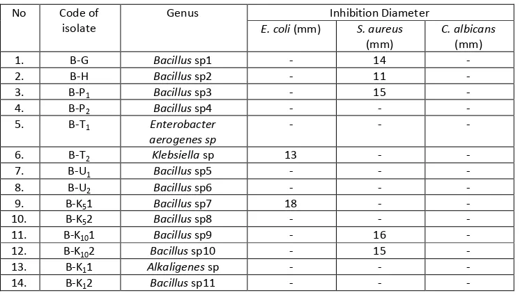
A research on antimicrobial activity testing of the endophytic bacteria which was symbiosis with mangosteen has been conducted. About fourteen isolates of bacteria from mangosteen have been isolated. Each bacterial isolates were fermented in aqueous media for 24 hours on a rotary shaker incubator at temperature of 30 oC and speed of 120 rpm. Each fermentation of bacterial isolates were tested their antimicrobial activity against microbes Staphylococcus aureus, Escherichia coli and Candida albicans with the agar diffusion method. There were two bacterial isolates that active against Escherichia coli and five bacterial isolates that active against Staphylococcus aureus.
Keywords:Garcinia mangostana L., endophytic bacteria, mangosteen, antimicrobial activity
INTRODUCTION
Search for the source of bioactive compounds is continued to be done along with the many new diseases are emerging. One source of bioactive compounds derived from microbes is endophytic microbes. Endophytic microbes can produce bioactive compounds that are potential to be developed into drugs. One of plant that is used to treat diarrhea, dysentery, and ulcers is mangosteen. The extract from mangosteen pericarp was effective against Staphylococcus aureus, Staphylococcus albus, and Micrococcus lutus. The strong anti bacterial activity of the extract suggests that it is a good drug of choice for which might be helpful in preventing the progress of various diseases and it can be used in alternative system of medicine [1]. There is a need to search new ecological niches for potential of natural bioactive agents for different pharmaceutical, agriculture and industrial application; these should be renewable, eco-friendly and easily obtainable natural products discovery in the search for new drugs, and is the most potent source for the discovery of novel bioactive compounds. Therefore, a large number of bioactive compounds are isolated from the plants, bacteria, fungi and many other organisms. Endophytic microbes being the most promising of these have been a source of various such bioactive compounds. Many of these compounds are being used for the treatment of a number of diseases [2]. The purpose of this study was to test the antimicrobial activity of the compounds isolated from endophytic microbes of mangosteen.
EXPERIMENTAL SECTION
Test drug and chemicals
The leaves, bark and pericarp of the mangosteen (Garcinia mangostana L.) were obtained from Lubuk Alung, West Sumatra, Indonesia. All other chemicals used were of analytical grade.
Isolation of endophytic bacteria
Each fresh plant was washed with running water. Then each part of the plant was cut to a size of 1 x 1 cm2. Furthermore, in the laminar air flow cabinet, each organ of the plant was disinfected of their surfaces by immersing the organ by successively in 70 % ethanol for 30 seconds, a solution of 5 % sodium hypochlorite for 5 minutes, 70 % ethanol for 30 seconds and rinse with distilled water for 3 minutes [3,4].
Each organ of the plant was planted using tweezers into a Petri dish containing media Nutrient Agar (NA) by splitting parts of the plant and put in prone position. Each Petri dish was planted 2-3 slices of the organ, incubation at temperature 37 oC for 18-24 hours. Bacteria that grow gradually were purified one by one. Colonies that have a different form with each other colonies can be considered different colonies [5, 6].
Antimicrobial activity test
Medium for production of antibiotics was prepared with the composition of corn soaking water (3 %), glucose (3 %), calcium carbonate (0.5 %), ferrous sulfate (0.1 %), magnesium sulfate (0.2 %), zinc sulfate (0.01%), and sterile distilled water added up to 100% [5]. The medium was heated to boiling and sterilized by autoclaving at 121 oC for 15 minutes. Each of isolates that had been purified in previous experiments was inoculated on the liquid medium that has been prepared. Each of the isolates was fermented. The fermentation process was done in 250-mL Erlenmeyer flask and incubated at temperature of 30 oC for 24 hours on a rotary shaker incubator at 120 rpm. Then the fermentation solution was centrifuged at a speed of 5000 rpm for 15 minutes. Supernatant was tested of antimicrobial activity against microbes using paper disc method. Paper discs was dipped in the supernatant and planted in the medium NA containing bacteria and in the medium PDA containing fungi. Then it was incubated at temperature of 37 oC for 18-24 hours. Barriers to growth were observed and measured its diameter by using a caliper. The results of centrifugation of each microbe were washed 3 times with distilled water, dried by air, so that the biomass was obtained [7].
Characterization of endophytic bacteria isolates
RESULTS AND DISCUSSION
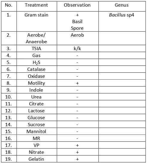
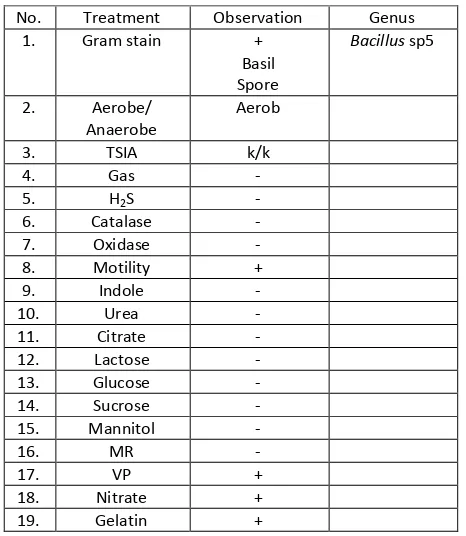
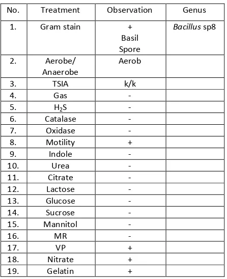
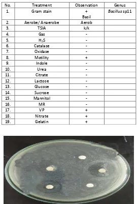
In the process of isolation of endophytic bacteria, it was used direct planting method in which pieces of plant organs which have been disinfected its surface were affixed to the media Nutrient Agar (NA) in the position of organ surface sticks to the media. This method is chosen because it is more practical and faster process [4, 6]. Bacteria that grow in media was purified and fermented to determine its ability to produce antimicrobial compounds. In this study, it was obtained 14 isolates of endophytic bacteria and biochemical test results were presented in Table11-14.
Table 1: Characteristics of endophytic bacteria isolated with BG code
No. Treatment Observation Genus
1. Gram stain +
Table 2: Characteristics of endophytic bacteria isolated with BH code
No. Treatment Observation Genus
Table 3: Characteristics of endophytic bacteria isolated with B-P1 code
No. Treatment Observation Genus
1. Gram stain +
Table 4: Characteristics of endophytic bacteria isolated with B-P2 code
No. Treatment Observation Genus
1. Gram stain +
Table 5: Characteristics of endophytic bacteria isolated with B-T1 code
No. Treatment Observation Genus
1. Gram stain -
Coccus
Enterobacter aerogenes
3. TSIA m/k
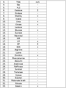
Table 6: Characteristics of endophytic bacteria isolated with B-T2 code
No. Treatment Observation Genus
25. Arabinose -
Table 7: Characteristics of endophytic bacteria isolated with B-U1 code
No. Treatment Observation Genus
1. Gram stain +
Table 8: Characteristics of endophytic bacteria isolated with B-U2 code
No. Treatment Observation Genus
Table 9: Characteristics of endophytic bacteria isolated with B-K51 code
No. Treatment Observation Genus
1. Gram stain +
Table 10: Characteristics of endophytic bacteria isolated with B-K52 code
No. Treatment Observation Genus
1. Gram stain +
Table 11: Characteristics of endophytic bacteria isolated with B-K101 code
No. Treatment Observation Genus
3. TSIA k/k
Table 12: Characteristics of endophytic bacteria isolated with B-K102 code
No. Treatment Observation Genus
1. Gram stain +
Table 13: Characteristics of endophytic bacteria isolated with B-K11 code
No. Treatment Observation Genus
14. Sucrose -
Table 14: Characteristics of endophytic bacteria isolated with B-K12 code
No. Treatment Observation Genus
1. Gram stain +
Table 15: Inhibition caused by fermentation liquid of endophytic bacteria
Table 16: pH of Media and Biomass of Bacteria after 48 hours cultivation
Isolate pH of Media Biomass (gram)
B-H 6.71 0.080
Table 15 showed that there were two bacterial isolates (B-T2 and B-K51) resulted in inhibition against
test bacteria E. coli and five isolates (BH, BG, B-P1, B-K101 and B-K102) resulted in inhibition against S. aureus.
In testing of antimicrobial activity of compound produced by endophytic bacteria against fungi
Candida albicans there was no inhibition due to Candida albicans including eukaryotic microorganisms, whereas pathogenic microbes used to test antimicrobial activity were Gram-positive and Gram-negative including prokaryotic microbes. There are difference of structure of cell wall components in eukaryotic especially Candida albicans. In eukaryotic the cell wall structural components contain chitin, cellulose or glucans, whereas in prokaryotic the cell wall component contain peptidoglycan. In addition metabolite produced was not suitable for eukaryotic microorganisms as secondary metabolites produced cannot damage the cells of Candida albicans [7]. In addition the limitations of antifungal drug discovery than antibacterial drugs this is due to the fungus associated with complex cell structure than bacteria [8].
do not have antimicrobial activity and the possibility of these isolates have other activities such as poison, giving color or pigment, growth agents, pesticides [7].
Table 16 showed that the pH of Media and Biomass of Bacteria after 48 hours cultivation. There was the influence of pH changes on filtrate endophytic microbes. Endophytic microbes decreasing in pH of the filtrate showed the role of the production of antimicrobial which can inhibit the growth of Escherichia coli and Staphylococcus aureus [9]. The addition of biomass of each endophytic microbe also has an influence on the growth of Escherichia coli and Staphylococcus aureus. The results showed that the antimicrobial synthesized in its infancy. Initial growth was seen when a microbial cell inoculated on nutrient agar was enlarging in size, volume and weight of the cell. The cells were continued to divide exponentially. As long as conditions allow, the growth and division of cells takes up a number of established cell population. The more cells that grow, the antimicrobial activity was increasing along with the metabolites excreted in the form of antimicrobial compounds out of the cell [9,10].
CONCLUSION
About fourteen isolates of bacteria from leaves, bark, and fruit peel of mangosteen (Garcinia mangostana L.) have been isolated. There were two bacterial isolates that active against Escherichia coli and five bacterial isolates that active against Staphylococcus aureus. There was no bacterial isolate that active against Candida albicans.
ACKNOWLEDGEMENTS
The authors thanks to all of participants for the help and concern for the realization of this paper. We also thanks to Balai Veteriner Bukittinggi, West Sumatra, Indonesia for the help of identification and characterization of the isolated bacteria.
REFERENCES
[1] Priya V, M Jainu, SK Mohan, P Saraswathi and SC Gopan. Int J Pharm Sci Res 2010;1(8):278-281. [2] Kumar S, RP Aharwal, H Shukla, RC Rajak and SS Sandhu. World J Pharm and Pharm Sci 2014;3(2):
1179-1197.
[3] Pradeepa V and M Jennifer. Advanced Bio Tech 2013;13(4):12-17.
[4] Aldi Y, Y Yuliandra, E Nasrul, Yanwirasti, D Handayani, A Bakhtiar. Res J Pharm Biol Chem Sci 2015; 6(4):1823-1829.
[5] Akmal D, MR Marjoni, I Friardi. Res J Pharm Biol Chem Sci 2015; 6(3): 583-591. [6] Sinaga E, Noverita and D Fitria. Jurnal Farmasi Indonesia 2009;4(4):161-170.
[7] Handayani D, N Sandrawaty, M Murniati, R Regina. J App Pharm Sci 2015; 5 (09): 139-142 [8] Cowen LE, JB Anderson and LM Kohn. Annu Rev Microbiol 2002;56:139-165.
[9] Radji M, A Sumiati, R Rachmayani, B Elya. Afr J Biotechnol 2011;10(1):103-107