Docking Simulation of Fevicordin into Human Receptor Estrogen α*
1
Muchtaridi, 1Jerry Voldo F.M., 2Abdul Mutalib, 1Jutti Levita
1
Faculty of Pharmacy, Universitas Padjadjaran, Bandung
2
Radioisotope and Radiopharmaca Centre of BATAN, Serpong
ABSTRACT
Fevicordin have similar structure to estradiol and cytotoxic in nature, thus can be developed as cancer drug. Docking method can be applied to simulate it interactions with
human estrogen receptor α which is known involve in growth of breast cancer.
Information from simulation can be used to optimize its activity on target. Docking method was validated with redocking of x-ray estradiol structure and modelled estradiol into x-ray estradiol cognate binding pocket. Fevicordin 3D structure modelled with molecular mechanic in MM+ force field, then its unique conformers modelled with molecular dynamic simulation in same force field. Each conformers docked into x-ray estradiol binding pocket (SP). Alternative pocket (AP) was searched using Q-SiteFinder software and each of fevicordin’s conformers was docked into that pocket. Validation of docking method showed that AutoDock was valid to predict binding mode of x-ray estradiol and modelled estradiol and value of both predicted Ki was in prediction range. Modelled fevicordin was a planar conformation relative to estradiol and it has three conformers which were K1, a planar form, K2 and K3, both were bend form. Conformer K2 bind well into SP relative to K1 and K3 with predicted binding free energy -9,80 kcal/mol and Ki 65,8 nM. Alternative pocket (AP) volume was 460 Å3 and located in groove formed by helix 3, helix 5 and loop 1 of hERα LBD. Conformer K3 bind well into AP relative to K1 and K2 with predicted binding free energy -8,48 kcal/mol and Ki 612 nM. Binding affinities comparison of fevicordin at SP and AP showed that fevicordin’s binding tendency into AP, thus fevicordin was predicted unable to compete with estradiol.
Key word : Fevicordin, estrogen receptor, docking, estradiol
Introduction
Fevicordin is steroidal compound isolated from mahkota dewa seed (Phaleria marcocarpa (Scheff) Boerl.) and had anticancer activity. The previous research, fevicordin compound was isolated from mahkota dewa (Phaleria macrocarpa Scheeft Boerl) seeds. The compound have citotoxicity activity. IC50 value of fevicordin on Hela
cell, CASKI, and TE-2 cell were 1,25 µg/mL, 12 µg/mL, and 40 µg/mL, respectively (Diantini, et al., 2007). On the other hand, fevicordin had citotoxicity on T47D (IC50 <
0.1 µg/ml) and on MCF7 (38.81 µg/ml) cell lines.
However, besides that compound is very toxic on normal cell of fibroblast (Diantini et al., 2007). Looking at the structure, this compound has –OH groups in C-2 position and what make it diferent is other estradiol derivate is the core structure of estradiol bounded in 3-heptenone (Figure 1.). estrogen receptor (ER) ligand pharmacophore which were hidroxy group substituted aromatic ring (Duax et al.., 1985) was present in this compound indicated ER binding ability. Its potentiality to cure breast cancer. ERα was known to involve in breast cancer growth (Fujita et al.., 2003).
One of the problem in this research is fevicordin has specificity and selectivity to estrogen receptor which is same or stronger if it is compared to estradiol, therefore further toxicity of this compound can be used as estradiol agonis to kill cancer cell in mamae tissue. CH3 HO OH Me HO HO OH Me O Me Me Me OAc OH O H Me Me
(a) (b)
Figure 1. Structure of estradiol (a) and fevicordin from mahkota dewa seeds (b)
Materials and Methods Materials
Ligand binding domain 3D structure, 2,9 angstrom of wild-type ERα, cocrystalized with estradiol by Eiler et al (2001), obtained from Protein Data Bank online database (www.rcsb.org) with access code 1G50. Fevicordin structure obtained from Diantini (Diantini et al., 2007).
Tools
was used to predict location and volume of binding pocket. OpenBabel v2.1.0 was used to convert HIN file format into PDB file format.
Methods
Docking Method Validation
Cocrytalized estradiol was separated from chain A of LBD ERα, polar hydrogens, partial charge and solvation parameter was assigned to chain A LBD ERα while hydrogen, partial charge was assigned to ligand; non polar hydrogens was merged. X-ray estradiol was docked into LBD ERα with run variation: 10, 30, 50, 70 run in (60)3 Å each grid dimension, and (40)3 Å, (60)3 Å, (80)3 Å, (100)3 Å grid dimension variation in 50 run each. Docking method valid if RMSD equal or less than 2 Å (Ali et al., 2007).
Fevicordin modeling
Fevicordin’s 3D structure built with Hyperchem v7.0 then minimized in MM+ force field to 0,1 kcal/mol convergention through Polak-Ribiere algorithm. Hyperchem format file of this model was converted into PDB file format with OpenBabel.
Fevicordin’s conformers modeling
Fevicordin’s model heated in 100 fs from 0 K to 300 K then assigned to 300 K’s dynamic molecular simulation for 4000 fs. Potential energy and total energy of system saved in 1 fs range. Conformers was sampled in each 100 fs then saved in separated file without energy minimization. Conformers was grouped based on torsional angle C10-C9-C17 and uniqueness ring B and C. Lowest energy conformers from each group was saved in separated file. Those conformers then called fevicordin’s unique conformers. Conformers file format then converted into PDB file format with OpenBabel.
Docking simulation of unique conformer into x-ray estradiol binding pocket
Unique conformers and LBD ERα prepared using same procedure in validation procedure. Grid was built in former binding pocket with dimension (70)3 Å to covered all important residues involved in binding process. AutoGrid 3.0.5 was used to generate grid maps of oxygen, polar hydrogen, aromatic and alifatic carbon. One hundred run of Lamarckian Genetic Algorithm was used to dock each of fevicordin unique conformers into LBD ERα thus most of ligand conformation in binding pocket could observed. Docking result saved in dlg file then analyzed to get informations about ligand orientation, binding energy value and Ki value.
Alternative binding pocket searching
Alternative pocket searching was done with Q-Site Finder online (www.bioinformatics.leeds.ac.uk/qsitefinder/). ERα PDB file 1G50 chain A without estradiol was used as searching input. Search result are biggest predicted pockets and volume after steroid pocket which would used in docking simulation.
Docking simulation of fevicordin conformers to alternative pocket (AP)
Results and discussion Docking method validation
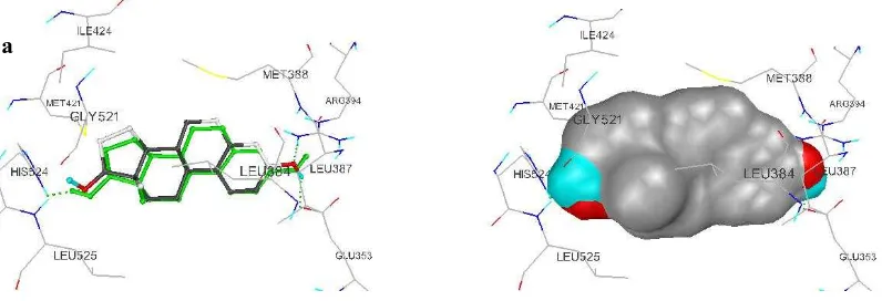
Estradiol binded to LBD hERα through van der waals interactions with Leu387, Phe404, Met388, Leu428, Met421, Ile424, Leu525 on ring A, B and D, hydrogen bond formed between oxygen OH3 and hydrogen NH Glu353 ((2,87 Å), oxygen OH3 with hydrogen NH Arg394 (2,72 Å) and nitrogen NH His524 (2,71 Å). All residues mentioned above form a pocket with volume 459 Å3.
Estradiol x-ray docking into its original binding pocket with run and grid dimension variations showed that AutoDock could predict estradiol x-ray original binding mode accurately with RMSD in all variation equal or less than 2 Å. Residue Phe404 and estradiol interaction can not identified in all run variation because their closest carbon distance is larger than 3,9 Å, even though, there are additional interaction between estradiol and Leu478 in 10 runs and 70 runs. Figure 1 showed example of x-ray estradiol docking with run variation.
Figure 1 (a) (right) Binding mode of estradiol at 30 runs. Estradiol x-ray colored green, (left) visualization of molecular surface with AutoDock Tools.
Figure 1 (b) (right) Binding mode of estradiol at grid dimension (80)3 Å, x-ray estradiol colored green , (left) visualization of molecular surface with AutoDock Tools.
a
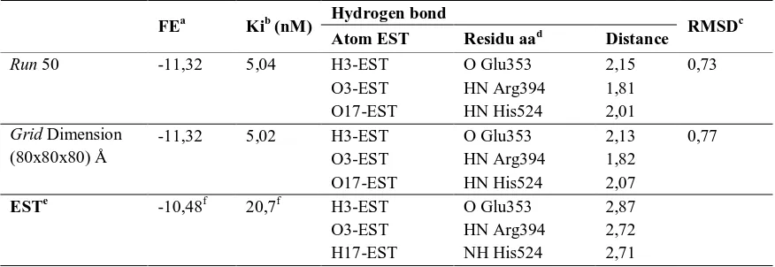
Table 1. Docking summarize x-ray estradiol at 50 runs and grid dimension (80)3 Å
All addtional interaction seems to decrease free binding energy compared to free binding energy calculated with AutoDock without docking process. All hydrogen bond distance less than 3 Å thus agree with good hydrogen bond proposed by Halperin (Halperin et al., 2002). This result showed that lamarckian Genetic Algorithm using in AutoDock 3.0.5 are efficient and efective to predict true binding mode of estradiol in grid dimension that cover all important residues with proper algorithm run amount, besides that in all variation, RMSD value are equal or less than 2 Å, then AutoDock are valid in docking simulation (Ali et al, 2007).
In grid dimension variation, most of true estradiol interactions can be observed with RMSD equal or less than 2 Å and increase of grid dimension equal to increase of binding free energy but doesn’t significant because average of increase is 0.02 kcal/mole compare to binding affinity. In grid dimension (80)3 interactions with Met421 doesn’t observed. Those result showed that docking simulation with AutoDock was valid (Ali et al., 2007). Example of docking result at 50 runs and grid dimension (80)3 Å listed in Table 2.
Hydrogen bond FEa Kib (nM)
Atom EST Residu aad Distance RMSD
c
Run 50 -11,32 5,04 H3-EST O Glu353 2,15 0,73
O3-EST HN Arg394 1,81
O17-EST HN His524 2,01
Grid Dimension -11,32 5,02 H3-EST O Glu353 2,13 0,77
(80x80x80) Å O3-EST HN Arg394 1,82
O17-EST HN His524 2,07
ESTe -10,48f 20,7f H3-EST O Glu353 2,87
O3-EST HN Arg394 2,72
H17-EST NH His524 2,71
a
Free energy (kcal/mol)
b
Inhibition constant
c Root mean square deviation d
Amino acid residues
e
x-ray estradiol bind to hERα LBD (PDB code:1G50)
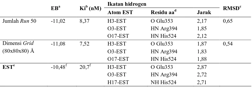
Table 2. Docking summarize modeled estradiol at 50 runs and grid dimension (80)3 Å
Fevicordin and its conformers modeling
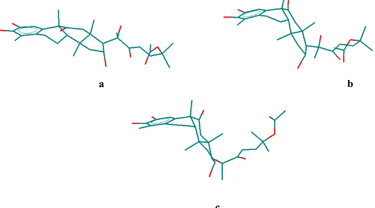
Fevicordin model was planar relative to estradiol shown in Figure 3, but there are bending in ring B and ring C junction because of substituent at C11. The angle of C10-C9-C17 is -32,135° while estradiol have angle 13,503°. Totalenergy of fevicordin model is 82,854 kcal/mole.
Dynamic molecular simulation is used to modeled fevicordin conformers because similar structure, which is estradiol 11Kβ, have four conformers using same method (Wiese dan Brooks, 1994). One of the predicted conformers of this estradiol analog is crystalography conformers. It is known that ER ligand structure affects its binding affinity and estrogenic activity (Palomino et al., 1994, Wiese dan Brooks, 1994).
Fevicordin have three unique conformers, coded K1, K2 and K3. K1 is relative planar (Figure 4 (a)) to K2 and K3 which are bended form with ”L” like structure (Figure 4 (b) and Figure 4 (c)).
Ikatan hidrogen EBa Kib (nM)
Atom EST Residu aad Jarak RMSD
c
Jumlah Run 50 -11,02 8,37 H3-EST O Glu353 2,17 0,65
O3-EST HN Arg394 1,85
O17-EST HN His524 2,12
Dimensi Grid -11,08 7,52 H3-EST O Glu353 1,87 0,54
(80x80x80) Å O3-EST HN Arg394 1,83
O17-EST HN His524 1,88
ESTe -10,48f 20,7f H3-EST O Glu353 2,87
O3-EST HN Arg394 2,72
H17-EST NH His524 2,71
a
Free energy (kcal/mol)
b
Inhibition constant
c
Root mean square deviation
d
Amino acid residues
e
x-ray estradiol bind to hERα LBD (PDB code:1G50)
f
Figure 4 (a) Conformation of K1, (b) Conformation of K2, (c) Conformation of K3.
Steroid bending on K2 and K3 caused by electrostatic interactions between C11 carbonyl group which have negative charge and C1 hydrogen atom which have positive charge.
Docking simulation of fevicordin’s unique conformation in steroid pocket (SP) Docking simulation of K1 on SP showed inverse orientation relative to estradiol (Figure 5). This orientation caused by unability of SP to accomadate ring D’s side chain of conformers, there are many steric clash in this region, such as O28 with Pro324 (2,75 Å) and OH20 (1,79 Å), C22 (2,37 Å) with Glu353, OH2 with Leu525 (2,55 Å), C28 with Ile424 (2,88 Å), OH16 with Leu387 (2,03 Å), because its shape match with ”benzene” like ring (Brzozowski et al., 1997). Conformers K1 formed four hydrogen bond with SP residues, listed in Table 3.
Binding free energy of K1 was predicted -2,85 kcal/mole with Ki value 0.01 M thus fevicordin not siutable to bind at SP. Figure 5 show K1 interaction at SP.
Conformers K2 bind with same orientation to K1. Steric clashes occur between K2 and residues of SP, such as Leu525 with oksigen OH2 (2,55Å), Ala350 with karbon
a b
C11 (2,39 Å), Arg 394 with carbonyl oxygen 22 (2,62 Å), side chain of ring D suffer same steric clash like K1. This steric clash show that complementary of SP with K2 is not good, even though, affinity of K2 is better than K1. Conformers K2 formed two hydrogen bond listed in Table 3. Figure 6 showed position of K2 in binding pocket.
Conformer K3 have same orientation and steric clashes relative to K2 but only one hydrogen bond formed because of slight shift of conformer relative to K2, thus decrease its stability in SP. Figure 6 showed K3 position in SP, Table 3 listed hydrogen bond formed.
Ka Ki (nM) EBb Residu-residu yang berkontak dekat
Ikatan hidrogen (donor→akseptor)
K1 107 -2,85 Gly521, Pro324, Ile386, Ile424, Leu387, Lys449, Met357, Leu384, Leu525, Met421, Glu353, His524, Phe404, Leu391, Arg394, Met388, Leu428
OH3→Gly521 (1,646 Å) Leu391→OH16 (1,646 Å) Arg394→O20(1,711 Å) Arg394→O22(1,754 Å)
K2 65,8 -9,80 Leu384, Leu387, Leu391, Phe402, Leu525, Gly521, Ile386, Arg394, Glu353, Trp383, Met388, Leu346, Met522, Lys449, Gly390, Pro324, Ala350
O23→Glu353 (1,94 Å) Arg394→H23 (2,22 Å)
K3 812 -6,94 Arg394, Leu387, Leu391, Met357, Thr347, Gly521, Leu346, Trp383, Ile386, Leu384, Phe404, Gly390, Met522, Glu353, Leu540, Leu525, Lys449, Ala350.
Arg394→OH16 (2,15 Å)
Conclusion
AutoDock was valid in this docking simulation with RMSD 2 Å. Fevicordin was planar relative to estradiol and predicted to bind at alternative pocket in binded conformation (K3).
Reference
Diantini, A.; D. Kurnia; A. Faried;L.S. Faried; A. Subarnas; T.H. Achmad; H. Hayashi; Supriyatna. 2007. Antiproliferative activity on hela and CASKI cells of fevicordin A isolated from the seeds of Phaleria macrocarpa. Proceeding International Conference On Traditional Medicine And Medicinal Plant. Surabaya 8-9 September 2007.
Ali, H. I. et al., 2007. Antitumor studies. Part 3: Design, synthesis, antitumor activity, and molecular docking study of novel 2-methylthio-, 2-amino and 2-(N-substituted amino)-10-alkyl-2-deoxo-5-deazaflavins. Bioorganic & Medicinal Chemistry.
Brzozowski et al. 1997. Molecular basis of agonism and antagonism in the oestrogen receptor. Letters to Nature 389:753-757.
Anstead, Gregory M., Kathryn E. Carlson, John A. Katzenellenbogen. 1997. The Estradiol Pharmacophore: Ligand structure-estrogen receptor binding affinity relationships and a model for the receptor binding site, Steroids 62:268-303.
Duax W.L., J.F. Griffin, C.M Weeks, K.S. Korach. 1985. Molecular Conformation, Receptor Binding, and Hormone Action of Natural and Synthetic Estrogens and Antiestrogens. Enviromental Health
Perspectives 61:111-121.
D’Ursi, Pasqualina, Erika Salvi, Paola Fossa, Luciano Milanesi, Ermanna Rovida. 2005. Modelling The Interaction Of Steroid Receptors With Encocrine Disrupting Chemicals, BMC Bioinformatics 6(Suppla 4):S10.
Fujita et al.., 2003. Full Activation of Estrogen Receptor α Activation Function-1
Induces Proliferation of Breast Cancer Cells. The Journal of Biological Chemistry Vol 278. 29:26704-26714
Good Sell, David S., Garrett M. Morris and Arthur J. Olson. 1996. Automated Docking of Flexible Ligands: Application of AutoDock, Journal of Molecular Recognition 9:1-5.
Hsieh, Robert W. et al., 2006. Identification of Ligands with Bicyclic Scaffolds Provides Insights into Mechanisms of Estrogen Receptor Subtype Selectivity. The Journal of Biological Chemistry 281(26):17909-17919.
Ikeda, Kazuhiro and Satoshi Inoue, 2004. Estrogen receptor and their downstream targets in cancer Arc Histol Cytol 67(5):435-442.
Jeyakumar et al., 2007. Raloxifene And ICI 182,780 Increase Estrogen Receptor Alpha Association with a Nuclear Compartment via Overlapping Sets Of Hydrophobic Amino Acids in AF2 Helix 12. Molecular Endocrinology
Katzenellenbogen, J.A., and B. Katzenellenbogen. 2000. Estrogen Receptor Transcription and Transactivation Estrogen Receptor Alpha and Eestrogen Receptor Beta: Regulation by Selective Estrogen Receptor Modulators and Importance in Breast Cancer. Breast Cancer Res 2:335-334.
Katzenellenbogen, John A., ‡, Rajeev Muthyala, and Benita S. Katzenellenbogen, 2003. Workshop 1.4. Nature of the ligand-binding pocket of estrogen receptor a and b: The search for subtypeselective ligands and implications for the prediction of estrogenic activity. Pure Appl. Chem. 75:2397-2403.
Lambrinidis, George et al., 2006. The Estrogen receptor and polyphenols:molecular simulation studies of their interaction, a review. Environ Chem Lett 4:159-174.
Morris et al. 1998. Automated Docking Using a Lamarckian Genetic Algorithm and an Empirical Binding Free Energy Function. Journal of Computational Chemistry 19:1639-1662.