The effect of organic selenium supplementation on the antioxidative
characteristics and lipid peroxidation of chicken blood during
fattening and after fasting
Jasna Piršljin*, Suzana Milinković-Tur, Blanka Beer Ljubić, and Maja Zdelar-Tuk
Department of Physiology and radiobiology, Faculty of Veterinary Medicine, University of Zagreb, Croatia
PiršLJin, J., S. MiLinković-Tur, B. Beer LJuBić, M. ZdeLar-Tuk: The
effect of organic selenium supplementation on antioxidative characteristics and
lipid peroxidation in chicken blood during fattening and after fasting. vet. arhiv 78, 187-196, 2008.
aBSTraCT
Selenium is an essential trace element in human and animal nutrition and an integral component of antioxidative proteins. Organic selenium, a natural form of selenium, has more beneficial effects in maintaining antioxidative system than its inorganic form.The objective of this study was to examine the effect of organic selenium food supplements on the level of glutathione peroxidase (GSH-Px), glutathione (GSH) and lipid peroxides (TBARS) in chicken whole blood. Chickens were randomly allocated into two groups: standard diets (<0.15 ppm sodium selenite) fed control group (n = 40) and Se+ group (n = 40), fed the same diets supplemented with 0.3 ppm organic selenium. Ten randomly selected birds from each group at the age of two, four and six weeks and after 48-hours of food deprivation at the end of the fattening period, were subjected to blood withdrawal from jugular vein. The GSH-Px activity and concentrations of GSH and TBARS were analyzed by spectrophotometry. A trend was observed toward increasing GSH-Px in the blood of the control group from two to six weeks of age (P<0.001), whereas in the Se+ group the increase was obtained in the first four weeks (P<0.01). GSH-Px activity after fasting was lower only in the control chickens (P<0.01). Organic selenium supplementation resulted in higher GSH-Px activity at two and four weeks of age (P<0.01), as well as after fasting (P<0.01). The GSH level in four week old Se+ chickens was lower than in two and six week old broilers (P<0.01). At the same time, in the two week old control chickens higher values were recorded in comparison with older birds (P<0.05). After fasting, the GSH was lower in both groups of chickens compared to values given after the end of the fattening period (P<0.001). The TBARS decreased in concentration in both groups at six weeks of age compared to younger chickens (P<0.05). Dietary organic selenium supplementation manifested higher activity of GSH-Px during fattening and maintaining its activity in stress conditions provoked by fasting.
key words: chicken blood, organic selenium, glutathione peroxidase, glutathione, lipid peroxide, fasting *Contact address:
Dr. Jasna Piršljin, PhD, DVM, Department of Physiology and Radiobiology, Faculty of Veterinary Medicine, University of Zagreb, Heinzelova 55, 10 000 Zagreb, Croatia, Phone: +385 1 2390 173; Fax: +385 1 2441 390; E-mail: pirsljin@vef.hr
Introduction
Selenium is an essential trace element in human and animal nutrition. The major form of selenium supplement for poultry feeds in the past 20 years has been inorganic selenium, as selenite and selenate. In contrast, in nature (cereals, forage) animals receive organic selenium that is bonded to the amino acids cysteine and methionine in the form of selenomethionine and selenocysteine (SURAI, 2002). Ingested selenomethionine is easily absorbed and incorporates into skeletal muscles, erythrocytes, pancreas, liver, kidney, stomach and gastrointestinal mucosa (SCHRAUZER, 2000). This allows selenium to be stored as a reserve during periods of increased demand or decreased selenium intake. On the other hand, inorganic selenium is capable of promoting superoxide radical formation and oxidative stress through its reductive reactions with reduced glutathione (SURAI, 2002) and provoking oxidative damages of DNA (WyCHERly et al., 2004).
More than 80% of the selenium in rat organs is present as selenocysteine (BURk, 1991). Selenocysteine in the active site of an enzyme increases enzyme activity from 100 to 1000 fold (BURk, 2002). Approximately half of this amino acid is in glutathione
peroxidase (GSH-Px). Until now five izoenzymes of GSH-Px have been isolated, four
members of the selenium containing-GSH-Px family and one non-selenium dependent form of GSH-Px. In blood cells citosolic GSH-Px and phospholipide hydroperoxide GSH-Px has been isolated (BROWN et al., 2000). In plasma extracellular GSH-Px was discovered, an isoenzyme which is synthesized in the kidneys and transported into the blood plasma (AVISSAR et al., 1994). The GSH-Px reduces lipidic and nonlipidic hydroperoxides as well as H2O2, while oxidizing two molecules of glutathione. The GSH-Px enzyme family also regulates prostaglandin and leukotriene synthesis, maintaining
cell redox potential, playing a role in signal transduction, inflammation and programmed
cell death (BRIGElIUS-FlOHE, 1999; IMAI and NAkAGAWA, 2003).
The reduced glutathione molecule (GSH) consists of three amino acids - glutamic acid, cysteine, and glycine. The GSH often attains milimolar level inside cells, which makes it one of the most highly concentrated intracellular antioxidants (MEISTER and ANDERSON, 1983). GSH is an electron donor in reactions catalyzed by GSH-Px, scavenges free radicals directly in nonenzymatic reactions and it is effective as a systemic antitoxin (kIDD, 1997). Glutathione reductase maintains a high concentration of reduced GSH in cell utilizing NADPH (MICHIElS et al., 1994).
The activity of GSH-Px as well as GSH concentration is species, strain and gender dependent (RIkANS and HORNBROOk, 1997; PIRšlJIN et al., 2006). In mammalian blood, the GSH concentration was found to be 2-3 (or more) times lower than that found in birds (SMITH, 1974; GRADINSkI-VRBANAC et al., 2002).
Synthesis of antioxidant molecules, which requires an adequate dietary supply of amino acids and dietary minerals, may well be impaired when animals are subjected to
prolonged stress, nutritional deficiency or starvation (GODIN and WOHAIEB, 1988). In rat food deprivation is associated with alterations in the free radical scavenging system that differ from tissue to tissue (WOHAIEB and GODIN, 1987) according to specific function
and tissue metabolism. Taking this into account, the purpose of the present study was to
determine the influence of organic selenium food supplements on the activity of GSH-Px,
GSH and lipid peroxide concentrations in the whole blood of broiler chickens, in response to age-related changes and 48 hour food deprivation at the end of the fattening period.
Materials and methods
animals and treatments. The experiment was performed on Ross 308 chickens. One hundred newly hatched broiler chickens were allocated in two pens (>200 cm2 per bird
from 1st to 14th day of fattening, 400 cm2 per bird from 15th to 28th day of fattening,
and >600 cm2 per bird from 29th day of fattening until the end of the experiment). The
ambiental temperature of the experimental room was set at 32 oC at the time of placement,
and over the 6 week growing period, the ambiental temperature was reduced to 20 oC.
lights were on continuously. Feed and water were provided for ad libitum consumption. The diets consisted of the starter diet - 12.85 MJ/kg ME, 22.5% CP (1-10 days of age)
grower diet - 12.85 MJ/kg ME, 18.5% CP (11-29 days of age), and finisher diet - 12 MJ/
kg 16% CP (30-42 days of age). Seven day old chickens were randomly allocated into two groups: standard diets (<0.15 ppm selenium as sodium selenite) fed control group and Se+ group, fed the same diets, supplemented with 0.3 ppm organic selenium (Sel Plex™, Alltech, Inc., ky).
Measurements and analyses. Ten randomly selected birds at the age of two, four and six weeks from each treatment were subjected to blood withdrawal from the jugular vein. Other chickens were deprived of food for 48 hours and blood was collected from ten broilers of each treatment group. Blood samples for GSH-Px ware frozen at -80 oC until
analyzed, whereas GSH, lipid peroxide and hemoglobin concentrations were determined immediately after sample collection. Blood for the GSH-Px activity (E.C. 1.11.1.9) was frozen and thawed three times, and enzyme activitywas measured using a commercial kit (Ransel, “Randox”, Uk). The concentration of GSH was determined by the method of BEUTlER et al. (1963). lipid peroxide concentration measured as thiobarbituric acid reactive substances (TBARS) was performed according to the method of TROTTA et al. (1982). Absorption peak was measured at 532 nm and concentration was calculated
using molar extinction coefficient of 1.5×10⁵ (PlACER et al., 1966). Haemoglobin was
measured spectrophotometricaly using commercially available kits from Herbos d.d. (Sisak, Croatia). All parameters were expressed per g of hemoglobin.
statistical analysis. All results are presented as mean ± SD. Inter-group comparisons were made by one-way analysis of variance fallowed by Tukey test using StatSoft, Inc.,
STATISTICA (data analysis software system), version 7. Student’s t-test was utilized to determine the effects of organic selenium supplementation and fasting. A probability level of P≤0.05 was considered statistically significant.
Results
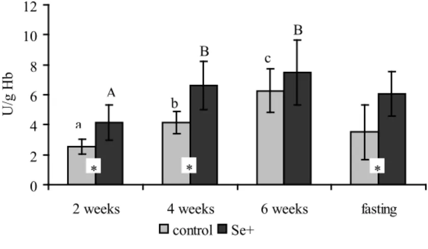
As can be seen from Fig. 2 and Fig. 3, organic selenium supplementation did not affect glutathione or lipid peroxide levels, whereas GSH-Px activity (Fig. 1) was higher
in the whole blood of Se+ broiler chickens both during the first four weeks of fattening
period (P<0.01), as well as after 48-hours of food deprivation (P<0.01). The GSH-Px
activity increases with age. Standard diet fed broilers show a significant increase of
GSH-Px activity during the six weeks of fattening (P<0.001). Simultaneously, in Se+ chickens,
a significant increase of GSH-Px activity was recorded until four weeks of age (P<0.01). Food deprivation resulted in significantly lower (P<0.01) GSH-Px activity only in the
control group of chickens (Fig. 1).
Fig. 1. Glutathione peroxidase activity in chicken blood during fattening and after 48 hours fasting for control group (standard diet) and Se+ group (organic selenium supplementation). Values are
expressed as means ± SD for n = 10. abcletters indicates significant differences among control
values (P<0.05), ABCletters indicate significant differences among Se+ values (P<0.05),
*Significant differences control vs. Se+ (P<0.05).
0 2 4 6 8 10 12
2 weeks 4 weeks 6 weeks fasting
U/g Hb control Se+ * * * a A b B c B
0 10 20 30 40 50 60
2 weeks 4 weeks 6 weeks fasting
nmol/g Hb
control Se+ a A
a A
b B
Fig. 2. Glutathione concentration in chicken blood during fattening and after 48 hours fasting for control group (standard diet) and Se+ group (organic selenium supplementation). See Fig. 1.
Fig. 3. lipid peroxide concentration in chicken blood during fattening and after 48 hours fasting for control group (standard diet) and Se+ group (organic selenium supplementation). See Fig. 1.
The GSH of Se+ chickens reached its lowest concentrations at the four weeks of age in whole blood (P<0.01). In the control broilers the highest GSH level was recorded
at two weeks of age (P<0.05). A significant decrease of the GSH occurred after food
The lipid peroxide concentration decreased in both groups at six weeks of age compared to younger chickens (P<0.05). Food deprivation did not affect lipid peroxide concentration either in the control or the Se+ group (Fig. 3).
discussion
Sel-Plex® is a selenized yeast product. It is an organic selenium, presented in the same
forms naturally present in plants (SCHRAUZER, 2000). The most important metabolic role of selenium is manifested in the activities of the selenoenzymes GSH-Px and thioredoxin reductase. The data from this study show a clear trend toward increasing blood GSH-Px in control and Se+ chickens during fattening. In contrast to our results, MAHMOUD and EDENS (2003) observed no effect of organic selenium supplementation in broiler chicken blood on GSH-Px activity during fattening, whereas in birds fed basal starter (26 ppm sodium selenite) activity decreased with age.
Organic selenium dietary supplementation in chicken increases GSH-Px activity in liver and blood plasma (ARAI et al., 1994) as well as erythrocyte (ARAI et al., 1994; AyDEMIR et al., 2000). In the present investigation, selenium supplementation caused an increase of GSH-Px activity in blood by about 60% in two and four week old chickens and about 20% at six weeks of age. Smaller elevation of GSH-Px activity in six week old chickens is possibly related to intensive increase of body mass and muscular tissue which is the main site where selenium is stored in the organism (SCHRAUZER, 2000). This is in
agreement with the previously published findings of DEAGEN et al. (1987). The authors
reported that 0.2 ppm selenomethionine as feed supplement resulted in 10-fold higher selenium concentration in the skeletal muscle of rats compared to selenite.
Food deprivation influences the cell oxidative balance. We report here that fasting
caused a decrease of GSH-Px activity in standard diets fed chicken, this decrease amounted to about 79%. In contrast to our results, WOHAIEB and GODIN (1987) observed
no influence of fasting on activity of that enzyme in rat erythrocytes.
Studies by ENkVETCHAkUl et al. (1995) on chicken showed that blood GSH
concentration is age-associated. They reported that in five week old chickens the GSH
is higher than in younger chickens. In the present study higher values in two week old chickens of both groups were recorded in comparison with older birds. These results are
in agreement with findings previously published by MAHMOUD and EDENS (2003). After
48-hours of food deprivation, GSH concentration decreased in both observed groups. Depletion of the GSH in starved chickens might have resulted from a decreased supply of NADPH from glucose in combination with a decreased glutathione reductase activity (WOHAIEB and GODIN, 1987). Furthermore, food deprivation increases peroxisomal oxidation of fatty acids and hydrogen peroxide production (GODIN and WOHAIEB, 1988)
as well as other reactive oxygen species (ROS), resulting in increased GSH utilization for direct or indirect antioxidative protection of cells.
lipid peroxidation is a complex process involving rearrangement and destruction of the double bonds of polyunsaturated fatty acids, which results in damage to lipid molecular structure and cell death. In the present study lipid peroxide concentration decreased during fattening in both groups. In contrast to our results, AyDEMIR et al. (2000)
observed age-associated increase of peroxidation in the chickens’ erythrocytes in spite of dietary selenium supplementation, whereas RIkANS and HORNBROOk (1997) showed 50% increase of lipid oxidation in the livers of male rats, but the effect of aging in female
rats was a 50% decrease in hepatic TBARS. This finding might suggest that differences in susceptibility of lipids to peroxidation are species, sex and tissue specific and that
increased lipid peroxidation is not an inevitable consequence of aging.
After 48-hours of food deprivation, TBARS concentration was similar to values recorded at the end of the fattening period. Fasting reduces chicken body temperature ( AIT-BOUlAHSEN et al., 1989) and during hypothermia the metabolic processes are retarded, oxygen consumption and TBARS concentration decreases (GRADINSkI-VRBANAC et al., 1999). Such changes of lipid peroxide concentration in the present experiment with starved chickens can also be explained by the way the rate of ROS production in cells is largely determined by the availability of mitochondrial energy substrates. Animals fed restricted amounts of diet show diminished accumulation of oxidative damage that presumably stems from a lower rate of ROS production in the mitochondria (kOIZUMI et al., 1987; SOHAl and WEINDRUCH, 1996).
According to the results of the present experiment, one may conclude that dietary organic selenium supplementation has a positive effect on the antioxidant system during the fattening period as well as after food deprivation. This positive effect manifested in the higher activity of GSH-Px during fattening, and its maintaining its activity in stress conditions provoked by fasting.
_________
acknowledgements
This research was supported by a grant from the Ministry of Science, Education and Sport of the Republic of
Croatia (No. 0053314). The authors thank ms. Jasna Sačer for her technical assistance. References
AIT-BOUlAHSEN, A., J. D. GARlICH, F. W. EDENS (1989): Effect of fasting and acute heat stress on body temperature, blood and acid-base and electrolyte status in chickens. Comp. Biochem. Physiol. 94A, 683-687.
AyDEMIR, T., R. ÖZTüRk, l. A. BOZkAyA, l. TARHAN (2000): Effects of antioxidant vitamins A, C, E and trace elements Cu, Se on CuZn SOD, GSH-Px, CAT and lPO levels in chicken erythrocytes. Cell Biochem. Funct. 2, 109-115.
ARAI, T., M. SUGAWARA, N. SAkO, S. MOTOyOSHI, T. SHIMURA, N. TSUTSUI, T. kONNO (1994): Glutathione peroxidase activity in tissues of chicken supplemented with dietary selenium. Comp. Biochem. Physiol. 107A, 245-248.
AVISSAR, N., D. B. ORNT, y. yAGIl, S. HOROWITZ, R. H. WATkINS, E. A. kERl, k. TAkAHASHI, I. S. PAlMER, H. J. COHEN (1994): Human kidney proximal tubules are the main source of plasma glutathione peroxidase. Am. J. Physiol., C367-C375.
BEUTlER, E., O. DURON, B. MIkUS kElly (1963): Improved method for the determination of blood glutathione. J. lab. Clin. Med. 61, 882-886.
BRIGElIUS-FlOHE, R. (1999): Tissue-specific functions of individual glutathione peroxidases. Free Radical Biol. Med. 9/10, 951-965.
BROWN, k. M., k. PICkARD, F. NICOl, G. J. BECkETT, G. G. DUTHIE, J. R. ARTHUR (2000): Effects of organic and inorganic selenium supplementation on selenoenzyme activity in blood lymphoctyes, granulocytes, platelets and erythrocytes. Clin. Sci. 98, 593-599. BURk, R. F. (1991): Molecular biology of selenium with implications for its metabolism. FASEB
J. 5, 2274-2279.
BURk, R. F. (2002): Selenium, an antioxidant nutrient. Nutr. Clin. Care, 2, 75-79.
DEAGEN, J. T., J. A. BUTlER, M. A. BEIlSTEIN, P. D. WHANGER (1987): Effects of dietary selenite, selenocysteine and selenomethionine on selenocysteine lyase and glutathione peroxidase in rat tissues. J. Nutr. 117, 91-98.
ENkVETCHAkUl, B., N. B. ANTONy, W. G. BOTTJE (1995): liver and blood glutathione in male broiler chickens, turkeys and quails. Poultry Sci. 74, 885-889.
GODIN, D. V., S. A. WOHAIEB (1988): Nutritional deficiency, starvation, and tissue antioxidant status. Free Radical Biol. Med. 5, 165-176.
GRAdiNSki-VRBANAC, B., d. EMANoVić, S. MiliNkoVić-TUR, Z. SToJEVić, Ž. ŽUPANčić, V. SUšić, J. GREGURić, V. doBRANić (1999): influence of hypothermia on chicken erythrocyte lipid peroxidation in vivo. Vet. Med.-Czech. 44, 129-132.
GRAdiNSki-VRBANAC, B., Z. SToJEVić, S. MiliNkoVić TUR, T. BAlENoVić, J. PIRšlJIN, M. ZDElAR-TUk (2002): in vitro susceptibility of duck, chicken, and pig erythrocyte lipids to peroxidation. Vet. Med.-Czech. 47, 303-308.
IMAI, H., y. NAkAGAWA (2003): Biological significance of phospholipid hydroperoxide glutathione peroxidase (PHGPx, GPx4) in mammalian cells. Free Radical Biol. Med. 2, 145-169.
kIDD, P. M. (1997): Glutathione: systemic protectant against oxidative and free radical damage. Altern. Med. Rev. 3, 155-176.
kOIZUMI, A., R. WEINDRUCH, R. l. WAlFORD (1987): influences of dietary restriction and age on liver enzymes activities and lipid peroxidation in mice. J. Nutr. 2, 361-367.
MAHMOUD, Z. k., F. W. EDENS (2003): influence of selenium sources on age-related and mild heat stress-related changes of blood and liver glutathione redox cycle in broiler chickens (Gallus domesticus). Comp. Biochem. Physiol. B 136, 921-934.
MEISTER, A., M. A. ANDERSON (1983): Glutathione. Ann. Rev. Biochem. 52, 711-760. MICHIElS, C., M. RAES, O. TOUSSAINT, J. REMAClE (1994): Importance of Se-glutathione
peroxidase, catalase and Cu/Zn-SOD for cell survival against oxidative stress. Free Radical Biol. Med. 17, 235-248.
PiRšlJiN, J., S. MiliNkoVić TUR, M. ZdElAR-TUk, B. BEER-lJUBić, Z. SToJEVić, B. GRADINSkI-VRBANAC (2006): Einfluss des Fastens und der erneuten Fütterung auf die Glutathion- und lipidperoxidblutspiegel bei Junghähnen und Junghennen. Dtsch. Tierärztl. Wochenschr. 113, 453-457
PlACER, Z. A., l. l. CUSHMAN, B. CONNOR JOHNSON (1966): Estimation of product of lipid peroxidation (malonyl dialdehyde) in biochemical systems. Anal. Biochem. 16, 359-364. RIkANS, l. E., k. R. HORNBROOk (1997): lipid peroxidation, antioxidant protection and aging.
Biochem. Biophis. Acta 1362, 116-127.
SCHRAUZER, G. N. (2000): Selenomethionine: a review of its nutritional significance, metabolism and toxicity. J. Nutr. 130, 1653-1656.
SMITH, J. E. (1974): Relationship of in vivo erythrocyte glutathione flux to the oxidized glutathione transport system. J. lab. Clin. Med. 3, 444-450.
SOHAl, R. S., R. WEINDRUCH (1996): Oxidative stress, caloric restriction, and aging. Science 273, 59-63.
SURAI, P. F. (2002): Selenium in poultry nutrition. 1. Antioxidant properties, deficiency and toxicity. World Poultry Sci. J. 58, 333-347.
TROTTA, R. J., S. G. SUllIVAN, A. STERN (1982): lipid peroxidation and hemoglobin degeneration in red blood cells exposed to T-butyl hydroperoxide. Biochem. J. 204, 405-415. WOHAIEB, S. A., D. V. GODIN (1987): Starvation-related alterations in free radical tissue defense
mechanisms in rats. Diabetes 36, 169-173.
WyCHERly, J. B., M. A. MOAk, M. J. CHRISTENSEN (2004): High dietary intake of sodium selenite induces oxidative DNA damage in rat liver. Nutr. Cancer 1, 78-83.
PiršLJin, J., S. MiLinković-Tur, B. Beer LJuBić, M. ZdeLar-Tuk: utjecaj dodavanja organskoga selena u hranu na antioksidativna svojstva i lipidsku peroksidaciju u krvi pilića tijekom tova i nakon gladovanja. vet. arhiv 78, 187-196, 2008.
SažeTak
Selen je esencijalni element u hranidbi životinja zbog svojih antioksidativnih značajki. organski selen, prirodni oblik selena, pokazuje bolje učinke u održavanju antioksidativnog statusa životinja nego anorganski selen. U radu je istražen utjecaj dodavanja organskoga selena u hranu na aktivnost glutation peroksidaze (GSH-Px), koncentraciju glutationa (GSH) i jačinu lipidske peroksidacije (TBARS) u krvi pilića tijekom tova i nakon
48-satnog gladovanja. kontrolna skupina (n = 40) hranjena je standardnom hranom (min. 0,15 ppm natrijeva Received: 6 March 2007
selenata), dok je Se+ skupini (n = 40) u standardnu hranu dodavano 0,3 ppm organskoga selena. Tijekom
tova pilića u dobi od dva, četiri i šest tjedana, te nakon 48-satnog gladovanja uzeta je krv iz jugularne vene s heparinom kao antikoagulansom od po deset pilića Se+ i kontrolne skupine. Spektrofotometrijski određena je aktivnost GSH-Px te koncentracije GSH i TBARS. Aktivnost GSH-Px u krvi kontrolnih pilića rasla je s dobi (P<0,001) tijekom cijeloga istraživanja, dok je u Se+ pilića porast zabilježen u prva četiri tjedna starosti (P<0,01). Gladovanje je samo u kontrolnih pilića izazvalo pad aktivnosti GSH-Px (P<0,01). dodatak organskoga selena doveo je do viših aktivnosti GSH-Px u Se+ pilića starih dva i četiri tjedna (P<0,01) kao i nakon 48-satnoga gladovanja (P<0,01) u odnosu na kontrolne piliće. koncentracija GSH u Se+ pilića starih četiri tjedna bila je niža nego u pilića starih dva i šest tjedana (P<0,01), dok se u kontrolnih smanjivala s dobi (P<0,05). Gladovanje je dovelo do pada GSH u obje skupine pilića (P<0,001). koncentracija TBARS u šest tjedana starih pilića obje skupine se smanjila u usporedbi s mlađim pilićima (P<0,05). dodatak organskoga selena doveo je do porasta aktivnosti GSH-Px sa starošću uz održavanje aktivnosti u stresu izazvanom gladovanjem.