In vivo stimulatory effect of
Cordyceps sinensis
mycelium and
its fractions on reproductive functions in male mouse
Yuan-Li Huang
a, Sew-Fen Leu
b, Bi-Ching Liu
a, Chia-Chin Sheu
c, Bu-Miin Huang
a,*
a
Department of Cell Biology and Anatomy, College of Medicine, National Cheng Kung University, #1, Ta-Hsueh Road, Tainan, Taiwan 70101, ROC
b
National Laboratory Animal Breeding and Research Center, National Science Council, Taipei, Taiwan, ROC c
Simpson Biotech Co. Ltd., Taipei, Taiwan, ROC
Received 17 November 2003; accepted 7 January 2004
Abstract
Cordyceps sinensis(CS), anAscomycetesfungus parasitic toLepidopteralarvae, has been traditionally used as nutritious food for the enhancement on sexual performance and the restitution of impairment in sexual function in Chinese society. We have previously demonstrated the stimulatory effect of CS and its fractions on steroidogenesis both on primary mouse Leydig cells and MA-10 mouse Leydig tumor cells. In the present studies, we determined the in vivo effects of CS and its fractions on steroidogenesis in mouse. Different concentrations of CS and CS fractions (0.02 and 0.2 mg/g body weight) were fed to immature or mature mice from 1 to 7 days. The plasma levels of testosterone were evaluated by radioimmunoassay. The weights of reproductive organs were also determined. Results illustrated that CS significantly induced plasma testosterone levels both in immature and mature mice in 3 and/or 7 days treatment (p < 0.05). F2 and F3 at 0.02 and/or 0.2 mg/g body weight for different feeding duration could also significantly stimulated plasma testosterone levels both in immature and mature mice (p < 0.05). In general, CS, F2 and F3 didn’t have considerable effect on the weights of reproductive organs. Taken together, these studies illustrate that CS and its fractions significantly stimulated in vivo mouse testosterone production.
D2004 Elsevier Inc. All rights reserved.
Keywords: Cordyceps sinensis(CS); Leydig cell; Testosterone; Mouse; In vivo
0024-3205/$ - see front matterD2004 Elsevier Inc. All rights reserved. doi:10.1016/j.lfs.2004.01.029
* Corresponding author. Tel.: +886-6-2089357; fax: +886-6-2093007.
E-mail address:[email protected] (B.-M. Huang).
Introduction
Testosterone, an essential steroid hormone controlling male reproductive function, is secreted from Leydig cells and finely regulated by luteinizing hormone (LH)(Saez, 1994). LH from the pituitary gland is further controlled by gonadotropin-releasing hormone from the hypothalamus. LH binds to Leydig cells to activate signal pathway and induce new protein synthesis for testosterone production(Moger, 1991; Stocco and Clark, 1996; Stocco, 2002). However, this hypothalamus-pituitary-gonad axis is profoundly influenced by physical factors, social and psychological factors, which always lead to infertility due to the insufficient secretion of testosterone (Sinclair, 2000; Roscoe et al., 2001). The injection of gonadotropin hormones and/or testosterone to restore the reproductive function has been applied to treat men with insufficient testosterone secretion by modern physicians for decades(Zitzmann and Nieschlag, 2000; Huff et al., 2001; Bouloux et al., 2002). In fact, alternative approaches, such as the intake of plants, fungi, and insects, or their extracts, have also been practiced to enhance sexuality and ameliorate illness with notable successes (Rege et al., 1997; Veal, 1998; Crimmel et al., 2001). However, the scientific evidence related to the mechanisms and efficacy of these alternative medicines is both scarce and all too often unconvincing.
CS is anAscomycetesfungus parasitic toLepidopteralarvae(Zhu et al., 1998a), and has long been used as medicine to treat many illnesses and promote longevity in Chinese society(Zhu et al., 1998b). Previous investigations have shown that CS has many pharmacological activities(Wang et al., 1998; Chiou et al., 2000; Yamaguchi et al., 2000; Kuo et al., 2001; Yang et al., 2003). Reports have also illustrate that CS can enhance libido, sexual performance and can restore impaired reproductive functions, such as impotency or infertility, in both sexes(Zhu et al., 1998a). In fact, we have previously demonstrated that CS and its fractions could stimulate in vitro steroid production both in mouse normal and MA-10 mouse Leydig tumor cells(Huang et al., 2000, 2001a,b; Hsu et al., 2003a)and in human granulosa-lutein cells(Huang et al., 2004). Beside, we also demonstrated one time point in vivo stimulatory effects by CS and its fractions (Hsu et al., 2003b). In the present study, the feeding of CS and its fractions for different time length was applied to the immature and mature mice for determining the in vivo effect on plasma testosterone levels and weights of reproductive organs.
Materials and methods
Chemicals
Culture mycelium of Cordyceps sinensis was supplied by the Simpson Biotech Co. LTD. Tris, sucrose, EDTA, Tris/HCl, mercaptoethanol, SDS, glycerol, bromophenol blue, glycine, polyvinylidene difluoride membranes, methanol, testosterone were purchased from Sigma Chemical Co. (St. Louis, MO).3H-testosterone was purchased from DuPont-New England Nuclear (Boston, MA). Antiserum to testosterone was a gift from Dr. Paulus S. Wang (National Yang-Ming University, Taipei, Taiwan).
Preparation of CS fractions
buffer at pH 6.0. Two peaks were collected; the first peak was designated F1 and the second peak, F2. The yield percentage of F1, F2, and F3 were 1.69%, 13.46%, and 84.85%, respectively. The main content of those CS fractions were F1 with soluble low molecular weight polysaccharides, F2 with water-soluble low molecular weight proteins and F3 with relatively poor water-water-soluble polysaccharides and proteins.
Animals
Male B6 (C57BL/6NCrj) mice were purchased form NCKUAC (National Cheng Kong University Animal Center). All animals were housed in groups of 4–6 in 29 18 13-cm polyethylene cages. The animal room was maintained at 22–24 jC on a 12:12 h light:dark cycle. Purina mouse chow (Ralston-Purina, St. Louis, MO) and water were always available. Animals were randomly divided into three groups, 5 in each group, with the infusions of water, 0.02 or 0.2 mg/g-body weight of CS, F2 or F3, respectively, for 1, 3 or 7 days. The age at the beginning of experiment was 5 weeks old for immature mice and was 10 weeks old for mature mice. Animals were killed by cervical dislocation and trunk blood was collected. The blood samples were kept on ice until the end of the study when they were centrifuged for 1 min at 12,000gto separate plasma. Plasma was collected and stored at 20jC until assayed for testosterone by radioimmunoassay. Reproductive organs, such as right testis, epidydimis, seminal vesicle and whole prostate gland were collected and weighted.
Radioimmunoassay
Testosterone levels in plasma were determined by established radioimmunoassay after 5 ul plasma samples were diluted to 250 ul and extracted with 5 ml ether (Sigma Chemical Co., St. Louis, MO). Recovery after ether extraction averaged 85%. Twenty-five ul of ether with extracted plasma was put into tube and blown try, and 100 ul of testosterone antiserum and 100 ul of3H-testosterone were added. Equilibrium reaction occurred at room temperature for 2 hr and was stopped by putting the tubes in ice. Charcoal was added and incubated for 15 min at 4jC and then centrifuged for 10 min to spin down the charcoal bound with free 3H-steroids. The supernatant was poured into 3 ml of scintillation fluid and samples were counted inh-counter for 2 min(Huang et al., 2001c).
Statistics
Each data point in the figures represents the mean F SEM of plasma testosterone production and weight of reproductive organs of at least 5 mice. Statistically significant differences between treatments and control were determined by one-way ANOVA and then the Fisher-PLSD multiple comparison procedure. Statistical significance was set at p < 0.05.
Results
The effect of CS on plasma testosterone levels and the weight of reproductive organs in immature mice
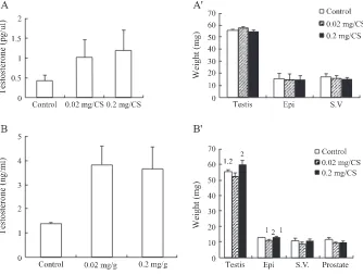
testosterone level in blood was used to simulate dysfunctional animal in reproduction, which was able to determine if CS could stimulate testicular functions.Fig. 1Aillustrates that CS could significantly induce testosterone release in plasma by the feeding of 0.02 and 0.2 mg/g-body weight of CS for 3 days in immature mice (P < 0.05). However, 3 day feeding of CS at both dosages didn’t have any effect on the weights of testis, epididymis and seminal vesicle in immature mice (p > 0.05)(Fig. 1AV). Similar to 3 days feeding, CS could significantly induce testosterone release in plasma by the feeding of 0.02 and 0.2 mg/g-body weight of CS for 7 days in immature mice (P < 0.05)(Fig. 1B). However, 7 days feeding of CS at both dosages didn’t have any effect on the weights of testis, seminal vesicle and prostate gland compared to control in immature mice (p > 0.05) (Fig. 1BV), whereas the weights of epididymis decreased with 0.02 mg/g-body weight of CS treatment for 7 days (P < 0.05).
The effect of F2 on plasma testosterone levels and the weight of reproductive organs in immature mice
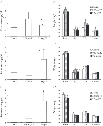
We have shown that F2 and F3 extracts, but not F1, were able to induce in vitro primary Leydig cell testosterone production, respectively(Huang et al., 2001b). Thus, F2 and F3 were used to feed mice for examining their biological function on testosterone production in the present study. Immature mice were fed with F2 mycelium extract for 1, 3 or 7 days, and testosterone levels and weights of reproductive organs were determined. Testosterone release in plasma could be significantly induced by the feeding of
F2 at 0.02 mg/g-body weight for 1 day(Fig. 2A), 0.2 mg/g-body weight for 3 day(Fig. 2B), and 0.2 mg/ g-body weight for 7 days(Fig. 2C)in immature mice, respectively (P < 0.05). Considering the effect of F2 on the weight of reproductive organs, feeding of F2 at 0.02 mg/g-body weight, but not 0.2, for 1 day
had stimulatory effect on the weights of testis, epididymis, seminal vesicle and prostate gland in immature mice, respectively (p < 0.05)(Fig. 2AV). Feeding of F2 at 0.02 and 0.2mg/g-body weight for 3 days had no effect on the weights of testis and prostate gland in immature mice, respectively (p > 0.05) G. However, the weight of epididymis and seminal vesicle increased by the treatment of F2 at 0.02 mg/
g-body weight and F2 at 0.2 mg/g-body weight for 3 days, respectively (p < 0.05) (Fig. 2BV). Interestingly, feeding of F2 at 0.02 and 0.2mg/g-body weight for 7 days had no effect on epididymis, seminal vesicle and prostate gland (p > 0.05), but the weight of testis was reduced (p < 0.05) in immature mice, respectively (p < 0.05)(Fig. 2CV).
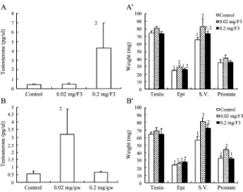
The effect of F3 on plasma testosterone levels and the weight of reproductive organs in immature mice
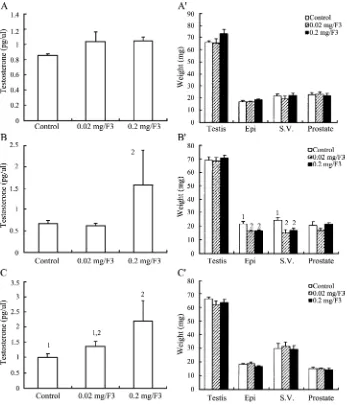
Immature mice were fed with F3 mycelium extract for 1, 3 or 7 days, and testosterone levels and weights of reproductive organs were determined. Feeding of F3 for 1 day at 0.02 and 0.2 mg/g-body weight dosages did not have any effect on testosterone production in immature mice(Fig. 3A)(P > 0.05). However, feeding of F3 for 3 days at 0.2 mg/g-body weight significantly induced testosterone release(Fig. 3B)(P < 0.05). Moreover, feeding of F3 for 7 days at 0.02 and 0.2 mg/g-body weight also significantly induced testosterone release in immature mice(Fig. 3C)(P < 0.05). F3 didn’t have any effect on the weights of reproductive organs in immature mice (p > 0.05) except that the feeding of F3 at both dosages for 3 days reduced the weight of epididymis and seminal vesicle (P < 0.05)(Fig. 3AV, 3BV, 3CV).
The effect of CS on plasma testosterone levels and the weight of reproductive organs in mature mice
In our previous in vitro studies, CS suppressed hCG-treated testosterone production in purified mouse Leydig cells(Huang et al., 2001b). In the present study, mature mice, whose LH levels are higher than immature mice, were used for in vivo study to examine if this suppressive phenomenon will also happen as in vitro study. Mature mice were fed with CS mycelium for 3 or 7 days, and testosterone levels and weights of reproductive organs were determined. CS didn’t have any inhibitory effect on testosterone release in plasma by the feeding of 0.02 and 0.2 mg/g-body weight of CS for 3 in mature mice (P > 0.05) (Fig. 4A). Conversely, CS at 0.2 mg/g-body weight by feeding 7 days did stimulate plasma testosterone release in mature mice (P < 0.05)(Fig. 4B). Neither CS at both dosages for 3 and 7 days had any effect on the weights of reproductive organs in mature mice (p > 0.05)(Fig. 4AVand 4BV).
The effect of F2 on plasma testosterone levels and the weight of reproductive organs in mature mice
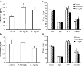
Mature mice were fed with F2 mycelium extract for 3 or 7 days, and testosterone levels and weights of reproductive organs were determined. Plasma testosterone was significantly induced by
F2 at 0.02 and 0.2 mg/g-body weight for 3 days (Fig. 5A) (P < 0.05), whereas there was no effect by 0.02 and 0.2 mg/g-body weight of F2 for 7 days in mature mice (Fig. 5B) (P > 0.05). F2 didn’t have any effect on the weights of reproductive organ compared to control in mature mice (p > 0.05) (Fig. 5AV and 5BV).
The effect of F3 on plasma testosterone levels and the weight of reproductive organs in mature mice
Mature mice were fed with F3 mycelium extract for 3 or 7 days, and testosterone levels and weights of reproductive organs were determined. Plasma testosterone was stimulated by F3 at 0.2 mg/g-body weight for 3 days and at 0.02 mg/g-body weight for 7 days in mature mice, respectively (P < 0.05)(Fig. 6A and 6B). Besides F3 at 0.02 mg/g-body weight for 3 days did stimulate the weight of epididymis and seminal vesicle (P < 0.05)(Fig. 6AV), there were no any effect of F3 on the weights of reproductive organs in mature mice (p > 0.05). Weights of epididymis were increased by F3 at 0.2 mg/g-body weight, and the weights of seminal vesicle and prostate gland were increased by F3 at 0.02 mg/g-body weight for 7 days in mature mice, respectively (p > 0.05) (Fig. 6AV and 6BV).
Discussion
In the present study, we have found that CS, F2 and F3 could induce plasma testosterone levels both in immature and mature mice in 1, 3 and/or 7 days feeding at the dosages of 0.02 and/or 0.2 mg/g-body weight. Indeed, there were divergent quantities in the stimulatory effects on mouse plasma testosterone production by CS and its fractions between immature and mature mice. CS at both dosages induced testosterone production at 3 and 7 days feeding in immature mice, whereas only CS at 0.2 mg/g-body weight for 7 days induced testosterone production in mature mice. This indicates that Leydig cells in immature mice have higher sensitivity to CS. In fact, it has been illustrated that Leydig cells in old mice have lower responsiveness to LH for testosterone production(Chen et al., 2002), and a high dose of ligand may inhibit target cell functions (Stojilkovic et al., 1994). Thus, CS with stronger effect on testosterone production in immature mouse than mature mice is a reasonable phenomenon in the present study.
Considering F2 and F3, only 7 days feeding of F2 in mature mice have no effect on testosterone production, whereas F2 induced significant testosterone levels from 1, 3 and 7 days treatment in immature mice and 3 days treatment in mature mice. Only 1 day feeding of F3 in immature mice have no effect on testosterone production, whereas F3 induced significant testosterone levels from 3 and 7 days treatment in mature and immature mice. However, it is not clear why only 0.02, but not 0.2, mg/ g-body weight of F2 for 1 day feeding and 0.2, but not 0.02, mg/g-body weight of F2 for 3 and 7 days feeding had stimulatory effect in immature mice, Also, it is not clear why only 0.2, but not 0.02, mg/g-body weight of F3 for 3 days and 0.02, but not 0.2, mg/g-body weight of F3 for 7 days had stimulatory effect in mature mice, respectively. However, there was a tendency that testosterone levels increased as the day length of feeding increased in both different age mice with the treatment of CS and its fractions.
We have previously demonstrated that CS suppressed hCG-treated testosterone production in purified mouse Leydig cells(Huang et al., 2001b). In the present study, CS and its fractions stimulated in vivo testosterone production not only in immature mice but also in mature mice, although mature mice have high plasma LH levels. Thus, the in vitro inhibitory phenomenon is not consistent to the present in vivo study. However, the in vivo study corroborates the physiological effect of CS and its fraction on Leydig cell function. Moreover, in the present study, CS, F2 and F3 did not profoundly affect the weight of reproductive organs. These data indicate that the mass of testis or even the number of Leydig cells might not change much. If it is so, the sensitivity of Leydig cells might increase to respond to the stimulatory effect of CS and its fractions. However, this speculation needs more study to be verified.
F3 after digestion are worth investigating. In conclusion, CS, F2 and F3 could significantly induce in vivo testosterone secretions. It is possible that CS, F2 and F3 might contribute to an alternative medicine for the treatment of reproductive problems caused by insufficient testosterone production in male human being.
Acknowledgements
Yuan-Li Huang and Sew-Fen Leu contribute equally to the study. This study was supported by the Department of Health Grant DOH92-TD-1005, Taiwan, Republic of China, to BMH.
References
Bouloux, P., Warne, D.W., Loumaye, E., 2002. Efficacy and safety of recombinant human follicle-stimulating hormone in men with isolated hypogonadotropic hypogonadism. Fertility and Sterility 77 (2), 270 – 273.
Chen, H.L., Hardy, M.P., Zirkin, B.R., 2002. Age-related decreases in Leydig cell testosterone production are not restored by exposure to LH in vitro. Endocrinology 143 (5), 1637 – 1642.
Chiou, W.F., Chang, P.C., Chou, C.J., Chen, C.F., 2000. Protein constituent contributes to the hypotensive and vasorelaxant activities of Cordyceps sinensis. Life Sciences 66 (14), 1369 – 1376.
Crimmel, A.S., Conner, C.S., Monga, M., 2001. Withered Yang: a review of traditional Chinese medical treatment of male infertility and erectile dysfunction. Journal of Andrology 22 (2), 173 – 182.
Hsu, C.C., Tsai, S.J., Huang, Y.L., Huang, B.M., 2003a. Regulatory mechanism ofCordyceps sinensismycelium on mouse Leydig cell steroidogenesis. FEBS Letters 543 (1 – 3), 140 – 143.
Hsu, C.C., Huang, Y.L., Tsai, S.C., Sheu, C.C., Huang, B.M., 2003b. In vivo and in vitro stimulatory effects ofCordyceps sinensison testosterone production in mouse Leydig cells. Life Sciences 73 (16), 2127 – 2136.
Huang, B.M., Chuang, Y.M., Chen, C.F., Leu, S.F., 2000. Effects of extracted Cordyceps sinensis on steroidogenesis in MA-10 mouse Leydig tumor cells. Biological and Pharmaceutical Bulletin 23 (12), 1532 – 1535.
Huang, B.M., Ju, S.Y., Wu, C.S., Chuang, W.J., Sheu, C.C., Leu, S.F., 2001a. Cordyceps sinensis and its fractions stimulated MA-10 mouse Leydig tumor cell steroidogenesis. Journal of Andrology 22 (5), 831 – 837.
Huang, B.M., Hsu, C.C., Tsai, S.J., Sheu, C.C., Leu, S.F., 2001b. Effect of Cordyceps sinensis on steroidogenesis in normal mouse Leydig cells. Life Sciences 69 (22), 2593 – 2602.
Huang, B.M., Leu, S.F., Yang, H.Y., Norman, R.L., 2001c. Testosterone effects on luteinizing hormone and follicle-stimulating hormone responses to gonadotropin-releasing hormone in the mouse. Journal of Andrology 22 (3), 507 – 513.
Huang, B.M., Hsiao, K.Y., Chuang, P.C., Wu, M.H., Pan, H.A., Tsai, S.J., 2004. Upregulation of steroidogenic enzymes and ovarian 17h-estradiol in human granulosa-lutein cells byCordyceps sinensismycelium. Biology of Reproduction (in press). Huff, D.S., Snyder III, H.M., Rusnack, S.L., Zderic, S.A., Carr, M.C., Canning, D.A., 2001. Hormonal therapy for the
subfertility of cryptorchidism. Hormone Research 55 (1), 38 – 40.
Kuo, Y.C., Tsai, W.J., Wang, J.Y., Chang, S.C., Lin, C.Y., Shiao, M.S., 2001. Regulation of bronchoalveolar lavage fluids cell function by the immunomodulatory agents from Cordyceps sinensis. Life Sciences 68 (9), 1067 – 1082.
Moger, W.H., 1991. Evidence for compartmentalization of adenosine 3V,5V-monophosphate (cAMP)-dependent protein kinase in rat Leydig cells using site-selective cAMP analogs. Endocrinology 128 (3), 1414 – 1418.
Pigny, P., Berault, A., Dewailly, D., Boersma, A., 1992. Glycoprotein hormones, glycosylation and biological activity. Annales de Biologie Clinigue 50 (8), 557 – 564.
Rege, N.N., Date, J., Kulkarni, V., Prem, A.R., Punekar, S.V., Dahanukar, S.A., 1997. Effect of Y virilin on male infertility. Journal of Postgraduate Medicine 43 (3), 64 – 67.
Roscoe, W.A., Barr, K.J., Mhawi, A.A., Pomerantz, D.K., Kidder, G.M., 2001. Failure of spermatogenesis in mice lacking connexin43. Biolology of Reproduction 65 (3), 829 – 838.
Saez, J.M., 1994. Leydig cells: endocrine, paracrine and autocrine regulation. Endocrine Review 15 (5), 574 – 626.
Stocco, D.M., 2002. Clinical disorders associated with abnormal cholesterol transport: mutations in the steroidogenic acute regulatory protein. Molecular and Cellular Endocrinology 191 (1), 19 – 25.
Stocco, D.M., Clark, B.J., 1996. Regulation of the acute production of steroids in steroidogenic cells. Endocrine Review 17 (3), 221 – 244.
Stojilkovic, S.S., Reinhart, J., Catt, K.J., 1994. Gonadotropin-releasing hormone receptors: structure and signal transduction pathways. Endocrine Review 15 (4), 462 – 499.
Ukai, S., Kiho, T., Hara, C., Morita, M., Goto, A., Imaizumi, N., Hasegawa, Y., 1983. Polysaccharides in fungi. XIII. Antitumor activity of various polysaccharides isolated from Dictyophora indusiata, Ganoderma japonicum,Cordyceps cicadae, Auricu-laria auricula-judae, and AuricuAuricu-laria species. Chemical and Pharmaceutical Bulletin 31 (2), 741 – 744.
Veal, L., 1998. Complementary therapy and infertility: an Icelandic perspective Complement. Therapies in Nursing and Midwifery 4 (1), 3 – 6.
Wang, S.M., Lee, L.J., Lin, W.W., Chang, C.M., 1998. Effects of a water-soluble extract of Cordyceps sinensis on steroido-genesis and capsular morphology of lipid droplets in cultured rat adrenocortical cells. Journal of Cellular Biochemistry 69 (4), 483 – 489.
Yamaguchi, Y., Kagota, S., Nakamura, K., Shinozuka, K., Kunitomo, M., 2000. Inhibitory effects of water extracts from fruiting bodies of cultured Cordyceps sinensis on raised serum lipid peroxide levels and aortic cholesterol deposition in atherosclerotic mice. Phytotherapy Research 14 (8), 650 – 652.
Yang, L.Y., Huang, W.J., Hsieh, H.G., Lin, C.Y., 2003. H1-A extracted from Cordyceps sinensis suppresses the proliferation of human mesangial cells and promotes apoptosis, probably by inhibiting the tyrosine phosphorylation of Bcl-2 and Bcl-XL. Journal of Laboratory and Clinical Medicine 141 (1), 74 – 83.
Yoshida, J., Takamura, S., Yamaguchi, N., Ren, L.J., Chen, H., 1989. Antitumor activity of an extract of Cordyceps sinensis (Berk.) Sacc. against murine tumor cell lines. Japanese Journal of Experimental Medicine 59 (4), 157 – 161.
Zhu, J.S., Halpern, G.M., Jones, K., 1998a. The scientific rediscovery of a precious ancient Chinese herbal regimen: Cordyceps sinensis: Part I. Journal of Alternative and Complementary Medicine 4 (3), 289 – 303.
Zhu, J.S., Halpern, G.M., Jones, K., 1998b. The scientific rediscovery of a precious ancient Chinese herbal regimen: Cordyceps sinensis: Part II. Journal of Alternative and Complementary Medicine 4 (3), 429 – 457.