ASTRAL-R score predicts non-recanalisation after intravenous
thrombolysis in acute ischaemic stroke
Peter Vanacker1,2,3; Mirjam R. Heldner4; David Seiffge5; Hubertus Mueller6; Ashraf Eskandari1; Christopher Traenka5; George Ntaios7; Pascal J. Mosimann8; Roman Sztajzel6; Vitor Mendes Pereira9*; Patrick Cras3; Stefan Engelter5,10; Philippe Lyrer5; Urs Fischer4; Dimitris Lambrou1; Marcel Arnold4; Patrik Michel1
1Department of Neurology, Centre Hospital Universitaire Vaudois and University of Lausanne, Lausanne, Switzerland; 2Department of Neurology, General Hospital Sint-Lucas, Bruges, Belgium; 3Department of Neurology, Born Bunge Institute, University and University Hospital, Antwerp, Belgium; 4Department of Neurology, University Hospital, Bern, Switzerland; 5Department of Neurology and Stroke Center, University Hospital, Basel, Switzerland; 6Department of Neurology, University Hospital, Geneva, Switzerland; 7Department of Medicine, University of Thessaly, Larissa, Greece; 8Institute for Diagnostic and Interventional Neuroradiology, Centre Hospital Universitaire Vaudois, Lausanne, Switzerland; 9Institute for Diagnostic and Interventional Neuroradiology, University Hospital, Geneva, Switzerland; 10Neurorehabilitation Unit, University Center for Medicine of Aging and Rehabilitation, Felix Plattee Hospital, Basel, Switzerland; *Current address: Department of Medical Imaging and Surgery, University of Toronto, Toronto, Ontario, Canada
Summary
Intravenous thrombolysis (IVT) as treatment in acute ischaemic strokes may be insufficient to achieve recanalisation in certain pa-tients. Predicting probability of non-recanalisation after IVT may have the potential to influence patient selection to more aggressive man-agement strategies. We aimed at deriving and internally validating a predictive score for post-thrombolytic non-recanalisation, using clini-cal and radiologiclini-cal variables. In thrombolysis registries from four Swiss academic stroke centres (Lausanne, Bern, Basel and Geneva), patients were selected with large arterial occlusion on acute imaging and with repeated arterial assessment at 24 hours. Based on a logistic regression analysis, an integer-based score for each covariate of the fitted multivariate model was generated. Performance of integer-based predictive model was assessed by bootstrapping available data and cross validation (delete-d method). In 599 thrombolysed strokes, five variables were identified as independent predictors of absence of recanalisation: Acute glucose > 7 mmol/l (A), significant extracranial
vessel STenosis (ST), decreased Range of visual fields (R), large Arterial occlusion (A) and decreased Level of consciousness (L). All variables were weighted 1, except for (L) which obtained 2 points based on
β-coefficients on the logistic scale. ASTRAL-R scores 0, 3 and 6 corsponded to non-recanalisation probabilities of 18, 44 and 74 % re-spectively. Predictive ability showed AUC of 0.66 (95 %CI, 0.61–0.70) when using bootstrap and 0.66 (0.63–0.68) when using delete-d cross validation. In conclusion, the 5-item ASTRAL-R score moderately pre-dicts non-recanalisation at 24 hours in thrombolysed ischaemic strokes. If its performance can be confirmed by external validation and its clinical usefulness can be proven, the score may influence patient selection for more aggressive revascularisation strategies in routine clinical practice.
Keywords
Cerebral infarction, thrombolytic therapy, decision support techniques, cerebral revascularisation, cerebrovascular occlusion
Correspondence to: Peter Vanacker
Centre Hospitalier Universitaire Vaudois 46, Rue de Bugnon
1011 Lausanne, Switzerland E-mail: [email protected]
Received: June 1, 2014
Accepted after major revision: November 21, 2014 Epub ahead of print: January 15, 2015
http://dx.doi.org/10.1160/TH14-06-0482 Thromb Haemost 2015; 113: ■■■
Introduction
Recanalisation of the occluded large vessels in acute ischaemic strokes (AIS) may occur spontaneously in a subset of acute is-chaemic stroke patients, but is more effectively achieved by intra-venous thrombolysis (IVT) (1–6). However, patients with strokes due to large vessel occlusions have low recanalisation rates with IVT alone, associated with poor functional outcome despite treat-ment (1, 5). More aggressive, endovascular treattreat-ment strategies are increasingly used to treat large vessel occlusive strokes, as they re-canalise these occlusions more effectively and rapidly. However, recent endovascular trials as IMS III, SYNTHESIS and MR-RES-CUE (7) did not show superiority of endovascular therapy, al-though a trend of effectiveness was present in severe strokes (NIHSS ≥ 20). Further, recanalisation by an acute endovascular
in-tervention is not always associated with reperfusion and better clinical outcome (8), given that multiple other factors (time, collat-erals, core and penumbra volumes, peripheral occlusions, site of is-chaemia, metabolic states etc.) interfere. A better selection of pa-tients who are most likely to benefit from aggressive (endovascu-lar) therapy is therefore necessary (9).
AS-PECTS scores (17), thrombus length (18) and thrombus density (19).
Patients and methods
Study design and patient selection
The study includes data from a prospective cohort of consecutive, is-chaemic stroke patients in four Swiss academic stroke centres (Uni-versity Hospitals of Lausanne: 2003–2012, Basel: 2007–2013, Berne: 2007–2012 and Geneva: 2007–2012). The coordinating centre (Lausanne) compiled the data of the different prospective registers and performed the analysis of the pooled data. The inclusion criteria for the analysis were A) acute noninvasive vascular imaging (CTA, MRA or duplex) before IVT showing B) arterial occlusion in cervi-cal and/or cerebral arteries in relation to the ischaemic territory, C) availability of a second arterial imaging (CTA, MRA, angiography or duplex) at 24 hours (h) (allowed range 12–48 h) permitting assess-ment of recanalisation and D) treatassess-ment with IVT alone within proven time windows (3 h after last proof of well-being up to 10/2008, 4.5 h thereafter). Arterial occlusions were defined accord-ing to standardised methodologies that were published elsewhere (20). Vascular imaging (CTA, MRA or duplex) was performed in all centres before or within minutes after administration of the IV thrombolysis bolus which should not influence arterial pathology. In the vast majority of patients (n=563, 94 %) CTangiography was the initial imaging technique. Indications for IVT with recombinant tis-sue plasminogen activator (rtPA) followed European Stroke Organi-sation guidelines (21). Patients with additional acute endovascular procedures after IVT were excluded.
Medical variables collected and analysed included demo-graphics, cardiovascular risk factors, co-morbidities, pre-stroke medication, type of clinical deficit, NIHSS at admission (with all items individually collected), onset-to-admission time, vital signs and radiological imaging at admission. Pretreatment NIHSS scores were assessed by certified stroke physicians. The clinical signs “decreased range of visual fields” and “loss of consciousness” were defined as in the NIHSS scale. On the acute vascular imaging, large vessels pathology was analysed in each centre by both a neur-oradiologist and stroke neurologist aware of clinical findings. As both raters collaborated as a team, inter-rater variability has not been assessed. The information on the presence and site of large vessel occlusions and significant intra- and extracranial stenoses were collected, based on pre-specified definitions. Intracranial oc-clusions in the ischaemic territory were categorised according to their site in large vs intermediate occlusions. Large intracranial oc-clusions were defined as an occlusion of the basilar artery (with our without intracranial vertebral artery occlusion), the intracran-ial carotid siphon (large and/or including carotid T) and large middle cerebral artery (M1) both with and without ipsilateral extracranial carotid occlusion. Intermediate intracranial occlu-sions were defined as occluocclu-sions in anterior cerebral artery (A1 or A2 segments), peripheral middle cerebral artery (M2), posterior cerebral artery (P1 or P2 segments), intracranial part of the verte-bral artery (V4) and siphon of the internal carotid artery without
distal T-occlusion; the latter two were considered “intermediate” because thrombus load and clinical symptoms are usually minor in the absence of extension into the basilar artery and the carotid T, respectively. Recanalisation of initially occluded arteries was de-fined for intra- and extracranial vessel occlusions and for the dif-ferent imaging techniques separately, and was classified as “absent” vs “partial or complete”. These criteria were published elsewehere (20) If several good quality studies are available at 12–48 h, priori-tiy was given to DSA > CTA > MRA > Doppler.
The collection, analysis and publication of data in the four stroke registries were performed according to the guidelines of the respective ethical boards of each centre.
Statistical analyses
Continuous variables are summarised using median and inter-quartile range, while categorical ones with count and percentage. Univariate comparisons for all the variables considered, between the patients who recanalise after intravenous thrombolysis and those who did not, were performed using logistic regression analy-sis, where the significance of each variable was assessed separately. Before starting the multiple analysis, imputation of the missing values of the covariates considered was performed using the method of chained equations (i. e. filling in the missing values on a variable-by-variable basis, by first specifying the imputation model for each incomplete variable and then using this model to fill in the gaps). Five imputed datasets were generated using this method. Multiple logistic regression analysis was performed on each im-puted dataset separately. Stepwise methods were implemented on each one of the five analysis, to identify significant main effects and interactions. Final results were derived as combinations of the outcome of the five imputed analysis. The log-odds of the final model were used to define the coefficients in the proposed score. Predictive ability of the score is measured via the area under the receiver-operating characteristic curve (AUC-ROC), calculated for the imputed datasets using bootstrapping and cross validation. Bootstrapping assesses the predictive ability of our model by creat-ing copies of the imputed datasets and recalculatcreat-ing AUC on these copies. Cross-validation splits the imputed dataset in two parts (not necessarily in half), fits a model to one part, and assesses its predictive ability using the other part. As the two methods might be considered as complementary to the other, both techniques were used to evaluate the predictive ability of models. The ROC analysis was carried out on the imputed datasets separately, and final results were derived as combination of the outcome of these analyses. All tests were carried out at 5 % significance level. All analyses performed using the statistical package R (version 3.0.2).
Results
shown partially in
▶
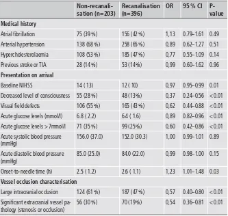
Table 1. Numbers of included patients per centre were as follows: Lausanne (n=210), Basel (n=217), Bern (n=136) and Geneva (n=35). Recanalisation was assessed by CTA in 82 % (n=491), by MRA in 12 % (n=72) and by duplex only in 6 % (n=36). Five clinical and radiological variables were identified as independent predictors of non-recanalisation: Acute glucose > 7 mmol/l (A), STenosis or occlusion of extracranial vessels (ST), Range of visual fields decreased (R), Arterial occlusion in a large site (A) and an decreased level of consciousness (L) (▶
Table 2). The cut-off value for acute glucose levels was chosen on a tree-based method, suggesting that a cut-off of 7 mmol/l would be the most adequate. As the initial multivariate analysis failed to ac-knowledge the role of time in relation to recanalisation, additional statistical analyses were performed to check the effect of different cut-off values for the onset-to-needle times (< 1.5 h, < 3 h, < 4.5 h) on the prediction of recanalisation. No significant correlation was detected between dichotomisation at 3 h (OR 1.2, 95 %CI 0.8–1.8)and at 4.5 h (OR 1.5, 95 %CI 0.5–4.9) with recanalisation after IV thrombolysis. Only, dichotomisation at 1.5 h was significantly as-sociated with recanalisation (OR 1.97, 95 %CI 1.1–3.5). This par-ameter, however, did not improve the predictive performance of the ASTRAL-R score in terms of AUC-ROC.
The ordering of the predictors used in our score, according to their significance in the final model, judged from the p-value of the imputation analysis and starting from the most significant to the least significant, is as follows: decreased consciousness, extra-cranial pathology, visual field defect, acute glucose level and proxi-mal intracranial occlusion. A predictive model with the item de-creased consciousness only has AUC 57.7 %, adding extracranial pathology the AUC raises to 62.1 %, adding further visual field de-fect AUC is estimated at 64.6 % and adding acute glucose level AUC goes to 65.0 % and finally adding proximal occlusion gives an AUC of 66.2 % (Suppl. Figure 1, available online at www.thrombo sis-online.com).
Acute glucose levels > 7mmol/l
Acute systolic blood pressure (mmHg)
Acute diastolic blood pressure (mmHg)
Table 1: Selection of baseline character-istics and radiological findings, dichotomis-ed according to presence or absence of post-thrombolytic recanalisation. Values are expressed as medians and interquartile range (IQR) for continuous variables unless stated otherwise, and as absolute counts and percen-tage for categorical variables.
Table 2: The ASTRAL-R Score: Independent predictors of absence of post-thrombolytic recanalisation at 24 h with the respective score points.
Covariates
Acute Glucose level > 7mmol/l
STenosis/Occlusion in extracranial vessel
Range of visual fields decreased
Arterial Occlusion: large size
Level of consciousness altered
▶
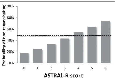
Figure 1 displays the predicted probability of non-recanali-sation after IV thrombolysis based on the ASTRAL-R score. The absolute risk of non-recanalisation for the corresponding AS-TRAL-R scores 0, 1, 2, 3, 4, 5 and 6 were 17.7 %, 24.8 %, 33.5 %, 43.6 %, 54.2 %, 64.4 % and 73.5 %, respectively. Patients with an AS-TRAL-R score more than 3 have a likelihood of absence of post-thrombolytic recanalisation of more than 50 %. Suppl. Figure 2 (available online at www.thrombosis-online.com) shows the dis-tribution of the overall ASTRAL-R score and its components among the population.The predictive ability of the model was assessed in two ways: a) by calculating the AUC of the ROC-curve using the original sample (599 patients after imputing for missing values) and attach-ing uncertainty usattach-ing bootstrap methodology (1,000 repetitions), and b) by cross-validation (deleted-d method repeated 1,000 times). The estimated AUC in both cases was 0.66 with a 95 %CI of 0.61–0.70 (Suppl. Figure 3, available online at www.thrombosis-on line.com) using the first method, while the corresponding 95 % CI for the cross validation was 0.63–0.68 (Suppl. Figure 4, available online at www.thrombosis-online.com).
Discussion
This study describes the derivation and internal validation of the ASTRAL-R score in a multi-centre cohort which estimates the likelihood of non-recanalisation 24 h after IVT. A linear relation-ship between the total score and non-recanalisation could be shown, and the performance of the model was assessed in a multi-centre cohort.
The score consists of five easily available variables already be-fore IVT: acute glucose (cut-off > 7 mmol/l), extracranial stenosis > 50 % or occlusion, visual field defects, decreased level of con-sciousness and a large artery intracranial occlusion. Hyperglycae-mia in acute stroke patients is common and may decrease plasma t-PA activity. Further, acute hyperglycaemia may be related to atherosclerotic disease, which has been shown to be more resistant
to recanalisation after tPA treatment (10, 22). The concomitant presence of extracranial arterial stenosis or occlusion may lead to less recanalisation because of decreased inflow of blood into the blocked arteries, leading to less arrival of IV rt-PA and less wash-out of clots (23). Occlusion of larger intracranial arteries is likely to be related to less recanalisation because of higher thrombus burden than in smaller arteries, as shown previously (13, 14). It is unclear why decreased level of vigilance and visual field defects were independent predictors of recanalisation, although these fac-tors have been associated with worse clinical outcome (24). One hypothesis may be that these signs of clinical severity may indicate poorer collaterals which could also contribute to recanalisation (25).
Some previously described predictors of recanalisation, such as onset-to-needle time (10), atrial fibrillation (12, 13), smoking (14) and metabolic syndrome (15) and ASPECTS scores (17) were not confirmed in our logistic regression analysis. This may be related to differences in the cohorts or the sample size.
Use of the ASTRAL-R score in clinical practice has the potential to improve acute stroke management. Given several negative inter-ventional revascularisation trials, a more careful selection of pa-tients is mandatory (9). A more individualised patient selection based not only on the severity of the stroke but also on the non-re-canalisation risk after IV thrombolysis could be promising. In in-dividual cases with a high likelihood of non-recanalisation after IVT (e. g. large vessel occlusions, extracranial stenosis, hypergly-caemia), endovascular intervention for non-recanalising patients may be of added benefit, in particular if salvageable tissue can be demonstrated by multimodal imaging (26, 27). In addition, suc-cessful recanalisation is associated with better outcome also in se-vere stroke patients (28).
Some items in the ASTRAL-R score (level of consciousness, vis-ual field defects and acute glucose level) are the same as in the prognostic ASTRAL score for clinical outcome (24). Both scores may be used as selection tools for future clinical trials on endovas-cular treatment after IVT.
One of the strengths of the ASTRAL-R score is the large number of patients and variables included and the multi-centre design including four sites with a longstanding experience in acute stroke research. Data were available on the specific site of arterial occlusions sites in all cohorts. Validation of the score was perform-ed by a bootstrap and cross validation (delete-d method).
Several limitations apply: First, implementation in routine clinical practice may be hampered by the moderate predictive abil-ity of the model with areas under the ROC curve of 0.66. This cor-responds to other predictive scores in acute stroke care (e. g. SEDAN score) which have similar c-statistic values (29). This can partially be explained by combing four different cohorts of throm-bolysed stroke patients. Adding factors such as collateral vessel status (25), perfusion imaging (30) and thrombus length (13) could further improve the score. However, such radiological data and imaging techniques are not routinely used in clinical practice and are hampered by standardisation issues. Secondly, external validation in independent cohorts will be necessary to confirm the performance of the score. The validity of the score in patients who Figure 1: Association between the ASTRAL-R score and the
Conflicts of interest
P. V., D. L., A. E., M. R. H. and C. T. report no disclosures. D. S. re-ceived a travel grant from Bayer. P. L. has served on scientific ad-visory boards for Bayer, Schering Pharma, BMS/Pfizer and Boehr-inger Ingelheim; has received funding for travel or speaker honor-aria from Bayer Schering Pharma, Boehringer Ingelheim, and Shire plc; he serves as Co-Editor for Neurologie und Psychiatrie and on the editorial board of Swiss Archives of Neurology and Psychiatry; and has received research support from AstraZeneca, Boehringer Ingelheim, Sanofi-aventis, Photo-Thera, the Swiss National Science Foundation, and the Swiss Heart Foundation. G. N. received consulting fees from Boehringer-Ingelheim; honor-arium from Medtronic; speaker fees from Boehringer-Ingelheim and Sanofi. P. M. received funding for travel or speaker honoraria from Shire, Bayer, Sanofi-Aventis; consulting fees from Lundbeck and Pierre-Fabre; honoraria for scientific advisory boards for Bayer and Boehringer-Ingelheim.
References
1. Rha JH, Saver JL. The impact of recanalisation on ischaemic stroke outcome: a meta-analysis. Stroke 2007; 38: 967–973.
2. Fiebach JB, Al-Rawi Y, Wintermark M, et al. Vascular Occlusion Enables Select-ing Acute Ischaemic Stroke Patients for Treatment With Desmoteplase. Stroke 2012; 43: 1546–1566.
3. Galimanis A, Jung S, Mono ML, et al. Endovascular therapy of 623 patients with anterior circulation stroke. Stroke 2012; 43: 1052–1057.
4. Mazighi M, Serfaty JM, Labreuche J, et al. RECANALISE investigators. Com-parison of intravenous alteplase with a combined intravenous-endovascular ap-proach in patients with stroke and confirmed arterial occlusion (RECANALISE study): a prospective cohort study. Lancet Neurol 2009; 8: 802–809.
5. Yeo LLL, Paliwal P, Teoh HL, et al. Timing of Recanalisation after Intravenous Thrombolysis and Functional Outcomes After Acute Ischaemic Stroke. J Am Med Assoc Neurol 2013; 70: 353–358.
6. Zaidat OO, Suarez JI, Sunshine JL, et al. Thrombolytic Therapy of Acute Is-chaemic Stroke : Correlation of Angiographic Recanalisation with Clinical Out-come. Am J Neuroradiol 2005; 26: 880–884.
7. Broderick JP, Palesch YY, Demchuk AM, et al; for the Interventional Manage-ment of Stroke (IMS) III Investigators. N Engl J Med 2013; 368: 893–903.
underwent endovascular treatment needs to be assessed, as these patients were excluded in the current analysis. Thirdly, large vessel occlusions were classified into large and intermediate arterial oc-clusions, mainly based on anatomical location. The likelihood of recanalisation between the location sites may be more variable than expected. This arbitrary classification is debatable, but clini-cally relevant in the absence of a standardised classification of the intracranial arteries. Fourthly, our definition of recanalisation was not based on a single imaging modality but on heterogeneous methods, the degree of recanalisation was dichotomised at an ar-bitrary point (between none and partial or complete) and the best timing for recanalisation assessment is still unknown. Fifthly, a pre-selection bias may have been introduced in the study by ex-cluding stroke patients with additional acute endovascular pro-cedures after insufficient IV t-PA recanalisation. Potentially, lower recanalisation rates and different predictive parameters would have been found in this subgroup of patients. And finally, reading of the vascular imaging data was not centralised in order to pre-vent high inter-rater variabilities.
Conclusions
In conclusion, the ASTRAL-R score is an easy-to-assess 5-item score moderately predicting non-recanalisation after IVT in acute ischaemic strokes.
Acknowledgements
The first and last author (P. Vanacker, P. Michel) had full access to all of the data in the study and takes responsibility for the integrity of the data and the accuracy of the data analysis. This research is supported by grants from the Swiss Cardiology foundation (P. M., P. V.), CardioMet-CHUV (P. M.) and a scholarship of the European Neurological Society (P. V.).
Author contributions
Vanacker P: Study concept and design, analysis, interpretation, preparation of the manuscript. Heldner MR: Data acquisition and analysis, critical revision of the manuscript for important intellec-tual content. Seiffge D: Data acquisition and analysis, critical revi-sion of the manuscript for important intellectual content. Mueller H: Data acquisition and analysis. Traenka C: Data acquisition and analysis. Ntaios G: Study concept and design, data acquisition and analysis, critical revision of the manuscript for important intellec-tual content. Stajzel R: Data acquisition and analysis. Lyrer P: Data analysis, critical revision of the manuscript for important intellec-tual content. Fischer U: Data acquisition, data analysis, critical revision of the manuscript for important intellectual content. Lam-brou D: Data analysis, statistical analysis. Arnold M: Data analysis, critical revision of the manuscript for intellectual content. Michel P: Study concept and design, data acquisition, analysis and inter-pretation, critical revision of the manuscript for important intel-lectual content, study supervision.
What is known about this topic?
•
A few clinical and radiological factors influencing the recanali-sation rate in IV thrombolysis-treated acute ischaemic strokes, are known from small single-centre studies.What does this paper add?
•
By performing a logistic regression analysis in a multi-centre co-hort, we could detect independent predictors of arterial recanali-sation after IV thrombolysis.•
For the first time, a diagnostic tool (ASTRAL-R score) has been shown to have a good predictive accuracy for recanalisation after IV thrombolysis.•
The ASTRAL-R score is an easy-to-use 5 items score (0-> 6).8. Soares BP, Tong E, Hom J, et al. Reperfusion is a more accurate predictor of fol-low-up infarct volume than recanalisation : a proof of concept using CT in acute ischaemic stroke patients. Stroke 2010; 41: 34–40.
9. Liebeskind DS. The Quest to Prove Endovascular Stroke Therapy: Searching for the “Sweet Spot” in Patient Selection. Mayo Clin Proc 2013; 88: 1039–1041. 10. Ribo M, Molina C, Montaner J, et al. Acute hyperglycemia state is associated
with lower tPA-induced recanalisation rates in stroke patients. Stroke 2005; 36: 1705–1709.
11. Zangerle A, Kiechl S, Spiegel M, et al. Recanalisation after thrombolysis in stroke patients: predictors and prognostic implications. Neurology 2007; 68: 39–44.
12. Kimura K, Iguchi Y, Yamashita S, et al. Atrial fibrillation as an independent pre-dictor for no early recanalisation after IV-rtPA in acute ischaemic stroke. J Neurol Sci 2008; 246: 57–61.
13. Mendonça N, Rodriguez-Luna D, Rubiera M, et al. Predictors of tissue-type plasminogen activator nonresponders according to location of vessel occlusion. Stroke 2012; 43:
417-14. Kufner A, Nolte CH, Galinovic I, et al. Smoking-Thrombolysis Paradox: Reca-nalisation and Reperfusion Rates After Intravenous Tissue Plasminogen Acti-vator in Smokers With Ischaemic Stroke. Stroke 2013 ; 44: 407–413.
15. Arenillas JF, Sandoval P, Pérez de la Ossa N, et al. The metabolic syndrome is as-sociated with a higher resistance to intravenous thrombolysis for acute is-chaemic stroke in women than in men. Stroke 2009; 40: 344–349.
16. Saqqur M, Uchino K, Demchuk AM, et al; CLOTBUST Investigators. Site of Ar-terial Occlusion Identified by Transcranial Doppler Predicts the Response to In-travenous Thrombolysis for Stroke. Stroke 2007; 38: 948–954.
17. Tsivgoulis G, Saqqur M, Sharma VK, et al; CLOTBUST Investigators. Associ-ation of pretreatment ASPECTS scores with tPA-induced arterial recanalisAssoci-ation in acute middle cerebral artery occlusion. J Neuroimag 2008; 18: 56–61. 18. Riedel CH, Zimmerann P, Jensen-Kondering U, et al. The importance of size:
succesful recanalisation by intravenous thrombolysis in acute anterior stroke depends on thrombus length. Stroke 2011; 42: 1775–1777.
19. Moftakhar P, English JD, Cooke DL, et al. Density of Thrombus on Admission CT Predicts Revascularisation in Large Vessel Occlusion Acute Ischaemic Stroke. Stroke 2013; 44: 243–245.
20. Vanacker P, Lambrou D, Eskandari A, et al. Improving Prediction of Recanali-sation in Acute Large Vessel Occlusive Stroke. J Thromb Haemost 2014; Epub ahead of print.
21. Wunderlich MT, Goertler M, Postert T, et al; Duplex Sonography in Acute Stroke (DIAS) Study Group; Competence Network Stroke. Duplex Sonography in Acute Stroke (DIAS) Study Group; Competence Network Stroke. Recanali-sation after intravenous thrombolysis: does a recanaliRecanali-sation time window exist? Neurology 2007; 68: 1364–1368.
22. Arnold M, Mattle S, Galimanis A, et al. Impact of admission glucose and dia-betes on recanalisation and outcome after intra-arterial thrombolysis for is-chaemic stroke. Intern J Stroke 2014; 9: 985-991.
23. Ntaios G, Faouzi M, Ferrari J, et al. An integer-based score to predict functional outcome in acute ischaemic stroke: the ASTRAL score. Neurology 2012; 78: 1916–1922.
24. Bhatia R, Hill MD, Shobha N, et al. Low Rates of Acute Recanalisation With In-travenous Recombinant Tissue Plasminogen Activator in Ischaemic Stroke. Real-World Experience and a Call for Action. Stroke 2010; 41: 2254–2258. 25. Campbell BC, Christensen S, Parsons MW, et al; EPITHET and DEFUSE
Inves-tigators. Advanced imaging improves prediction of hemorrhage after stroke thrombolysis. Ann Neurol 2013; 73: 510-519.
26. Mayza MV, Bovi U, Castillo J, et al. External validation of the SEDAN score for prediction of intracerebral hemorrhage in Stroke Thrombolysis. Stroke 2013; 44: 1595–1600.
27. Miteff F, Levi CR, Bateman GA, et al. The independent predictive utility of com-puted tomography angiographic collateral status in acute ischaemic stroke. Brain 2009; 132: 2231–2238.
28. Bill O, Zufferey P, Faouzi M, Michel P. Severe stroke: patient profile and predic-tors of favorable outcome. J Thromb Haemost 2013; 11: 92–99.
29. Liebeskind DS. Trials of endovascular therapies or collaterals? Int J Stroke 2013; 8: 258–259.