RADIO PROTECTIVE EFFECTS OF GINSENG
EXTRACT IN GAMMA-RAYS INDUCED
CHROMOSOMAL DAMAGES OF
HUMAN LYMPHOCYTE
M. Syaifudin1, Jie-Young Song2, Yun-Sil Lee3, and Chang-Mo Kang4
1.Center For Technology of Radiation Safety and Metrology
National Nuclear Energy Agency (BATAN), Jakarta, Indonesia.
2.
Laboratory of Radiation Immunology, Korea Institute of Radiological and Medical Sciences (KIRAMS), Seoul, Republic of Korea.
3.
Laboratory of Radiation Effects, KIRAMS, Seoul, Republic of Korea.
4.Laboratory of Radiation Cytogenetics and Epidemiology KIRAMS, Seoul, Republic of Korea.
ABSTRACT
RADIOPROTECTIVE EFFECTS OF GINSENG EXTRACT IN GAMMA-RAYS INDUCED CHROMOSOMAL DAMAGES OF HUMAN LYMPHOCYTE.
Ginsan, a polysaccharide extracted from Panax ginseng and subsequently referred as ginseng, posses various biological properties as an anticancer and antioxidant agent. Ginseng also approved effective against radiation effects through its immunomodulating actions in whole body irradiated mouse. But its protective effects on radiation induced DNA damage are not thoroughly investigated, mainly in human. This experiment aimed to assess the effects of ginseng at 2 working doses in suppressing radiation effects of human peripheral blood lymphocyte (PBL) i.e. chromosome aberration and micronuclei yields. The treatment times were 24 hours before, subsequently (0 hour) or 3 hours after and irradiation with gamma rays at doses of 0.5 - 2.0 Gy (dose rate of 3.16 Gy/min). Treated and untreated blood cultivation and metaphase spreading technique was done according to standard procedures. Results showed that without ginseng treatments, radiation significantly increased dicentrics and micronuclei frequencies. Different with the results in mouse study, however, our results indicated that none of the experimental concentrations of ginseng crude water extract tested had an effect on baseline chromosomal aberration and micronuclei (MN) yields in PBL. A protective effect was only seen in chromosome aberration yields of sample irradiated with 2.0 Gy and treated with ginseng 3 h post irradiation rather than 24 h pre-irradiation in one volunteer. Opposite results that ginseng suspected to be a weak radiosensitizer was found in some cases. This may be due to discrepancies exist in route of treatment and its fundamental mechanisms of protective action between both studies. Even though in general it was not
effective, the possible mechanism involved in radioprotective influence of ginseng is discussed.
Key words : Radioprotective Effect, Ginseng Extract, Gamma-Rays, Human Lymphocyte
INTRODUCTION
Ginseng that has been used as a medicine for at least 2,000 years, currently is being cultivated throughout the world [1]. Numerous biochemical and pharmacological studies reveal that ginseng possess various biological
ginseng root, while polysaccharides have been observed to have immunomodulating and antiproliferative effects in certain tumor
cell lines [2-6].
In more detail studies ginseng was known to increase the rate of production of serum albumin and gamma globulin as well as DNA and RNA protein, and lipid synthesis in bone marrow cells. In radiation therapy adjunct, several studies reported that water-extracted polysaccharides of ginseng injection into mice with 100 mg/kg before exposure to ionizing radiation could survived at given radiation dose 45% more intense than control. This is due to cytokines that are required for hematopoietic recovery was induced with enhanced T-helper 1 function. Moreover, the pretreated cells had a significantly increased number of bone marrow, spleen cells, granulocyte-macrophage colony-forming cells, and circulating neutrophils
[4,7]. The antioxidant activities of ginseng also help explain its DNA-preserving qualities with respect to some carcinogens and
inflammation. Ginseng extracts have been shown to scavenge reactive oxidative species, a typical product induced by ionizing radiation [8-10].
Zhang et al [11] have revealed that the water-soluble extract of whole ginseng injected intraperitoneally 24 h before irradiated gave the best protection against gamma radiation in mice in term of LD50/30. Treatment with ginseng for 24h before radiation exposure, resulted in a significant linear decline of MN yields as ginseng concentration increases. These results indicated that ginseng crude water extract exerts no apparent cytogenetic effect on human PBL at concentrations up to 2000 microgram/ML as evaluated by the CBMN assay and the protection of ginseng water extract against 137Cs-induced MN in human PBL is concentration-dependence. Therefore, their findings indicated that ginseng may have therapeutic value as a possible radioprotector for normal tissue during radiotherapy of cancer patients. Preliminary clinical observations suggest that following radiotherapy or chemotherapy, partially purified ginseng components may reduce therapeutic-related side effects and stimulate recovery of hematopoietic functions in cancer patients. It is well known that the G0 stage
PBL are particularly suitable for quantifying the effects of radioprotective chemicals in modifying radiation response, however, information regarding the effect of ginseng on human PBL is still need to be elucidated [6].
spindle fiber defects and it can be detected in the cytoplasm besides the cell nucleus as small nucleus-like particles [15]. In additional, the formation of MN in cytokinesis-blocked PBL is one of the most sensitive biomarkers for assessing genotoxicity or radiation damage in situ [16,17]. The aim of the present study is to evaluate the possible propertiesof ginseng extract against radiation-induced chromosomal damage.
EXPERIMENTAL METHODS
Plant Materials and Treatment Procedure
In this experiment, ginseng was obtained from Laboratory of Radiation
Immunology, KIRAMS, Seoul, Korea. As reported, ginseng (a polysaccharide) preparation was started by purification from the ethanol
insoluble fraction of the aqueous Panax ginseng extract, as described previously [19,20]. Ginseng was dissolved in phosphate buffer saline (PBS, pH 7.4) and filtered through a 0.25 micrometer Millipore membrane
(Millipore, USA). The concentration of ginseng used were 100 and 1000 µg/ML working doses.
Blood Collection and Irradiation
Peripheral blood samples were collected in sterile heparinised vacutainers (Becton Dickinson) from three healthy volunteers (all males) aged between 30 and 47 years old (mean 39.67 y). No medicines or drugs were taken by all volunteers for at least six months prior to sampling and free from any diseases for more than 12 months. The treatment time in this experiment was similar with others [11,18,19] i.e. 24 hour before, immediately and 3 hours after irradiation. For 24 h treatment, blood was mixed with culture medium in conical tube and keep in incubator at 37oC for 24 hours. Two other blood samples from the same volunteer were irradiated with ionizing radiation doses and then mixed with culture medium, followed by treatment with ginseng immediately and 3 hours post exposure. All three treatments wereirradiated at a dose rate of 3.16 Gy/minin Gammacell 3000 Elam Nordion International machine located in KIRAMS. The doses of gamma rays irradiation were 0.5-2.0 Gy.
Culture Set up For Unstable Chromosomal Aberration
The procedure of analysis was according to standard procedures given by IAEA [21] with slight modifications. Two milliliters of irradiated and ginseng treated and control bloods were cultured at 37oC for 48 h in a humidified atmosphere containing 5% CO2. The culture medium consisted of
medium were obtained from Gibco. Besides this, 3.0% ML of phytohemagglutinin (PHA-Gibco BRL, Grand Island, NY) was added to stimulate cell division. After cultured for 48 hours, the contents of the tube were then centrifuged for ten minutes at 1500 rpm and re-suspend in 8 ML of 75 mM KCl (pre-warmed to 37oC) for twenty minutes. At this stage, 2 ML of -20oC fresh fixative solution (3:1 = methanol : acetic acid glacial) was added into the tube, and this fixation step was repeated four times (until white sediment was obtained).
Scoring the Metaphases and Imaging
Two slides were prepared for each sample, encoded, stained with 10% Giemsa (Merck) and mounted. The number of aberrations was observed under an Nikon microscope, using the 100x oil immersion objective, and a cell was considered as aberrant if it had one or more unstable CA (dicentric,
rings and chromosome fragments) in complete metaphases with 46 centromers or more only asper the scoring criteria described in IAEA
[21]. At least500 first division metaphase cells were scoredper sample. In the control samples 1000-2000 metaphase cells were analysed per donor. Tricentrics and tetracentrics were considered as one and two dicentric equivalents, respectively. Light microscopy was coupled to animage analysis system using a CCD (Char Coupled Device) camera (Model DP70, Olympus) to facilitate scoring and imaging. Digitized data were stored into a personal computer and processed using Adobe Photoshop.
Culture Set up For Micronucleus Assay
One and half milliliter of whole blood of the same volunteers was put into culturescontaining 8.5 ML of RPMI 1640 medium with L-glutamineand 25 mM HEPES buffer (Gibco Laboratories) supplementedwith 10% fetal calf
serum (Gibco) and antibiotics. Purified phytohaemagglutinin (PHA P, 30 µg/ML; Sigma Chemical Co., St Louis, MO) was added as mitogen.
Cytochalasin B (6 µg/ML, stock solution2 mg/ML in dimethylsulphoxide; Sigma Chemical Co.) was added 43 h after culture initiation to block cytokinesis. After an incubation period of 72 h the cells were collected, treated with a hypotonic solution of 0.075 M KCl (cooled at 4oC) and prefixed with 2 ML of cold pre-fixative solution (3% formaldehyde in fresh fixative solution). Fixative was done with a mixture of methanol/glacial acetic acid as described [21,22]. After fixation the cells were dropped onto clean slides and allowedto dry. After mounted, MN was scored according to IAEA [21].
Scoring Micronuclei (MN)
proposed by IAEA [21] and Fenech, M. [23] : the diameter of an MN is less than one thirdof the diameter of the macronucleus, it is non-refractile andis not linked to the macronucleus by a nucleoplasmic bridge. MN partly overlapping with the nucleus or with each other werealso taken into account. Onethousand binucleated (BN) cells were scored on one or two slides per individual. Imaging was done as chromosomal aberration.
RESULTS AND DISCUSSION
The frequencies of chromosomal aberrations (dicentrics and rings) for different doses of gamma irradiation in control and ginseng treated cultured
human blood lymphocytes (24 h before-, immediately after- and 3 h after-irradiation) were presented in Table 1. Whereas the results of experiment for micronuclei yield in the same lymphocyte cells were presented in Table 2. An increase in the frequencies of dicentrics and rings were observed in a dose dependent manner. Maximum number of dicentric (as high as 362 in 1000 cells counted) was observed at highest dose (2.0 Gy). Results showed that without ginseng treatments, radiation significantly increased dicentrics and micronuclei frequencies. Different with the results in mouse study, however, our results indicated that none of the experimental concentrations of ginseng crude water extract tested had an effect on baseline chromosomal aberration yields in PBL. A protective effect was only seen in chromosome aberration yields of sample irradiated with 2.0 Gy and treated with ginseng 3 h post irradiation rather than 24 h pre-irradiation. Opposite results showed that ginseng tended to be a radiosensitizer was found in some cases. This may be due to discrepancies exist in route of treatment and its fundamental mechanisms of protective action between both studies.
All concentration of ginseng assessed (100 and 1000 µg/ml) was not clearly decreased the dicentric frequencies when the lymphocytes were exposed to 1 Gy, moreover to 2.0 Gy. At 2.0 Gy irradiation, 100 µg/ML of ginseng did not brought the dicentric frequencies still in normal level (Figure 1). Only 100 µg/ML of ginseng brought down the dicentric frequency to near normal level when the lymphocytes were irradiated at 2.0 Gy irradiation and this only found in one sample. Even 1000 µg/ML of ginseng pretreatment exhibited no any decreased levels of chromosome aberration in gamma-irradiated lymphocytes for all doses of irradiation. Ginseng pretreated (100 µg/ML) lymphocytes (24 h pre-treated) showed
slightly decreased frequencies of dicentric+rings when compared with gamma-irradiated cells only. Our very simple study also shows that gamma-irradiation caused a significant decrease in the levels of CA when compared with the normal lymphocytes. Ginseng pretreated groups did not
did not decreased frequencies of these radiation induced chromosomal alterations.
Figure 1. Three dicentric chromosomes induced by gamma rays with dose of 2.0 Gy and ginseng treatment 24 h before irradiation.
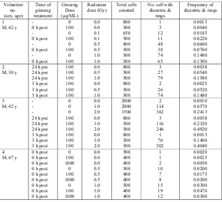
Table 1. Frequencies of chromosome aberrations induced by exposure to 0.1-2.0 Gy of gamma rays in blood sample treated with ginseng 24 pre-, 0 h post- and 3 h post-irradiation.
Across the 1.0 - 2.0 Gy in vitro radiation dose range used in this study, the yields of MN in PBL without ginseng treatment were consistently higher for higher doses of radiation. We found that a 24 h pre-treated ginseng crude water extract appeared to have very weak radioprotective effects as evidenced by MN analysis (Table 2). However, in general the addition of ginseng at the concentrations used in this study could not clearly decreased the 137Cs-induced MN in PBL.
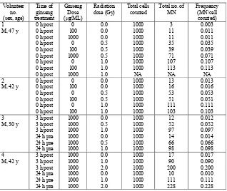
Table 2. Effects of ginseng treatment on MN yields in human irradiated and non-irradiated peripheral blood lymphocytes.
Volunteer
Different with study on mice, this study could not revealed a radioprotective effect of ginseng. In assessing the effect of ginseng extract in modifying the radiation-induced MN yield in human Go PBL with
immediately after or 3 hour after radiation exposure, resulted in no reduction in MN yields for all ginseng concentrations tested.
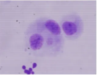
Figure 2. Micronuclei (small round shape besides binucleated cells in cytoplasm) induced by 0.5 Gy of gamma rays and treated with ginseng of 100 µg/ML 3 hour post irradiation.
These results indicated that ginseng crude water extract exerts no apparent cytogenetic effect on human PBL at all concentrations assessed as evaluated by the CBMN assay; and the protection of ginseng water extract against 137Cs-induced MN in human PBL is not known. Therefore, our findings could not clearly indicate that ginseng may have therapeutic value as a possible radioprotector for normal tissue during radiotherapy of cancer patients. Based on the limited quantitative information in the literature, this simple research addresses the issue of the radioprotective effects of ginseng on mammalian cells obtained from three human peripheral blood lymphocytes. The significant radioprotective effects of different ginseng preparations were not demonstrated in vitro by cytokinesis-blocked micronuclei assay. Results of research did not clearly indicate that the water-soluble extract of whole ginseng appears to give a protection against radiation - induced damage. However, although the exact underlying radioptrotective mechanism of ginseng is unclear, it could be through its
antioxidantative capability by scavenging free radicals responsible for DNA damage.
protection against radiation-induced lethality due to hematopoietic or gastrointestinal injury, other specific tissue damage, apoptosis, mutagenesis, and carcinogenesis.
Since the 1980’s, the radioprotective effects of Panax ginseng
(P. ginseng) and its partially purified constituents have been documentedin experimental models [3,18]. Ben-Hur and Fulder [3] demonstrated that in the presence of partially purified P. ginseng saponin mixture, Chinesehamster V79 fibroblasts are significantly more resistant tosubsequent -irradiation as determined by the ex vivo colonyforming assay. In the studies of cultured spleen lymphocytesfrom mice, application of P. ginseng water extract 48 h before -ray-irradiation has been shown to reduce the frequency of DNA double strand breaks, and it also has been demonstratedto reduce the degree of radiation-induced apoptosis in bothjejunal crypt cells and hair follicles [25]. In addition,Hsu et al. [26] found in ICR mice that the intraperitoneal injectionof a Chinese herbal medicine that contains 25% of P. ginsengroot before whole-body X-ray exposure (0–5 Gy) markedly enhanced the radiotolerance of bone marrow stem cells and peripheralhematocytes.
In contrast to the research in rodents, however, reports on the radioprotective effects of ginseng in human populations are very limited,
although one clinical study from Korea suggests that partially purified
P. ginseng componentsmay reduce morbidities and stimulate the recoveryof
hematopoietic functions in cancer patients [27]. Moreover,since unrepaired or misrepaired DNA damage in PBL may be responsiblefor micronuclei (MN) formation, Lee et al [18] recently assessed theeffect of P. ginseng
dried root crude water extract on the radiation-induced MN formation in human G0 PBL ex vivo using the cytokinesis-blockedmicronuclei assay and
found that treatment with ginseng24 h before 137Cs exposure (2 Gy) resulted in a linear declineof MN yields as ginseng concentration increased). These findings suggest thatthe ginseng crude water extract may contain a potential radioprotectiveconstituent with therapeutic value in dampening the damaging effects of ionizing radiation on normal tissues without exhibitingany negative effects on PBL. Obviously, experimental studies on the radioprotective potential of this important ginseng compound are criticallyneeded.
Song et al [19] reported that ginseng polysaccharide isolated from ginseng had a mitogenic activity and was also found to significantly increase the number of bone marrow cells, spleen cells, granulocyte-macrophage colony-forming cells, and circulating neutrophils, lymphocytes and platelets in irradiated mice. Pretreatment with ginseng protected mice from the lethal effects of ionizing radiation more effectively than when it was given immediately after or at various times after irradiation. A significant increase in the LD50/30 of mice pretreated with ginseng was also observed. These
It has long been known that ionizing radiation can interact directly with biological chromophores such as deoxyribonucleic acid (DNA) and in so doing, can damage those molecules. The use of ionizing radiation has become an integral part of modern medicine. It is used for diagnostic as well as therapeutic purposes. The therapeutic differentiation may be achieved with chemical radiation sensitizers or protectors. The development of radiation protectors is important not only to enhance the effectiveness of cancer treatment, but also for the study of the underlying mechanisms of radiation cytotoxicity [27]. A wide variety of compounds have been tested for radioprotective activity, including ginseng extract and many antioxidants [28,29]. Of all the compounds studied as potential radioprotective agents, unfortunately, available radioprotective substances possess unacceptable toxicity limiting its clinical usefulness and the precise mechanisms
responsible for radiation-induced cell death remain uncertain. Therefore, it is necessary to develop protectors that will minimize toxicity while
maintaining efficacy [30]. This was assessed in this very simple and preliminary experiment.
In our study, we found that ginseng did not renders protection against gamma-radiation induced DNA damage expressed as chromosomal aberration and micronuclei in human lymphocyte. This is different with recent studies which are demonstrate that ginseng exhibits protective activity against oxygen species in whole body irradiated mouse. The baseline of protecting action of plant extract is discussed in the following paragraphs. It seems that the strong free radical scavenging effects of P. ginseng have been extensively documented by many studies [8, 31, 32] where the radioprotective effect of P. ginseng has been closely linked to its antioxidativecapability through both the chelating of transition metal ions and the scavenging of free radicals responsible for DNA damage [33]. Lipid peroxidation leads to altered lysosomal membranepermeability and results in the release of hydrolytic enzymesin response to radiation-induced damage
in vivo: P. ginseng extract has been shown to inhibit lipid peroxidation
through transition metal chelation and scavenging of hydroxyl and superoxide radicals [31]. Recently, most effects of ginseng have been attributed to its antioxidant action. Moreover, ginseng has been shown to be strongly radioprotective through its ability to stimulate hematopoietic stem cells (colony-forming unit spleen) and produce a battery of cytokines such IL-1, IL-6, IL-12 and TNF-α. Its was also to be proven in study with mice that ginseng exerted its radioprotective effect through activation of antioxidant defense systems [4,25]. However, this was not seen in PBL cells irradiated in vitro with gamma rays as revealed by this study.
radiation cytotoxicity [35]. Hence search for an ideal radioprotector without side effects like hypotension, nausea, vomiting etc. and toxicity is a compelling urgency. These side effects limit its clinical usefulness. An ideal radioprotector also should be free from side effects, should be long acting, less expensive and capable of long term storage without change in action and constitution. Moreover, the most effective in vivo radioprotectors, however, like plant flavanoids and thiol compounds studied so far are effective when administered before irradiation, as they must be present in the system at the time of irradiation [35]. Hence they can be used only when the eventuality of the exposure is known and are not suitable against unplanned exposures, e.g. accidents, spillage, warfare and terrorist attack. Free radical scavenging may be a likely mechanism of action as the extract was found to possess significant hydroxyl radical scavenging activity.
This research focused on the effectivity of ginseng to attenuate the radiation effects expressed as chromosomal damages. Ginseng is typical Korean and Chinese plant and also cultivated in USA. In fact, Indonesia also has many types of plant such as Buah Merah, of which their extracts can be used as radioprotector. Buah merah or Red fruit (pandanous conoideus), an original traditional medicine of Papua Indonesia, was approved for healing for any degenerative diseases, such as cancer, tumor, hepatitis, lever disease, prostate, diabetes, gout, cholesterol, hypertension, stroke, and can be used for a healing of HIV [36,37]. Buah merah contains high concentration of active and important compounds such as beta-caroten, tocopherol, oleic acid, linoleic acid, and dekanoic. The research should be undertaken to check the
in vivo and in vitro radioprotective property of the aqueous extract of
Buah merah which remain unexplored for their effectiveness in suppressing
radiation damages especially chromosomal aberration/instability either by using rodent as experimental animal or pheripheral blood of Indonesian people. Further investigations are necessary to identify the active component(s) responsible for protection and to study their mechanism of action. This plant is widely available in our country, it is worthwhile to conduct detailed studies in order to explore the full potential of this plant in human radiation protection. Other plants, temulawak or wild ginger
(curcuma xanthorrhiza Roxb) and Brotowali (Tiospora rumpii boerl) is also
has prospective availability for cancer treatment, hence for suppressing chromosomal instability.
CONCLUSION
was only seen in chromosome aberration yields of sample irradiated with 2.0 Gy and treated with ginseng 3 h post irradiation rather than 24 h pre-irradiation. Opposite results that ginseng suspected to be a radiosensitizer
was found in some cases. This may be due to discrepancies exist in route of treatment and its fundamental mechanisms of protective action between both studies. Most effects of ginseng have been attributed to its antioxidant action and strongly radioprotective through its ability to stimulate hematopoietic stem cells.
ACKNOWLEDGEMENTS
The authors are greatly obliged to KIRAMS, Republic of Korea for providing all chemicals and instrumentations needed for this experiment. The valuable technical assistance of member staff of Laboratory of Radiation Cytogenetics and Epidemiology (KIRAMS) is gratefully acknowledged.
REFERENCES
1. KENNEDY, DO. and SCHOLEY, AB., Pharmacol. Biochem. Behav., 75, 687–700 (2003).
2. KITTS, D.D. and HU, C. Pub. Health Nut., 4, 473–485 (2000).
3. BEN-HUR, E. and FULDER, S., Am. J. Chin. Med., 14, 48–56 (1981).
4. SONG, J.Y., HAN, S.K., BAE, K.G., LIM, D.S., SON, S.J., JUNG, I.S., YI, S.Y. and YUN, Y.S., Radiat. Res., 159, 768-774 (2003).
5. CHANG, T.K.H., CHEN, J. and BENETTON, S.A., Drug Metabol. Dis., 30, 378–384 (2002).
6. LEE, T.K., JOHNKE, R.M., ALLISON R.R., O'BRIEN, K.F. and DOBBS, Jr, L.J., Mutagenesis, 20 (4), 237-243 (2005).
7. LEE, Y., JIN, Y., LIM, W. et al., J. Steroid Biochem Mol Biol, 84, 463-468 (2003).
8. ZHANG, D., YASUDA, T., YU, Y., et al., Free Radio. Biol. Med., 20, 145-150 (1996).
9. LIU, Z.Q., LUO, X.Y., SUN, Y.X., et al., Biochim. Biophys. Acta, 1572, 58-66 (2002).
10.LIU, Z.Q., LUO, X.Y., LIU, G.Z., et al., J. Agric. Food Chem., 51, 2555-2558 (2003).
11.ZHANG, J.S., SIGDESTAD, C.P., GEMMELL, M.A., GRDINA, D.J.,
Radiation Reserach, 112 (1) 156-63 (1987).
13.MONTORO, A., ALMONACID, M., SERRANO, J., SAIZ, M., BARQUINERO, J.F., BARRIOS, L., VERDU, G., PEREZ, J. and
VILLAESCUSA, J.I., Radiation Protection Dosimetry, 115 (1-4), 461-464 (2005).
14.HOFER, M., MAZUR, L., POSPISIL, M., WEITEROVA, L. and
ZNOJIL, V., Radiation Research, 154, 217-221 (2000).
15.MULLER, WU., and STREFTER, C., Adv. Mutagen. Res., 5, 1–133 (1994).
16.FENECH, M., BONASSI, S., TURNER, J., LANDO, C., CEPPI, M.,
CHANG, WP., HOLLAND, N., KIRSCH-VOLDERS, M. et al.,
Mutation Research, 534, 45–64 (2003).
17.CATENA, C., CONTI, D., PARASACCHI, P., MARENO, P. and
BORTOLATO, B., Int. J. Radiat. Biol., 70, 301–308 (1996).
18.LEE, T.K., ALLISON, R.R., O'BRIEN, KF., KHAZANIE, P.G., JOHNKE, R.M., BROWN, R., BLOCH, R.M., TATE, M.L., DOBBS, L.J., and KRAGEL, P.J., Mutation Research, 557, (1) 75-84 (2004).
19.SONG, J.Y., HAN, S.K., SON, E.H., PYO, S.N., YUN, Y.S. and YI, S.Y., Int. Immunopharmacol., 2, 857–865 (2002).
20.AHN, J.Y., CHOI, I.S., SHIM, J.Y., YUN, E.K., YUN, Y.S., JEONG, G.J. and SONG, J.Y., Eur. J. Immunol., 36, 37–45 (2006).
21.INTERNATIONAL ATOMIC ENERGY AGENCY, Cytogenetic
Analysis for Radiation Dose Assessment A Manual, Technical Reports Series No. 405, Vienna, Austria, (2001).
22.VRAL, A., VERHAEGEN, F., THIERENS, H. and DE RIDDER, L.,
Mutagenesis, 9, 439–443 (1994).
23.FENECH, M., Mutat. Res., 285, 35–44 (1993).
24.WEISS, JF. and LANDAUER, MR., Annals of the New York Academy of Sciences, 899, 44-60 (2000).
25.HAN, Y.S., SON, S.J., AKHALAIA, M., PLATONOV, A., SON, H.J., LEE, K.H., YUN, Y.S. and SONG, J.Y., Modulation of Radiation-Induced Disturbances of Antioxidant Defense Systems by Ginsan, Advance Access Publication, 4 October (2005).
26.HSU, M.J., LEE, S.S., LEE, S.T. and LIN, W.W., Ganoderma lucidum,
Br. J. Pharmacol., 139, 289-298 (2003).
27.HAHN, S.M., KRISHNA, M.C., SAMUNI, A., DEGRAFF, W.,
CUSCELA, D.O., JOHNSTONE, P. and MITCHELLl, JB., Cancer Res., 54, 2006s–2010s (1994).
29.PARK, Y.S., KIM Y.G., CHANG, J.C., and KIM, DY., J. Biochem. Mol. Biol., 26, 184–191 (1993).
30.WEISS, J.F., KUMAR, K.S., WALDEN, T.L., NETA, R., LANDAUR, M.R. and CLARK, E.P., Int. J. Radiat. Biol., 57, 709–718 (1990).
31.KITTS, D.D., WIJEWICKREME, A.N. and HU, C., Mol. Cell. Biochem., 203, 1–10 (2000).
32.KIM, Y.K., GUO, Q. and PACKER, L., Toxicology, 172, 149–156, (2002).
33.KIM, S.H., CHO, C.K., YOO, S.Y., KOH, K.H., YUN, H.G. and KIM, T.H., In Vivo, 7, 467–470 (1993).
34.NAIR, C.K.K., PARIDA, D.K. and NOMURA, T., Journal of Radiation Research, 42, 21-37 (2001).
35.HAHN, S.M., KRISHNA, M.C., SAMUNI, A., DEGRAFF, W.,
CUSCELA, D.O., JOHNSTONE, P. and MITCHELL, J.B.,
Cancer Research, 54, 2006-2010 (1994).
36.MUN’IM, A., ANDRAJATI, R., and SUSILOWATI, H., Majalah Ilmu
Kefarmasian, 3 (3), 153-161 (2006).