P-ISSN.2503-0817, E-ISSN.2503-0825
CrossMark
Abstract
Objective: Periodontal therapy success is dependent on proper diagnosis and removal of sub gingival tooth–borne deposits such as acquired pellicle, calculus and bacterial plaque biofilm besides, proper case selection and patient cooperation. From clinical perspective, enhanced visualization during the diagnostic and therapeutic periods has been given away to produce better results when matched to old-style approaches. Minimally invasive periodontal therapy appraises the benefits of using minimal invasive techniques, the knowledge available for improving visualization during therapy. This review reports the benefits and drawbacks of minimally invasive periodontal surgery and validation for current approach.
Methods: Keywords such as minimally invasive periodontal surgery, periodontal surgery and regeneration was used to search in Google and PubMed. Full text articles in English that were published from 1995 to 2016 are presented. Only case and control studies and randomized clinical trials were included in this review.
Results: Abstracts and articles published in other languages were not included in the review. This review makes available an evidence–based assessment of the knowledge and procedures.
Conclusion: Minimally invasive periodontal surgery might be considered a true reality in the field of periodontal regeneration. Clinical improvements seen were consistently associated with very limited morbidity.
Keywords: Minimally invasive periodontal surgery, Periodontal surgery, Regeneration
Cite this Article: Reddy SM, Abdelmagyd HAE, Shetty SR, Khazi SS, Vannala VR. 2017. Minimal invasive periodontal surgery-a review. Journal of Dentomaxillofacial Science 2(2): 81-85. DOI: 10.15562/jdmfs.v2i2.522
Minimal invasive periodontal surgery
:
a review
Sesha M. Reddy,1* Hossam A.E. Abdelmagyd,1 Shishir R. Shetty,2
Shakeel S. Khazi,3 Venkata R. Vannala4
Introduction
Minimally Invasive Surgery (MIS) was established based on the idea of using small incisions to complete surgical techniques until that time had been performed through bigger surgical access. The term MIS was first introduced to periodontal surgical procedures in 1995. The MIS is a name that refers to the presentation of clear-cut and gentle invasive procedures that necessitate the usage of amplifying maneuvers, alike dental operating microscopes or dental magnifying lenses and periodontal micro-surgical instruments and materials.1
Methods
An internet search using Google and PubMed search engine and key words was carried out. Full text articles in English that were published from 1995 to 2016 are presented. Only case and control studies and RCT were included in this review. The systemic features, study design, time period, treat-ment rendered and level of evidence are presented in table 1.
Results
Regeneration of periodontal intra-bony defects has been achieved through different therapeutic methodologies such as barrier membranes,
demineralized freeze-dried bone allograft (DFDBA) and GTR with grafts and enamel matrix derivative (EMD), EMD and rhPDGF-BB with β-tricalcium phosphate.2
Information attained from numerous well-ordered clinical trials, meta-analyses and methodical reviews disclose additional benefits in relation to clinical attachment level (CAL), decrease as well as gain in probing pocket depth when matched with open flap debridement alone.3-4 Most recent specific importance remained committed towards the surgical plan and execution of the surgical procedures for periodontal regen-eration. Definite invasive periodontal methods are presently recommended to handle the soft tissues and to attain stable flap closure in order to avoid tissue collapse and cover the region of periodontal defects from exposure to oral environment.5
Subsequently, cutting-edge techniques have been used to further upsurge periodontal surgical efficacy, by means of operating microscopes and microsurgical instruments in terms of MIPS has been put forward, and the practice of a microsur-gical method now amalgamation through diverse regenerative materials lead to appropriate initial flap approximation in more than 92% of the treated defects for the total healing phase.6–7
The MIPS technique allows for minimization of soft tissue trauma and the removal of granula-tion tissue from periodontal defects using a much smaller surgical incision than that used in standard 1Department of Periodontics,
College of Dentistry, Gulf Medical University, Ajman, UAE
2Department of Oral Medicine & Radiology, College of Dentistry, Gulf Medical University, Ajman, UAE
3Department of Prosthodontics, College of Dentistry, Gulf Medical University, Ajman, UAE
4Department of Orthodontics, College of Dentistry, Gulf Medical University, Ajman, UAE
*Correspondence to: Sesha M. Reddy,
Department of Periodontics, College of Dentistry, Gulf Medical University, Ajman, UAE
Table 1 Clinical studies using minimal invasive periodontal surgery technique
No. Authors Study design Time Treatment Patient group level of Evidence Results
1 Harrel SK et al 11
Cohort Study 11 months EMP + MIST 160 sites in 16 patients.
Level II Combination of MIS and EMP yields significant reductions in probing depths (PD) and improvements in clinical attachment levels (CAL), while producing little or no increase in recession.
2 Cortellini P et al14 Case, cohort 1 year MIST + EMD 13 intra-bony defects + MIST.
Level III MIST associated with EMD resulted in excellent clinical improvements while limiting patient morbidity.
3 Cortellini P et al15 Case cohort study
1 year MIST + EMD + deep intra-bony defects.
40 intra-bony defects + MIST.
Level III MIST with EMD, resulting in excellent clinical outcomes with very limited intra- and post-operative morbidity.
4 Cortellini P et al16 Case cohort study
1 year MIST + EMD + deep intra-bony defects.
44 intra-bony defects + MIST along with EMD.
Level III MIST + EMD showed excellent clinical outcomes and very limited patient morbidity.
5 Cortellini P et al17 Cohort Study 1 year Modified minimally invasive surgical technique (M-MIST) + deep intra-bony defects + EMD
20 + intra-bony defects
Level II M-MIST resulted in very limited patient morbidity and excellent clinical improvements.
6 Cortellini P et al8 Case cohort study
1 year Evaluate the healing response of MIST + EMD + deep intra-bony defects.
40 intra-bony defects + MIST + EMD, microscope and microsurgical instruments used.
Level III Defect morphology and bleeding tendency seem to influence clinical outcomes from the use of MIST in combination with EMD.
7 Ribeiro FV et al18 Cohort Study 6 months MIST + EMD + intra-bony defects.
20 intra-bony defect + MIST + EMD.
Level II MIST + EMD promoted significant improvements in clinical parameters
8 Harrel SK et al 19
Cohort study 6 years 6-year results of prospective study.
142 sites in 13 patients.
Level II MIST + EMD yielded significant reductions in PD and improvement in CAL while producing no detectable recession. 11-month results remained stable at 6 years.
9 Cortellini P et al20
RCT 1 year M-MIST alone and
combined with EMD or EMD + xenograft (BMDX) + intra-bony defects.
45 deep isolated intra-bony defects
Level I M-MIST with or without regenerative materials resulted in significant clinical and radiographic improvements.
10 Ribeiro FV et al21 RCT 6 months EMD + MIST + intra-bony defects.
30 patients with intra-bony defects were randomly assigned to 1) MIST plus EMD or 2) MIST alone.
bone graft techniques. Periodontal surgery has been enhanced with this atypical and pioneering methodology in recent past. The objective of minimally invasive surgery was to en route for appropriate wound healing, minimal flap reflection and precise management of both soft and hard tissues during periodontal surgical procedures. Authors recom-mending MIST, also stressed upon characteristics of wound healing, clot stability besides initial flap closure for clot protection. These ideas indicated further for modified minimally invasive surgical technique (M-MIST), which suggested integrating the theory of space maintenance for periodontal regeneration.8
Authors have recommended the practice of an operative microscope in periodontal regenerative
surgery and suggested an improved capability to handle the soft tissues that give rise to enhanced prospective aimed at primary wound stability for an average 70% of the cases obtained through regu-lar surgery in comparison to an exceptional 92% achieved with the use of microsurgery technique.9
Authors have proposed two dissimilar MIST, one technique that results in the reflection of a minimal facial flap, interdental papilla as well as palatal flap. Second procedure flap reflection is limited to the facial flap only. These techniques are intended for the management of shallow as well as deep intra-bony periodontal defects.10
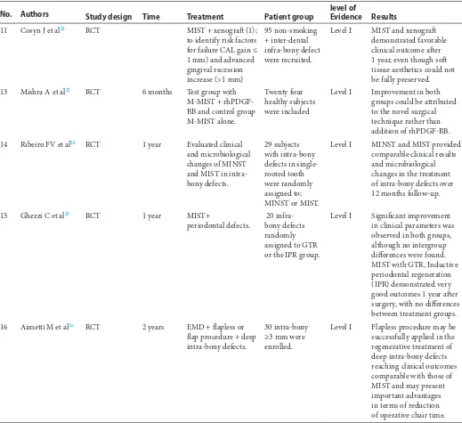
Review of the current scientific literature was undertaken to evaluate the efficacy of MIPS in the treatment of periodontal Table 1 Continued
No. Authors Study design Time Treatment Patient group level of Evidence Results
11 Cosyn J et al22 RCT MIST + xenograft (1);
to identify risk factors for failure CAL gain ≤ 1 mm) and advanced gingival recession increase (>1 mm)
95 non-smoking + inter-dental infra-bony defect were recruited.
Level I MIST and xenograft demonstrated favorable clinical outcome after 1 year, even though soft tissue aesthetics could not be fully preserved.
13 Mishra A et al23 RCT 6 months Test group with
M-MIST + rhPDGF-BB and control group M-MIST alone.
Twenty four healthy subjects were included
Level I Improvement in both groups could be attributed to the novel surgical technique rather than addition of rhPDGF-BB.
14 Ribeiro FV et al24 RCT 1 year Evaluated clinical
and microbiological changes of MINST and MIST in intra-bony defects.
29 subjects with intra-bony defects in single-rooted tooth were randomly assigned to; MINST or MIST.
Level I MINST and MIST provided comparable clinical results and microbiological changes in the treatment of intra-bony defects over 12 months follow-up.
15 Ghezzi C et al25
RCT 1 year MIST+
periodontal defects.
20 infra-bony defects randomly assigned to GTR or the IPR group.
Level I Significant improvement in clinical parameters was observed in both groups, although no intergroup differences were found. MIST with GTR, Inductive periodontal regeneration (IPR) demonstrated very good outcomes 1 year after surgery, with no differences between treatment groups.
16 Aimetti M et al26
RCT 2 years EMD + flapless or
flap procedure + deep intra-bony defects.
30 intra-bony ≥3 mm were enrolled.
defects and its impacts on clinical outcomes, surgi-cal chair-time, side effects, and patient morbidity.
Advantages of MIPS: less operative pain and trauma, no scarring, speeds recovery and reduces the incidence of postsurgical complications. Thermal sensitivity is rare because incisions are limited to anatomical areas. Post-operative gingi-val recession is minimal or non-existing, there is no tendency for deeper probing depths to reoccur over 6 or more years postoperatively, thus, although techniques of MIPS may encounter further advan-tages, other disadvantages of such methods should also be taken into consideration.
Disadvantages of MIPS: it requires special equipment, specialist training is probably required, some additional equipment’s could be more expen-sive, and some procedures may take longer than usual, compared with conventional surgeries. Armamentarium used: mostly, a useful microsurgi-cal tray for the routine use in MIPS should include: A. Micro-periosteal elevator, B. Bone scraper, C. 12b blade, D. Micro-scalpel holder, E. Needle holder, F. Micro-scissor, G. A dental micro-forceps, modified orban knife, monofilament suturing materials and use of microscopes has added advantage.
Features of minimally invasive periodontal surgery: diagnosis of intra-bony defects, incision places, flap elevation, preservation of papilla, suture as well as suturing technique used and use of microscopes, microsurgical instruments, and post-operative healing all are discussed.
Incisions used in MIPS are intended to preserve soft tissue as much as possible. Incisions used in for interproximal defects are sulcular incision which are separate and not continuous. By doing so it retains interproximal papilla and its height. Later horizontal incisions are used to connect the instar sulcular incisions.
Flap elevation is done by using modified orban knife as it creates sharp dissections which even-tually preserves interproximal papilla, soft tissue high, and retains good blood supply to flap.11
Papilla preservation: in aesthetic areas can be preserved by giving horizontal incisions in the pala-tal area this will help to preserve the shape of the papilla. In non-aesthetic areas horizontal incisions can be places buccally or lingually base on the need to cover the graft material.12
Visualization and Debridement: is best done by using magnifications. For debridement of granu-lation tissue use of slim line inserts with after five curettes are used.9
Suture technique: in the anterior areas, it is recommended to use the vertical matrix suture. In the premolar and/or molar areas, the use of modified matrix suture is a better choice. These techniques help removing the collapse of gingiva
and enhancing optimal adaptation of wound edges. Continuous suturing may be achieved wherever releasing-incisions have been done.13
Discussion
The difference in MIPS lies in the technique of accessing the periodontal defects, handling of soft tissue, method of debridement, flap closure, reduce surgical chair time and minimize patient discom-fort and side effects. These technical differences are significant compared to the tradition periodontal surgical techniques.
In reality tradition periodontal surgical tech-niques relies on wide incision and wide access to the root and bone in order for proper visualization of bone and root surface and with advancement in periodontal surgery with the use of MIPS, the same can be accessed by smaller opening so the need for wide access has to be justified or reassessed.
Lastly patient acceptance and satisfaction for MIPS has been excellent. It is difficult to quantify but periodontal treatment done by MIPS technique appears to be more frequently and easily accepted by patient. Minimally invasive periodontal surgery provide an additional tool to periodontist to treat periodontal disease more effectively. Whatever said and done MISP require a specific training and the use of dedicated instruments and materials are to be successfully applied to the treatment of intra-bony defects.
Conclusion
Applying MISP concepts in the field of periodontal surgery is found to be of great importance. The main advantages of MIPS are the following: A. Reduce surgical trauma, patient discomfort, and time spent on the procedure, B. Enhances flap/ wound stability, C. Provide primary flap closure, and D. Minimize unwanted complications and side effects. Disadvantage of MIPS is that it cannot be applied to all cases. A stepwise decisional algorithm must be drawn first to support clinician’s judgement while indicating this treatment approach.
Conflict of Interest
The authors report no conflict of interest.
References
1. Harrel SK, Rees TD. Granulation tissue removal in routine and minimally invasive procedures. Compendium of Continuing Education in Dentistry 1995;16: 960–967. 2. Mark AR, Richard TK, Paulo MC et al. Periodontal
the AAP regeneration workshop. J Periodontol 2015;86: 105–107.
3. Murphy KG, Gunsolley JC. Guided tissue regeneration for the treatment of periodontal intrabony and furcation defects: a systematic review. Ann Periodontol 2003;8: 266–302.
4. Needleman I, Tucker R, Giedrys LE, et al. Guided tissue regeneration for periodontal intrabony defects: a cochrane systematic review. Periodontol 2000 2005;37: 106–123.
5. Cortellini P, Prato GP, Tonetti MS. The simplified papilla preservation flap: a novel surgical approach for the management of soft tissues in regenerative procedures. Int J Periodontics Restorative Dent 1999;19: 589–599. 6. Cortellini P, Tonetti MS. Microsurgical approach to
periodontal regeneration: Initial evaluation in a case cohort. J Periodontol 2001;72: 559–569.
7. Cortellini P, Tonetti MS. Clinical performance of a regen-erative strategy for intrabony defects: Scientific evidence and clinical experience. J Periodontol 2005;76: 341–350. 8. Cortellini P, Tonetti MS. Improved wound stability with
a modified minimally invasive surgical technique in the regenerative treatment of isolated interdental intrabony defects. J Clin Periodontol 2009;36: 157–163.
9. Leonard ST, Dennis S. Principles and practice of periodontal microsurgery. Int J Microdent 2009; 1:13–24.
10. Dannan A. Minimally invasive periodontal therapy. Journal of Indian Society of Periodontology 2011;15: 338–343.
11. Harrel SK. A minimally invasive surgical approach for periodontal regeneration. Surgical technique and observation. J Periodontal 1999;70: 1547–1557.
12. Harrel SK, Wilson TG, Nunn ME. Prospective assessment of the use of enamel matrix proteins with minimally invasive surgery. J Periodontol 2005;76: 380–384.
13. Cortellini P, Tonetti MS. A minimally invasive surgical technique with an enamel matrix derivative in the regenerative treatment of intra-bony defects: a novel approach to limit morbidity. J Clin Periodontol 2007;34: 87–93.
14. Cortellini P, Tonetti MS. Minimally invasive surgical technique and enamel matrix derivative in intra-bony defects. I: clinical outcomes and morbidity. J Clin Periodontol 2007;34: 1082–1088.
15. Cortellini P, Nieri M, Prato GP, et al. Single minimally invasive surgical technique with an enamel matrix derivative to treat multiple adjacent intra-bony defects: clinical outcomes and patient morbidity. J Clin Periodontol 2008;35: 605–613.
16. Cortellini P, Pini-Prato G, Nieri M, et al. Minimally invasive surgical technique and enamel matrix derivative in intrabony defects: 2. Factors associated with healing
outcomes. Int J Periodontics Restorative Dent 2009;29: 257–265.
17. Ribeiro FV, Nociti JFH, Sallum EA, et al. Use of enamel matrix protein derivative with minimally invasive surgical approach in intra-bony periodontal defects: clinical and patient-centered outcomes. Braz Dent J 2010;21: 60–67. 18. Harrel SK, Wilson TGJR, Nunn ME. Prospective
assessment of the use of enamel matrix derivative with minimally invasive surgery: 6-year results. J Periodontol 2010;81: 435–441.
19. Cortellini P, Tonetti MS. Clinical and radiographic outcomes of the modified minimally invasive surgical technique with and without regenerative materials: a randomized-controlled trial in intra-bony defects. J Clin Periodontol 2011;38: 365–373.
20. Ribeiro FV, Casarin RC, Junior FH, et al. The role of enamel matrix derivative protein in minimally invasive surgery in treating intrabony defects in single-rooted teeth: a randomized clinical trial. J Periodontol 2011;82: 522–532. 21. Cosyn J, Cleymaet R, Hanselaer L, et al. Regenerative
periodontal therapy of infrabony defects using minimally invasive surgery and a collagen-enriched bovine-derived xenograft: a 1-year prospective study on clinical and aesthetic outcome. J Clin Periodontol 2012;39: 979– 986.
22. Mishra A, Avula H, Pathakota KR, et al. Efficacy of modified minimally invasive surgical technique in the treatment of human intrabony defects with or without use of rhPDGF-BB gel: a randomized controlled trial. J Clin Periodontol. 2013;40: 172–179. 23. Ribeiro FV, Casarin RC, Palma MA, et al. Clinical and
microbiological changes after minimally invasive therapeutic approaches in intrabony defects: a 12-month follow-up. Clin Oral Investig. 2013;17: 1635– 1644.
24. Ghezzi C, Ferrantino L, Bernardini L, et al. Minimally invasive surgical technique in periodontal regeneration: a randomized controlled clinical trial pilot study. Int J Periodontics Restorative Dent 2016;36: 475–482.
25. Aimetti M, Ferrarotti F, Mariani GM, et al. A novel flapless approach versus minimally invasive surgery in
periodontal regeneration with enamel matrix derivative proteins: a 24-month randomized controlled clinical trial. Clin Oral Investig 2016;21: 327–337.