Journal of Biology, Agriculture and Healthcare www.iiste.org ISSN 2224-3208 (Paper) ISSN 2225-093X (Online)
Vol.6, No.2, 2016
AQUEOUS EXTRACT OF PURPLE SWEET POTATO
INCREASED SOD-2, AND SOD-3 EXPRESSION ON HUMAN
UMBILICAL VEIN ENDOTHELIAL CELLS
IN VITRO
I Made Jawi1, Wiwiek Indrayani1, I G K Arijana2, A A N Subawa3,Dewa Ngurah Suprapta4* 1. Department of Pharmacology, 2. Department of Histology,
3 Department of Clinical Pathology, Faculty of Medicine,
Udayana University, Jl. PB. Sudirman Denpasar Bali Indonesia
4.Faculty of Agriculture, Udayana University Jl. PB. Sudirman Denpasar Bali Indonesia.
Abstract
Oxidative stress is thought to be one of the causes of impaired function of vascular endothelial cells, causing a variety of cardiovascular disorders. Studies in various animal experiments proved the aqueous extract of purple sweet potato tuber has antioxidant potential, through the mechanism of enhancement of endogenous antioxidants such as superoxide dismutase (SOD), that protects endothelial cells from oxidative stress. Effect of aqueous extract of purple sweet potato tuber in increasing SOD has not been proven in human vascular endothelial cells. This study aims to prove whether aqueous extract of purple sweet potato tuber may increase the expression of SOD-2 and SOD-3 in human vascular endothelium. This research was an experimental study on umbilical vein endothelial cells experiencing oxidative stress created by administering H2O2 in vitro, and protected with aqueous extract of purple sweet potato tuber at various concentrations. Observation of the SOD-2 and SOD-3 on endothelial cells was done by using immunohystochemestry with monoclonal antibodies. The results showed a significant (p < 0.05) increase in SOD-2 and SOD-3 on endothelial protected with aqueous extract of purple sweet potato tuber at concentration of 1.5625- 3.125 µg/ml for SOD-2 and at concentration of 1.5625- 25µg/ml for SOD-3. From these results it can be concluded that the aqueous extract of purple sweet potato tuber could protect endothelial cells from oxidative stress through increasing expression of SOD-2 and SOD-3.
Keywords: Aqueous extract of purple sweet potato, SOD, HUVEC.
1. Introduction
Cardiovascular disease has been known as a leading cause of the death until now. The disease is thought to be closely related to the oxidative stress. Vascular endothelium is highly sensitive to the oxidative stress, particularly caused by superoxide ions. These ions should be neutralized with antioxidants. Otherwise they will cause endothelial dysfunction and ultimately will lead to various cardiovascular disorders (Lin et al., 2005). The main sign of endothelial dysfunction is a disturbance in the process of endothelium-dependent vasodilation (Stangl and Stangl, 2010), which is determined by the presence of nitric oxide (NO) in the endothelium through the activity of endothelial nitric oxide synthase (eNOS). Superoxide ions will decrease the bioavailability of NO and inhibit the synthesis of NO, and increase eNOS uncoupling, and consequently will exacerbate oxidative stress (Droge, 2002). The use of a variety of both exogenous and endogenous antioxidants can be expected to be addressed to reduce oxidative stresses. Endogenous antioxidants are believed to be able to protect the endothelial function through ion superoxide i.e. superoxide dismutase (SOD). SOD is an antioxidant enzymes that can be divided into three namely; SOD-1 (CuZn SOD, cytosolic), SOD-2 (Mn SOD, found in mitochondria) and SOD-3 (extracellular SOD). In previous study it was found that SOD-2 has an important role on endothelial function (Ohashi et al., 2006; Miller et al., 2009), in addition to the role of SOD-3 (Iida et al., 2005).
Vascular endothelial function can also be improved by consuming foods that contain flavonoids as exogenous antioxidants (Middleton et al., 2000). Flavonoids may also reduce LDL sensitivity to the effects of free radicals through its activity as an antioxidant (Kelley et al., 2006). Anthocyanin pigment is one of important flavonoids with quite high concentration in the purple sweet potato tubers in Bali (Suprapta et al., 2004), and has been studied in vivo to posses antioxidant effects in various experimental animals (Jawi et al., 2008; Jawi et al., 2011), as well as can increase the expression of eNOS in hypertensive rats (Jawi et al., 2012). Antioxidant efficacy of the purple sweet potato tubers, especially in human endothelial was unknown so that needs to be investigated, whether the water extract of purple sweet potato tuber can increase the expression of superoxide dismutase in particular SOD-2 and SOD-3 in human vascular endothelium exposed by H2O2in vitro.
2. Materials and Method
Journal of Biology, Agriculture and Healthcare www.iiste.org ISSN 2224-3208 (Paper) ISSN 2225-093X (Online)
Vol.6, No.2, 2016
potato tuber (Ipoema batatas L.) as the test material. Observations on the expression of SOD-2 and SOD-3 was performed based on immunohistochemical method with monoclonal antibodies of SOD- 2 and SOD-3. Aqueous extract of purple sweet potato tuber was obtained from the juice of the purple sweet potato tubers obtained from the farmers in Marga, Tabanan, Bali. Aqueous extracts were made by the following procedure: purple sweet potato tubers were washed with clean water and then peeled out, and cut into pieces crosswise with a thickness of 2-2.5 cm.
The sweet potato chunks were mixed with water (1 kg chunks : 1 liter water) and then blended in a blender. Filtrate was obtained by filtering the mixture through three layers of cheese cloth. The filtrate was then boiled for 30 minutes, for sterilization. This filtrate was dried up with a rotary evaporator until dryness to get an extract to be used to study its effect on human endothelial cells cultured in vitro. The dried extract was diluted by RPMI 1640 medium to obtain concentrations of 50, 25, 12.5, 6.25, 3.125, 1.5625µg/ml. The expression of SOD-2 and SOD-3 was performed by using a single 96 micro plate wells. The HUVECs were cultured in RPMI1640 medium supplemented with 10% FBS, 100 U/mL penicillin and 100 µg/mL streptomycin in a humidified incubator under 5% CO2 at 37°C. Briefly, cells at the mid-log phase were seeded in a 96-well plate at a density of 104 cells per well in 100 µL medium. RPMI1640 medium was added to the control and model groups. Then the original culture medium was removed and cells were washed with PBS twice. Antioxidant activity was evaluated by using HUVEC injury model induced by H2O2. Cells were treated with H2O2 (1.250 µmol/L) for 2 hours. Then, 20 µL of MTT solution (5 mg/mL) was added to each well. After the evaluation of cell culture, the cells lived on cultures taken back by the media given the concentration of H2O2 with 1.250 mol/L as a model of oxidative stress and given the test material with a concentration of aqueous extract of purple sweet potato tuber, respectively 50, 25, 12.5, 6.25, 3.125, 1.5625µg/ml, then incubated for 24 hours. After 24 h, live cells from each of the wells were taken with a special pipette and dripped and blotted on glass object to be painted with immunohistochemical techniques.
The slides were fixed for immunocytochemistry staining performed with the following steps. First the slides were washed with PBS and let stand for 5 minutes and the PBS was discarded and added with H2O2 and kept for 5 minutes. The slides were washed with PBS 4 times and then were dropped into 100 mL Ultra V Block, and incubated for 5 minutes. The slide was washed with PBS, and droped with eNOS antibody, SOD-2 and SOD-3 (BIOS, USA) which were diluted 1: 200, respectively and incubated for 1.5 h in an incubator with a temperature of 250C. The remaining antibody was removed and washed with PBS 4 times. This slide was added with 100 µl Biotinylated Goat Anti-Polyvalent and incubated for 5 minutes at room temperature. The slide was washed with PBS 4 times and added with 100 µl Streptavidin Peroxidase and incubated for 5 minutes. Afterward, it was washed with PBS 4 times and added to the DAB for 5 minutes and then washed with aqua bides. The last step was staining with Meyer’s Hematoxyline as counterstaining and covered with a coverslip. Slides are ready to be observed under microscope ( Olympus CX41, Tokyo, Japan). On each slide 5 fields of views were observed with 40 times magnification. The data obtained were tested by ANOVA to determine the differences between groups. To prove the existence of the endothelium in the smear, observation was done in an inverted microscope with camera (Olympus DP 12, Tokyo, Japan).
3. Results
The examination of SOD-2 and SOD-3 showed a significant difference between the oxidative stress group with the treatment group, that were treated with aqueous extract of purple sweet potato tuber at concentrations 50, 25, 12.5, 6.25, 3.125, 1.5625µg / ml respectively. The results of the calculation of the average endothelial cells overexpressing SOD-2 and SOD-3 are presented in Figures 1 to 4.
0
Journal of Biology, Agriculture and Healthcare www.iiste.org ISSN 2224-3208 (Paper) ISSN 2225-093X (Online)
Vol.6, No.2, 2016
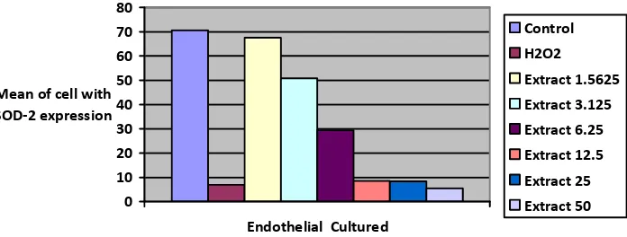
Figure 1 description:
Controls were culture of endothelial cell without treatment/in normal medium.
H2O2 were the culture endothelial cells treatment with H2O2 in the medium (Oxidative Stress). Extract 1.5625 were the culture of endothelial cells with H2O2 and aqueous extract of purple sweet potato tuber with a concentration of 1.5625 µg/ml and incubated for 24 hours. Extract 3.125 were the culture of endothelial cells with H2O2 and aqueous extract of purple sweet potato tuber with a concentration of 3.125 µg/ml and incubated for 24 hours. Extract 6.25 were the culture of endothelial cells with H2O2 and aqueous extract purple sweet potato tuber with a concentration of 6,25 µg/ml and incubated for 24 hours. Test showed average difference between the groups was: Oxidative Stress vs. Control, p <0.001. Control vs. Extract 1.5625, p> 0.05. Extract 6.25 vs. control p <0.05. Oxidative stress, vs. Extract 12.5, 25, and 12.5 p> 0.05.
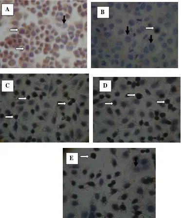
Figure 2 Endothelial cells with SOD-2 positive by immunohistochemical method
Figure 2 description:
A. Control endothelial cells culture. The endothelial cells express the SOD-2 (white arrows), seen as brown color in the cytoplasm, because of the SOD-2 localization in cytoplasm of the cell. Endothelial cells that do not express SOD-2 have pale color ( black arrows).
B. Treatment with H2O2, a decrease in the number of endothelial expression of SOD-2, and more endothelial cells without SOD-2 expression (black arrows).
A
B
D
C
Journal of Biology, Agriculture and Healthcare www.iiste.org ISSN 2224-3208 (Paper) ISSN 2225-093X (Online)
Vol.6, No.2, 2016
C. Treatment 1(15625), endothelial cells appear to express SOD-2 with higher intensity than the treatment with H2O2 only. D. Treatment 2 ( 3.125), endothelial cells appear to express SOD-2 with higher intensity than the treatment with H2O2 only. E. Treatment 3 (6.25), endothelial cells appear to express SOD-2 with higher intensity than the treatment with H2O2 only, but lower than the treatment 2
0 20 40 60 80 100
Cuture of endothelial
Mean of cells
with SOD-3
expression
Control
H2O2
Extract1.5625
Extract 3.125
Extract 6.25
Extract 12.5
Extract 25
Extract 50
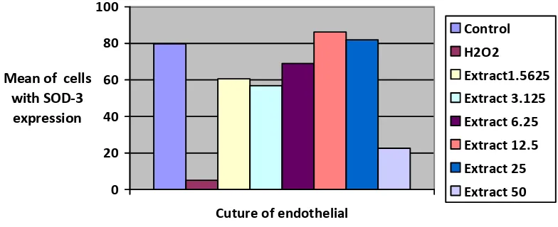
Figure 3
Average expression of SOD-3 on Endothelial Cells by Immunohistochemistry method with SOD-3 Antibody
Figure 3 description:
Controls were the culture of endothelial without treatment / in normal media.
H2O2 are the culture of endothelial that given H2O2 in the medium (as a Oxidative Stress ). Extract 1.6525 are the culture of endothelial that given H2O2 and purple sweet potato tuber aqueous extract at concentration of 1.6525 µg/ml and incubated for 24 hours. Extract 3.125 were the culture of endothelial that given H2O2 and purple sweet potato tuber, aqueous extract at concentration of 3.125 µg/ml and incubated for 24 hours. Extract 6.25 were the culture of endothelial that given H2O2 and purple sweet potato tuber aqueous extract at concentration of 6.25 µg/ml and incubated for 24 hours. Extract 12.5 were the culture of endothelial that given H2O2 and purple sweet potato tuber aqueous extract at concentration of 12.5 µg/ml and incubated for 24 hours. Extract 25 were the culture of endothelial that given H2O2 and purple sweet potato tuber aqueous extract at concentration of 25 µg/ml and incubated for 24 hours. Extract 50 were the culture of endothelial that given H2O2 and purple sweet potato tuber aqueous extract at concentration of 50 µg/ml and incubated for 24 hours. Test showed average difference between the groups was: Oxidative Stress vs. Control, and all of extract p
Journal of Biology, Agriculture and Healthcare www.iiste.org ISSN 2224-3208 (Paper) ISSN 2225-093X (Online)
Vol.6, No.2, 2016
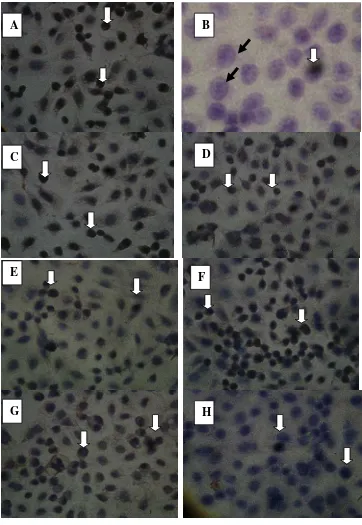
Figure 4
Endothelial cells with SOD-3 positive by immunohistochemical method
Figure 4 description:
A. Control endothelial cells culture. The endothelial cells express the SOD-3 (white arrows), seen as brown color in the cytoplasm and the membrane of the cells, because of the SOD-3 localization in cytoplasm and membrane of the cell/extracellular compartment.
B. Treatment with H2O2, a decrease in the number of endothelial expression of SOD-3, and more endothelial cells without SOD-3 expression (black arrows).
C. Treatment 1, endothelial cells appear to express SOD-3 with higher intensity than the treatment with H2O2 only. D. Treatment 2, endothelial cells appear to express SOD-3 with higher intensity than the treatment with H2O2 only. E. Treatment 3, endothelial cells appear to express SOD-3 with higher intensity than the treatment
A
E
C
B
D
F
Journal of Biology, Agriculture and Healthcare www.iiste.org ISSN 2224-3208 (Paper) ISSN 2225-093X (Online)
Vol.6, No.2, 2016
with H2O2 only, but lower than the treatment 4 (Concentration 12.5). F . Treatment 4, endothelial cells appear to express SOD-3 with highest intensity. G and H (treatment 5 and 6) , endothelial cells appear to express SOD-3 with lower intensity than treatment 4.
4. Discussion
Administration of H2O2 for 24 hours on vascular endothelium cells cultured in vitro significantly (p<0.05) reduced the expression of SOD-2 and SOD-3. Aqueous extract of purple sweet potato tuber could maintain the expression of SOD-2 and SOD-3 at concentrations 1.5625µg/ml-6.25µg/ml for SOD-2 and at concentrations 1.5625µg/ml-25µg/ml for SOD-3.
Treatment with H2O2 resulted in endothelial oxidative stress, and caused a decrease in SOD. H2O2 will cause activation of NADPH oxidase, thus causing an increase in endothelial cell superoxide ions (Coyle et al., 2006). The provision of H2O2, increase of superoxide ions and accompanied by the activation of oxidant-generating enzymes that would otherwise be causing endothelial dysfunction (Coyle et al., 2007).
Aqueous extract of purple sweet potato tuber can maintain the expression of SOD-2 and SOD-3 due to the antioxidant properties of anthocyanin/flavonoid contained in the extract. The content of anthocyanins from purple sweet potato tuber variety, in Switzerland ranging from 0.7 mg / 100 g up to 74.3 mg / 100 g (Lachman et al., 2009). Anthocyanin content of purple sweet potato from China 132 mg / 100 g (Jiao et al., 2012) whereas in Bali ranges from 110mg / 100 g up to 210mg / 100 g fresh tuber (Suprapta et al., 2004). Anthocyanin content of the aqueous extracts of purple sweet potato tuber in Bali can be a powerful antioxidant that can increase SOD on cultured endothelial given H2O2. Thus, the increase of SOD-2 and SOD-3 in the endothelium given aqueous extract of purple sweet potato tubers in this study, is due to the antioxidant properties of anthocyanins. The extract containing anthocyanin, can capture H2O2 and the lead levels would decrease oxidative stress of H2O2 (Gould et al., 2002).. In cultured endothelial given aqueous extract of purple sweet potato tuber, SOD were significantly increased (p <0.05).\ Aqueous extract of purple sweet potato tubers can reduce oxidative stress, because it contains anthocyanin (Suprapta et al., 2004; Lachman et al., 2009; Jiao et al., 2012) as a powerful antioxidant in vitro (Lachman et al.,2009; Jiao et al., 2012; Padda, 2006) and in vivo (Jawi et al., 2008; Kano et al., 2005; Garcia-Alonso et al., 2009). Anthocyanins can be as an antioxidant with a variety of mechanisms, for example through the effects of free-radical scavenging (Lila, 2004), by increasing the variety of antioxidant enzymes such as SOD, through activation of the antioxidant response element (Shih et al., 2007). Anthocyanins may also interact with transition metal ions (chelating) such as with Fe and Cu, thus preventing the formation of free radicals (Gomes et al., 2008).
This result consistent with studies in rats given a purple potato extract increased expression of SOD in rat liver tissue (Kyu-Ho et al., 2006; Chen et al., 2011). These results are also consistent with studies conducted by anthocyanins from black rice can reduce superoxide ions and increase SOD in cells in vitro (An-Na et al., 2006). Several studies that examining the benefits of anthocyanins from various plants, show the similar result. Research conducted in experimental animals, proving anthocyanins can reduce oxidative stress in various tissues such as the brain tissue (Shan et al., 2009), the liver (Kyu-Ho et al., 2006), and vascular endothelial (Speciale et al., 2010). Research on tissue culture with anthocyanin extracts of black rice was significant decrease in oxidative stress through a decrease in the formation of superoxide ions and increase in SOD (An-Na et al., 2006).
The results of this study is somewhat different from other studies because the type of anthocyanins in purple sweet potato tuber aqueous extract has not been identified. Specific anthocyanin antioxidants have different effects, so as to improve the ability of different SOD (Khoo et al., 2013).
Based on the results it can be concluded that the aqueous extract of purple sweet potato tubers can maintain the expression of superoxide dismutase (SOD-2 and SOD-3) on human endothelium in vitro that exposed by H2O2. Further studies are needed as clinical trial on the efficacy of antioxidant of aqueous extract of purple sweet potato tubers in healthy and disease people.
Acknowledgement
The authors extend their high appreciation to The HPEQ program of the Faculty of Medicine, Udayana University for providing research grant to support this study in the year 2013.
References
An-Na, C., Hua-Lin, W., Hung-I, Y., Chi-Shuen, C., Hui-Chiao, L., and Wei-Chin, L. (2006). Antioxidant Effect of Black Rice Extract through the Induction of Superoxide Dismutase and Catalase Activities. Lipid, 41, 797-803.
Journal of Biology, Agriculture and Healthcare www.iiste.org ISSN 2224-3208 (Paper) ISSN 2225-093X (Online)
Vol.6, No.2, 2016
Coyle, C.H., Martinez, L.J., Coleman, M.C., Spitz, D.R., Weintraub, N.L., Kader, K.N. (2007). Mechanisms of H2O2-induced oxidative stress in endothelial cells. Free Radic. Biol. Med. 40, 2206–2213.
Coyle, C.H. and Kader, K.N. (2007) Mechanisms of H2O2-induced oxidative stress in endothelial cells exposed to physiologic shear stress. ASAIO J., 53, 17-22.
Droge, W. (2002). Free Radicals in the Physiological Control of Cell Function. Physiological Reviews. 82, 47-95.
Garcia-Alonso, M.; Minihane, A.M.; Rimbach, G.; Rivas-Gonzalo, J.C.; de Pascual-Teresa, S. Red wine anthocyanins are rapidly absorbed in humans and affect monocyte chemoattractant protein 1 levels and antioxidant capacity of plasma. J. Nutr. Biochem., 20, 521-529.
Gomes, A., Fernandes, E., Lima, J.L.F.C., Mira,L., and Corvo, L. (2008). Molecular Mechanisms of AntiInflammatory Activity Mediated by Flavonoids. Current Medical Chemistry, 15, 1586-1605. Gould, K. S., McKelvie, J., and Markham, K. R. (2002) “Do anthocyanins function as antioxidants in leaves?
Imaging of H2O2 in red and green leaves after mechanical injury,” Plant, Cell and Environment, 25, 1261–1269.
Iida, S., Chu, Y., Francis, J., Weis R.M., Gunnett,C.A., Faraci, F.M., Heistad, D.D. (2005). Gene Ttransfer of Extracellular Superoxide Dismutase Improves Endothelial Function in Rats with Heart Failure. Am. J. Physiol. Heart Circ. Physiol., 289, 525-532.
Jawi, I M., Suprapta, D. N., Dwi, S. U., Wiwiek, I. (2008) Ubi Jalar Ungu Menurunkan Kadar MDA dalam Darah dan Hati Mencit setelah Aktivitas Fisik Maksimal. Jurnal Veteriner Jurnal Kedokteran Hewan Indonesia,9, 65-72.
Jawi, I M., and Budiasa K. Ekstrak air umbi ubi jalar ungu menurunkan total kolesterol serta meningkatkan total antioksidan pada darah kelinci. Jurnal Veteriner, Jurnal Kedokteran Hewan Indonesia, 12, 120-125. Jawi, I M., Sutirta-Yasa I W. P., Suprapta D. N., Mahendra A. N. (2012) Antihypertensive effect and eNOS
expressions in nacl-induced hypertensive rats treated with purple sweet potato. Universal Journal of Medicine and Dentistry, 1,102-107.
Jiao, Y., Jiang,Y., Zhai, W., and Yang, Z. (2012). Studies on antioxidant capacity of anthocyanin extract from purple sweet potato (Ipomoea batatas L). African Journal of Biotechnology, 11,7046-7054.
Kano, M., Takayanagi, T., Harada, K., Makino, K., and Ishikawa, F.(2005). Antioxidative Activity of Anthocyanins from Purple Sweet Potato, Ipomoea batatas Cultivar Ayamurasaki. Biosci Biotechnol. Biochem., 69, 979-988.
Kelley, D. S., Rasooly, R., Jacob, A., Kader, A.A., Mackey, B.E. (2006). Consumption of Bing Sweet Cherries Lowers Circulating Concentrations of Inflammation Markers in Healthy Men and Women. J Nutr., 136, 981-986.
Khoo, H.E., Azlan, A., Nurulhuda, M. H., Ismail, A., Abas, F., Hamid, M., and Roowi,R.(2013).Antioxidative and Cardioprotective Properties of Anthocyanins from Defatted Dabai Extracts. Evidence-Based Complementary and Alternative Medicine.
Kyu-Ho, H., Sekikawa, M., Ken-ichiro, S., Hashimoto, M., Hashimoto, N., Noda, T., Tanaka, H., Fukushima, M. (2006).Anthocyanin-rich purple potato flake extract has antioxidant capacity and improves antioxidant potential in rats. British Journal of Nutrition, 96, 1125-1133.
Lachman, J., Hamouz, K., Sulc, M., Orsak, M., Pivec, V., Hejtmankova, A., Dvorak, P., Cepl, J. (2009). Cultivar differences of total anthocyanins and anthocyanidins in red and purple-fleshed potatoes and their relation to antioxidant activity. Food Chemistry, 144, 836-843.
Lila, M. A. (2004). Anthocyanins and Human Health: An In Vitro Investigative Approach. Journal of Biomedicine and Biotechnolology, 5, 306-313
Lin, S.J., Shyue, S.K., Hung, Y.Y., Chen,Y,H., Ku,H.H., Chen, J.W., Tam,K.B., Chen, Y.L. (2005). Superoxide Dismutase Inhibits the Expression of Vascular Cell Adhesion Molecule-1 and Intracellular Cell Adhesion Molecule-1 Induced by Tumor Necrosis Factor-Alpha in Human Endothelial Cells Through The JNK/p38 Pathways .Arterioscler Thromb. Vasc. Biol., 25, 334-40.
Middleton, E., Jr.Kandaswami, C., and Theoharides, T. C. (2000). The Effects of Plant Flavonoids on Mammalian Cells:Implications for Inflammation, Heart Disease, and Cancer. Pharmacol. Rev., 52, 673-751.
Miller, J.D., Peotta,V.A., Chu, Y., Weiss, R.M., Zimmerman, K., Brooks, R.M., and Heistad D.D. (2009). MnSOD Protect Against COX1-Mediated Endothelial Dysfunction in Chronic Heart Failure. Am. J. Physiol. Heart Circ. Physiol., 298, 1600-1607.
Ohashi, M., Runge, M.S., Faraci, F.M., Heistad, D.D. (2006).MnSOD Deficiency Increases Endothelial Dysfunction in ApoE-Deficient Mice. Arterioscler Thromb. Vasc. Biol., 26, 2331-2336.
Journal of Biology, Agriculture and Healthcare www.iiste.org ISSN 2224-3208 (Paper) ISSN 2225-093X (Online)
Vol.6, No.2, 2016
in The Department of Horticulture.
Shan, Q., Lu, J., Zheng, Y., Li, J., Zhou, Z., Hu, B., Zhang,Z., Fan, S., Mao, Z., Yong-jian, W., and Ma, D. (2009). Purple Sweet Potato Color Ameliorates Cognition Deficits and Attenuates Oxidative Damage and Inflammation in Aging Mouse Brain Induced by D-Galactose. Journal of Biomedicine and Biotechnology.Volume 2009 (2009), Article ID 564737, 9 pages doi:10.1155/2009/564737
Shih, P. H., Yeh, C.T., and Yen, G.C. (2007). Anthocyanins induced the activation of phase II enzymes through the antioxidant response element pathway against oxidative stress-induced apoptosis. J. Agric. Food Chem., 55, 9427-9435.
Speciale, A. , Canali, R. , Chirafisi,J. , Saija, A. , Virgili,F. , and Cimino, F. (2010). Cyanidin-3-O-glucoside Protection against TNF-α-Induced Endothelial Dysfunction: Involvement of Nuclear Factor-κB Signaling. J. Agric. Food Chem., 58, 12048–12054.
Stangl, K and Stangl, V. (2010) The ubiquitin-proteasome pathway and endothel (dys)function. Cardiovascular Research, 85, 281-290.