E D I T O R I A L B O A R D
Editor in Chief Richard Robinson [email protected]
Tucson, Arizona
Advisory Editors
Peter Bruns, Howard Hughes Medical Institute
Rex Chisholm, Northwestern University Medical School
Mark A. Davis, Department of Biology, Macalester College
Thomas A. Frost, Trout Lake Station, University of Wisconsin
Kenneth S. Saladin, Department of Biology, Georgia College and State University
Editorial Reviewer
Ricki Lewis, State University of New York at Albany
Students from the following schools participated as consultants: Pocatello High School, Pocatello, Idaho
Eric Rude, Teacher
Swiftwater High School, Swiftwater, Pennsylvania Howard Piltz, Teacher
Douglas Middle School, Box Elder, South Dakota Kelly Lane, Teacher
Medford Area Middle School, Medford, Wisconsin Jeanine Staab, Teacher
E D I T O R I A L A N D P R O D U C T I O N S T A F F Linda Hubbard, Editorial Director
Diane Sawinski, Christine Slovey, Senior Editors
Shawn Beall, Bernard Grunow, Michelle Harper, Kate Millson, Carol Nagel, Contributing Editors
Kristin May, Nicole Watkins, Editorial Interns
Michelle DiMercurio, Senior Art Director
Rhonda Williams, Buyer
Robyn V. Young, Senior Image Editor
Julie Juengling, Lori Hines, Permissions Assistants
Deanna Raso, Photo Researcher
Macmillan Reference USA Elly Dickason, Publisher
Hélène G. Potter, Editor in Chief
biolog
y
V O L U M E
2
E – H
Copyright © 2002 by Macmillan Reference USA
All rights reserved. No part of this book may be reproduced or transmitted in any form or by any means, electronic or mechanical, including photo-copying, recording, or by any information storage and retrieval system, with-out permission in writing from the Publisher.
Macmillan Reference USA Gale Group 300 Park Avenue South 27500 Drake Rd.
New York, NY 10010 Farmington Hills, 48331-3535
Printed in the United States of America 1 2 3 4 5 6 7 8 9 10
Library of Congress Catalog-in-Publication Data Biology / Richard Robinson, editor in chief.
p. cm.
Includes bibliographical references and index.
ISBN 0-02-86551-6 (set: hardcover) — ISBN 0-02-86-5552-4 (vol. 1) — ISBN 0-02-865556-7 (vol. 2) — ISBN 0-02-865554-0 (vol. 3) — ISBN 0-02-865555-9 (vol. 4)
1. Biology. I. Robinson, Richard, 1956– QH07.2.B556 2001
For Your Reference
The following section provides information that is applicable to a num-ber of articles in this reference work. Included are a metric measurement and conversion table, geologic timescale, diagrams of an animal cell and a plant cell, illustration of the structure of DNA nucleotides, detail of DNA nucleotides pairing up across the double helix, and a comparison of the mol-ecular structure of DNA and RNA.
METRIC MEASUREMENT
To convert Into Multiply by
STARTED
Precambrian time: 4500–570 millions of years ago
A TYPICAL ANIMAL CELL
Smooth endoplasmic reticulum
Rough endoplasmic reticulum Golgi apparatus
Ribosomes
Vacuole
Lysosome Plasma membrane Nuclear membrane Nucleolus Nucleus Chromosome Centrioles Mitochondrion Peroxisome Stalk
Basal body Rootlet
Cilium
A TYPICAL PLANT CELL
Endoplasmic reticulum
Golgi apparatus
Chromosome
Nucleolus Nucleus
Nuclear membrane
Chloroplast
Ribosomes
Vacuole
Cell wall
Plasma membrane Mitochondrion
O two DNA strands connected
by hydrogen bonds
Sugar-phosphate backbone of complementary DNA strand
DNA NUCLEOTIDES PAIR UP ACROSS THE DOUBLE HELIX
3'
5'
H
O
H H
H H
HOCH
2 OH
H
OH
Deoxyribose
O
H H
OH H
HOCH2 OH
H
OH
Ribose
O
C
C N
C
N C
O H
3C H
H H
Thymine
O
C
C N
C
N C
O
H
H
H H
Uracil
DNA RNA
A T
C G
G C
T A
A
C
G
U
Table of Contents
F
Genetic Control of Development . . . 131
Genetic Counselor . . . 135
History of Biology: Cell Theory and Cell Structure . . . 186
History of Biology: Inheritance . . . 189
History of Evolutionary Thought . . . 192
History of Medicine . . . 196
Lamarck, Jean-Baptiste . . . 23
Mimicry, Camouflage, and Warning Coloration . . . 93
Mitochondrion . . . 94
Mitosis . . . 98
Model Organisms: Cell Biology and Genetics . . . 101
Model Organisms: Physiology and Medicine . . . 102
Pedigrees and Modes of Inheritance . . . 186
Peripheral Nervous System . . . 189
Peroxisomes . . . 191
Pharmaceutical Sales Representative . . . 192
Pharmacologist . . . 192
Pheromone . . . 193
Photoperiodism . . . 195
Photosynthesis . . . 196
Physical Therapist and Occupational Therapist . . . 200
PHOTO ANDILLUSTRATION CREDITS . . . 243
Separation and Purification of Biomolecules . . . 93
Sex Chromosomes . . . 94
Sex Determination . . . 96
Sexual Reproduction . . . 98
Sexual Reproduction, Evolution of . . . . 101
Sexual Selection . . . 104
Sexually Transmitted Diseases . . . 106
Shoots . . . 110
Signaling and Signal Transduction . . . . 112
Skeletons . . . 118
Skin . . . 120
Sleep . . . 121
Slime Molds . . . 124
Smoking and Health . . . 126
Social Behavior . . . 127
Sociobiology . . . 131
Soil . . . 132
Speciation . . . 134
Species . . . 136
Spinal Cord . . . 137
Stress Response . . . 139
Structure Determination . . . 141
Symbiosis . . . 142
Synaptic Transmission . . . 145
T
T Cells . . . 148Taxonomy, History of . . . 151
Temperature Regulation . . . 154
Theoretical Ecology . . . 157
Thyroid Gland . . . 158
Tissue . . . 159
Torrey, John . . . 160
Touch . . . 161
Transcription . . . 162
Transfer RNA . . . 166
Transgenic Techniques . . . 167
Translocation . . . 168
Transplant Medicine . . . 172
Transposon . . . 174
Tropisms and Nastic Movements . . . 175
Tuatara . . . 176
Tundra . . . 177
Tunicate . . . 178
Turtle . . . 179
V
Vaccines . . . 180Vacuole . . . 182
van Helmont, Jan . . . 183
Vavilov, Nikolay . . . 183
Vesalius, Andreas . . . 184
Veterinarian . . . 185
Viral Diseases . . . 186
Virus . . . 187
Vision . . . 188
Vitamins and Coenzymes . . . 190
von Humboldt, Alexander . . . 192
W
Water . . . 192Water Cycle . . . 193
Water Movement in Plants . . . 193
Watson, James . . . 196
Wetlands . . . 197
Wildlife Biologist . . . 199
Wine-making, Botany of . . . 200
Wood and Wood Products . . . 201
Z
Zoology . . . 204Zoology Researcher . . . 204
PHOTO ANDILLUSTRATION CREDITS . . . 207
GLOSSARY . . . 215
TOPICOUTLINE . . . 235
CUMULATIVEINDEX . . . 245
E
Echinoderm
The echinoderms (echinomeans “spiny;” dermmeans “skin”) are large, con-spicuous, entirely marine invertebrates. Today, this group inhabits virtually every conceivable oceanic environment, from sandy beaches and coral reefs to the greatest depths of the sea. They are also common as fossils dating back 500 million years. These less-familiar fossil types are represented by a bizarre variety of animals, some of which reveal their relationship to the liv-ing echinoderms only at close inspection.
Diversity
The species living today are generally regarded as belonging to five sub-groups: sea lilies and feather stars (Crinoidea, 650 species); starfish (Aster-oidea, 1,500 species), brittlestars and basket stars (Ophiur(Aster-oidea, 1,800 species), sea cucumbers (Holothuroidea, 1,200 species); and sea urchins and sand dollars (Echinoidea, 1,200 species).
Sea lilies have a central body, or calyx, surrounded by feathery, usually heavily branched arms. This whole arrangement sits at the end of a stem-like stalk attached to the sea bottom. The feather stars lack this stalk. Starfish (also called sea stars) have a central disk that is not marked off from the un-branched arms, of which there are usually five. Occasionally, one will en-counter starfish species with more than five arms. Brittlestars also typically have five relatively long, flexible arms, but these are well differentiated from the central disk.
Sea cucumbers are soft-bodied and wormlike, with a cluster of tentacles around the mouth at one end. Sea urchins usually have a rigid body of joined plates upon which is mounted a dense forest of spines. The sea urchin body can be almost spherical, with long spines, or flattened to varying degrees with very short spines in types such as the sand dollars.
Anatomy and Physiology
In general, echinoderms are characterized by several unique features not found in any other animal phylum. They have a limestone (calcium carbonate) skele-tal meshwork called “stereom” in their tissues, especially the body wall. The porous structure of stereom makes the skeleton light yet resistant to break-age. Echinoderms possess a special kind of ligament that can be stiffened or
loosened at will so that these animals can maintain a posture without expending energy by muscular contraction. Echinoderms have an internal set of plumb-ing tubes, the “water vascular system” that manipulate flexible external tube feet. Tube feet are the “hands” and “feet” of echinoderms, and are involved in sensory, locomotory, feeding, and respiratory activities.
Males and females are separate, and fertilized eggs develop into a typi-cally free-swimming larva that changes (or “metamorphoses”) from a bilat-erally symmetricform to an adult possessing a body structure with the five radiating rays that makes adult echinoderms so distinctive. Even the worm-like sea cucumbers and sea lilies show this five-part structure because the feeding tentacles and arms are usually present in multiples of five.
Echinoderms are relatives, although distant ones, of the vertebrates. Like vertebrates, and unlike other animal phyla, echinoderms are “denteros-tomes,” meaning the mouth pore forms after the anal pore during early de-velopment. This makes them ideal subjects for studies that shed light on human development and evolution. In addition, the ecological importance of echinoderms, combined with their sensitivity to environmental degrada-tion, gives them a key role to play in environmental research. S E E A L S O
An-imalia; Coral Reef; Development
Richard Mooi
Echinoderm
Brittlestar (ophiuroid) Starfish (asteroid)
Sea lily (crinoid)
Sea cucumber (holothuroid) Sea urchin (echinoid)
Echinoderms are marine invertebrates that inhabit every conceivable ocean environment. They are divided into five subgroups: Crinoidea, Asteroidea, Ophiuroidea, Holothuroidea, and Echinoidea.
Bibliography
Hyman, Libbie Henrietta. The Invertebrates, Vol. 4: Echinodermata.New York, Mc-Graw-Hill, 1955.
Lawrence John M. A Functional Biology of Echinoderms.Baltimore, MD: Johns Hop-kins, 1987.
Mooi, R., and B. David. “Skeletal Homologies of Echinoderms.” Paleontological Soci-ety Papers3: 305–335.
Nichols, David. Echinoderms.London: Hutchinson, 1962.
Ecological Research, Long-Term
Many ecological studies last just one or a few years. There are many rea-sons for this. Sometimes people are doing the study as part of their research in graduate school and they want a project they can finish in a few years. Much ecological research is funded by various federal and state agencies, and these grants are normally for only one to three years. The problem with this approach is many important ecological processes occur over longer time frames than this. For example, droughts and fires play a very important role in determining what trees can grow in certain environments, such as savannas. If one studied a savanna for three years, and no drought or fire occurred during this time, one would never discover the importance of fire and drought in that habitat. Some animals such as snow shoe hares and ruffed grouse experience dramatic fluctuations in the size of their popula-tions. If one conducted a study of just a few years on these species, one would never learn the fascinating fact that these populations experience reg-ular population cycles approximately ten years in length.
Thus, although much important ecological information can be learned from short-term studies, long-term studies are essential to understanding many processes that occur over a longer period of time. Fortunately, orga-nizations like the National Science Foundation (NSF), a federal agency that funds much ecological research, have recognized the need to support some long-term ecological studies. In 1980, the NSF instituted a special funding program called the Long Term Ecological Research (LTER) program. In-stead of funding projects for just one to three years, this program funds re-search for at least five years and usually for much longer. Some projects have been funded for as long as twenty years, and funding is expected to continue for these projects into the future. More than twenty LTER research sites are located throughout North America in almost all the major habitats, in-cluding prairies, forests, deserts, mountains, tundra, freshwater lakes, and ocean coastal environments. This funding has enabled scientists to study such important issues as the long-term effects of acid rain on forests and aquatic organisms, the long-term effects of pollution on native prairie plants, and the possible impacts of rising atmospheric carbon dioxide levels on for-est growth.
Ecologists are particularly interested in the possible ecological effects of global warming. Since this is a process that occurs over decades, and even centuries, very long studies are needed. Some of these studies are now un-derway and are expected to continue for decades. In other cases, ecologists have made use of data collected in the past to answer certain questions involving global warming. For example, century-old scientific notes and
Ecological Research, Long-Term
journals containing the spring arrival dates of migrating birds and bloom-ing dates of wildflowers have shown that sprbloom-ing is occurrbloom-ing about ten days earlier in Europe and North America than it was 150 years ago. Some churches in Europe have recorded the dates of ice-out in nearby lakes for several hundred years. These continuous monitoring efforts represent some of the longest ecological data sets in existence. S E E A L S O Community;
Ecology; Ecosystem; Fire Ecology; Global Climate Change; Landscape Ecology
Mark A. Davis
Bibliography
Bowman, W. D., and T. R. Seastedt. Structure and Function of an Alpine Ecosystem. Oxford: Oxford University Press, 2001.
Knapp, A. K., J. M. Briggs, D. C. Hartnett, and S. L. Collins. Grassland Dynamics: Long-Term Ecological Research in Tallgrass Prairie.Oxford: Oxford University Press, 1998.
Ecology
Ecology is the study of how plants, animals, and other organisms interact with each other and with their environment, or “home.” The word “ecol-ogy” comes from the Greek word oikos, which means “home.” Ecology is also the study of the abundance and distribution of organisms. An ecologist, for example, might try to find out why a species of frog that used to be com-mon is now rare, or why fir trees are rare in a dry pine forest but comcom-mon in a moister habitat.
Ecologists study living organisms in different ways. One might study a population, a group of individuals that can interbreed with each other; a community, the many species that inhabit an area; or an ecosystem, a com-munity of organisms along with the nonliving parts of their environment. The nonliving parts, which ecologists refer to as “abiotic” components, in-clude air, water, soil, and weather.
Population ecologists study what makes populations go extinct, what regulates populations at intermediate densities, and what makes populations increase in size. A major cause of extinction is loss of habitat or the break up of habitat into patches. Community ecologists study the relationships among different species; for instance, how groups of predators and prey af-fect one another.
The study of ecosystems means examining how all the parts fit together. An example of this is carbon in the atmosphere, which is taken up by plants during photosynthesis. Animals eat the plants, or eat the animals that ate the plants, and then exhale the carbon as carbon dioxide. The carbon cy-cles through networks of organisms, the atmosphere, and the Earth itself. Another example are shellfish, which make their shells from carbon. These shells drop to the bottom of the ocean to form thick sediments. Millions of years later, geological processes lift them up as mountains. The study of ecosystems is truly the study of life on the Earth. S E E A L S O Community;
Ecology, History of; Ecological Research, Long-Term; Ecosystem; Plankton; Population Dynamics; Theoretical Ecology
Jennie Dusheck
Ecology
ice-out a thawing of ice covering a lake or other body of water
Bibliography
Ecological Society of America.<http://esa.sdsc.edu/>.
Molles, Manuel C. Ecology, Concepts and Applications.3rd ed. Boston: McGraw-Hill, 1999.
Ecology, History of
Ecology descended from a tradition of natural history beginning in antiq-uity. What has been called protoecologyis seen in the writings of Caro-lus Linnaeus, a Swedish botanist, who, in the eighteenth century, wrote of interactions of plants and animals, which he called The Economy of Nature. In the early nineteenth century a German biogeographer, Alexander von Humboldt, stimulated the study of the distribution of vegetation as com-munities of plants and their environment that was pursued into the twen-tieth century by such European botanists as Oscar Drude and Eugene Warming. Edward Forbes, a British marine biologist, studied seashore communities early in the nineteenth century and was among the first to use quantitative methods for measuring water depth and counting individ-ual organisms.
Early Roots
The name ecology, however, was coined in 1866 by German biologist Ernst Haeckel, a prominent proponent of Darwinism. In 1870 Haeckel wrote, “Ecology is the study of all those complex interactions referred to by Dar-win as the conditions of the struggle for existence.” (DarDar-win himself figures prominently in protoecology.) Ecology emerged as a recognized science in the 1890s and early 1900s as a mix of oceanography, its freshwater coun-terpart limnology, and plant and animal ecology. It departed from the late-nineteenth-century emphasis on laboratory studies of physiology and genetics to return to the field emphasis of traditional natural history. Pre-mier British animal ecologist Charles Elton defined ecology as scientific nat-ural history.
In the United States, ecology flourished particularly in the Midwest. S. A. Forbes of the Illinois Laboratory of Natural History initiated studies of lakes and streams in the 1880s. In the 1890s Edward A. Birge pioneered lake studies at the University of Wisconsin. Frederic Clements initiated veg-etation studies at the University of Nebraska and formulated ideas of eco-logical communities in the 1890s that dominated American ecology for fifty years. In the same decade Henry C. Cowles, from the University of Chicago, studied the vegetation of the dunes of Lake Michigan.
Clements and Cowles, among the first to earn advanced degrees in ecol-ogy, examined the changes of plant species populations, communities, and environments over time, a process they called succession, adapting the term from poet-naturalist Henry D. Thoreau. Clements’s concept of succession, which dominated ecology until the 1950s, was of communities developing progressively to a relatively stable state, or climax, that he said had proper-ties of a superorganism. Ecology became institutionalized in British and American ecological societies in 1913 and 1915, respectively.
Ecology, History of
Integration and Quantification
Charles Elton wrote the first book on animal ecology in 1927 and provided organizing ideas that served to integrate population and community ecol-ogy and remain as key concepts. These were:
1. Food chain or cycle (later called food web or trophic structure): the sequence by which nutrients and energy passed from plants to her-bivores to predators then to various forms of decomposers and back to the inorganic environment.
2. Niche: Each species had adaptations that fitted it to a particular sta-tus in a community.
3. Pyramid of numbers: More small animals are required to support fewer large organisms in a food chain because some nutrients and en-ergy are lost from the food chain.
The 1920s and 1930s also produced early developments in quantitative ecology and mathematical theory. Ecological studies increasingly used quan-titative samples of populations and communities to assess the numbers and kinds of organisms in a habitat and to measure the physical environment. Theoretical, mathematical, population ecology was an attempt, particularly by a physicist, Alfred Lotka, and a mathematical biologist, Vito Volterra, to extend principles of physical chemistry into ecology in the form of a dif-ferential equation, the logistic, that describes the growth of a population over time.
Ecological theory flourished in the 1950s in the work of George Eve-lyn Hutchinson and Robert MacArthur, who formulated a niche theory of animal communities predicated on competition among species. Also in the 1950s, the long-ignored, individualistic concept of community of Henry A. Gleason, which held that organisms responded individualistically to the physical environment and other organisms, was resurrected and became widely accepted as alternative to the superorganism theory of Clements. Ecologists became increasingly aware of the significance of historical and chance events for developing ecological theory.
Ecosystems and Human Influences
British ecologist Sir Arthur Tansley recognized that it was not possible to consider organisms apart from their physical environment, as ecologists con-ventionally did, and in 1935 coined the term “ecosystem.” Ecosystems are integrated systems of living organisms (biotic) and inorganic (abiotic) con-ditions. The ecosystem concept was integrated with the trophic concept and succession in 1942 by a young American limnologist, Raymond Lindeman. Ecosystem ecology focused on the movements of matter and energy through the food web. Partly through the influence of American ecologist Eugene Odum, ecosystem ecology became one of the principal forces in ecology in the 1960s and 1970s and the basis of a new theoretical ecology termed “sys-tems ecology.”
As ecology developed as a science it became evident that its concepts of population, community, environment, and ecosystem must incorporate hu-man beings and their effects on Earth. This, too, had antecedents in nine-teenth-century natural history. In 1864 George Perkins Marsh argued that
Ecology, History of
human actions have profound, reciprocal, and commonly destructive effects on the earth on which humanity depends. Early ecologists were acutely aware of the implications of ecology for human environments and worked on agri-cultural, fisheries, wildlife, disease, and conservation problems. This insight became widely evident to the American public and politicians with the recog-nition in the 1970s of the environmental crisis. In 1962 marine biologist Rachel Carson provided an early warning of the threat of herbicides and pesticides to the environment, a warning for which she was castigated by the chemical industry that produced them and the agricultural industry that used them injudiciously.
Aldo Leopold, an American forester turned animal ecologist, published the Sand County Almanac in 1949 as a plea for an ecological view of the earth and of humanity. Leopold wrote: “That land is a community is the basic concept of ecology, but that land is to be loved and respected is an extension of ethics.” Leopold’s ideas influenced conservationists and philosophers, especially ethicists, and extended ecological ideas to a con-cerned public. S E E A L S O Biogeochemical Cycles; Biogeography;
Car-son, Rachel; Community; Ecology; Ecosystem; Linnaeus, Carolus; Theoretical Ecology; von Humboldt, Alexander
Robert P. McIntosh
Bibliography
Carson, Rachel. Silent Spring.Boston: Houghton Mifflin, 1962.
Kingland, Sharon E. Modeling Nature: Episodes in the History of Population Ecology. Chicago: The University of Chicago Press, 1985.
Leopold, Aldo. Sand County Almanac.Oxford: Oxford University Press, 1949. McIntosh, Robert P. The Background of Ecology: Concept and Theory.Cambridge:
Cam-bridge University Press, 1985.
Worster, Donald. The Wealth of Nature: Environmental History and the Ecological Imag-ination.Oxford: Oxford University Press, 1993.
Ecosystem
An ecosystem is all the living organisms in an area along with the nonliv-ing, or abiotic, parts of their environment. The abiotic parts of an ecosys-tem include physical substances such as soil, air, and water; forces such as gravity and wind; and conditions such as temperature, light intensity, hu-midity, or salinity.
Components and Boundaries
Physical substances can include organicmaterials that were once alive, such as bits of wood from trees, rotting plant material, and animal wastes and dead organisms. The physical substance of an ecosystem also includes in-organicmaterials such as minerals, nitrogen, and water, as well as the over-all landscape of mountains, plains, lakes, and rivers.
The organisms and the physical environment of an ecosystem interact with one another. The atmosphere, water, and soil allow life to flourish and limit what kind of life can survive. For example, a freshwater lake provides a home for certain fish and aquatic plants. Yet, the same lake would kill plants and animals adapted to a saltwater estuary.
Ecosystem
organic composed of carbon, or derived from living organisms
inorganic not bonded to carbon
Just as the environment affects organisms, organisms affect their envi-ronment. Lichens break down rock. Trees block sunlight, change the acidity and moisture content of soil, and release oxygen into the atmosphere. Ele-phants may uproot whole trees in order to eat their leaves, beavers dam streams and create meadows, and rabbits nibble grasses right down to the ground.
Ecosystems are not closed; in fact, an ecosystem’s boundaries are usually fuzzy. A pond, for example, blends little by little into marsh, and then into a mixture of open meadow and brush. A stream brings nutrients and or-ganisms from a nearby forest and carries away materials to other ecosys-tems. Even large ecosystems interact with other ecosysecosys-tems. Seeds blow from place to place, animals migrate, and flowing water and air carry organisms— and their products and remains—from ecosystem to ecosystem.
All ecosystems taken together make up the biosphere, all living organ-isms on the earth and their physical environment. The biosphere differs from other ecosystems in having fixed boundaries. The biosphere covers the whole surface of the earth. It begins underground and extends into the high-est reaches of the atmosphere.
Feeding Relations
Ecologists divide the living, biotic part of an ecosystem into two groups of organisms: the autotrophs and the heterotrophs. Autotrophs, also called pri-mary producers, are organisms that make their own food. The vast major-ity of autotrophs (literally self-nourishers) are either plants, algae, or bacteria that use sunlight to make sugars from carbon dioxide in the air through pho-tosynthesis.
Heterotrophs (which means “nourished by others”), also called con-sumers, are organisms that consume other organisms. Heterotrophs include animals, protists, and bacteria, or fungi. Animals that eat plants, such as deer and caterpillars, are called herbivores. Animals that eat other animals, such as mountain lions and wasps, are called carnivores.
Decomposers are heterotrophs that feed from the carcasses of dead an-imals or dead plants. If they are anan-imals, such as millipedes, lobsters, starfish, clams, and catfish, scientists sometimes call them scavengers. Many animals, including starfish, lions, hyenas, and humans, change from carnivore to scav-enger and back, depending on what food source is available.
Some of the most important decomposers are nearly invisible. These are the detritivores: fungi, bacteria, and other organisms that feed on the remains of dead plants and other organisms. Each year, detritivores break down the remains of millions of tons of dead plant and animal material, re-cycling nutrients back into ecosystems around the world.
Because animals eat one another, they can be linked in food chains, where, for example, a hawk eats a snake, which has eaten a ground squirrel, which has eaten a seed. Every ecosystem has numerous food chains that in-terlink to form a food web. A food web can change over time. In one year, a population explosion of oak moths means that insect predators focus on oak moth caterpillars. In another year, oak moths are rare, and predators eat a diversity of other herbivores.
Ecologists assign the organisms in a food web to different trophic lev-els, depending on where they get their energy. Plants, which get their
en-Ecosystem
food web set of feed-ing relations in an ecosystem
ergy directly from the sun, are in the first trophic level; caterpillars, which get their energy from plants, are in the second; birds that eat caterpillars are in the third. Predators that eat the birds would be in a fourth trophic level. Predators may eat at more than one level. (Humans are an example.)
Productivity and Nutrient Cycling
Every ecosystem is unique, yet similar ecosystems share fundamental char-acteristics, including climate, productivity, total mass of living organisms, and numbers of species. For example, tropical rain forests have higher species diversity than temperate forests.
In the same way, marshes all have high productivity and deserts all have low productivity. Primary productivity is the amount of energy captured by primary producers during photosynthesis on a square meter of land each year. One factor that determines productivity is latitude and its effect on sunshine. A square meter of land near the North Pole, for example, receives about 700,000 kcals (kilocalories) of sunshine per year, while the same area at the equator receives nearly 2.5 times that much sunshine. So all things
Ecosystem
being equal, the tropical region has the potential for higher productivity. However, even in the same latitude, primary productivity varies enormously from ecosystem to ecosystem. A marsh, for example, is twice as productive as a temperate forest, four times as productive as a wheat field, and thirty-five times as productive as a desert.
Another important characteristic of ecosystems is total biomass, the dry weight of all the organisms living in it. Rain forests have more organisms per square meter and therefore more total biomass than other ecosystems, more even than the superproductive marshes.
On land, the biomass of plants is usually greater than the biomass of herbivores, which is greater than the biomass of carnivores. The reason for this is that every chemical process releases energy in the form of heat. So producers can use only part of the energy from the sun to build their bod-ies; the rest is lost as heat. In the same way, consumers can use only part of the energy in plants to build their own bodies; the rest is lost as heat. Each trophic level passes along only about 10 percent of the energy from the one below. This generalization is called the 10 percent law.
The 10 percent law explains why ecosystems have so few trophic levels and so few individuals at the highest trophic levels. If on a square meter of land, primary consumers store 15,000 kcal/year, herbivores will be able to consume only about 1,500 kcal/year from that meter, and herbivore-eating carnivores will only get 150 kcals, about as many calories as are in a cup of spaghetti. Car-nivores must, therefore, roam over large areas to obtain enough to eat.
All sunlight energy eventually escapes from the biosphere in the form of heat. In contrast, the biosphere constantly recycles water, carbon, and other materials. As materials move from one trophic level to another, they may change form, but they rarely escape from the biosphere entirely. A sin-gle carbon atom in a fingernail may have been, at different times, part of an apple, part of a trilobite in the ocean, part of a mountain range, part of a dinosaur, or part of the oil in a Texas oil well. Carbon, oxygen, nitrogen, phosphorus, and other materials all pass through many forms—both biotic and abiotic—in a system called a biogeochemical cycle. The biogeochemi-cal cycles of materials such as carbon and oxygen involve the whole bios-phere. SEE ALSO Biogeochemical Cycles; Community; Desert; Estuaries;
Forest, Boreal; Forest, Temperate; Forest, Tropical; Landscape Ecol-ogy; Plankton; Population Dynamics
Jennie Dusheck
Bibliography
Brewer, Richard. The Science of Ecology,2nd ed. Philadelphia, PA: W. B. Saunders, Co., 1988.
Kareiva, Peter M., ed. Exploring Ecology and Its Applications: Readings from American Scientists.Sunderland, MA: Sinauer Associates, Inc., 1982–97.
Molles, Manuel C. Ecology: Concepts and Applications.Boston: WCB/McGraw-Hill, 1999.
Electron Microscopy
The light microscope (LM) is limited in its resolution to about 0.25 mi-crometers. If two objects are closer together than that, they blur together
and cannot be distinguished by the LM. The electron microscope (EM) overcomes this limitation and achieves resolutions down to 0.2 nanome-ters, allowing useful magnifications of biological material up to several hun-dred thousand times, and even more for nonbiological specimens. The EM achieves this by using a beam of electrons instead of visible light. Resolu-tion is governed by the wavelength of illuminaResolu-tion, and an electron beam has a much shorter wavelength (about 0.005 nanometers) than visible light (about 400 to 750 nanometers). Electron microscopes can therefore resolve objects as small as individual proteinand deoxyribonucleic acid (DNA) mol-ecules and pores in cell membranes.
The electron beam of an EM is generated by a heated tungsten wire (cathode) and accelerated down an evacuated column by a charge difference of typically 60,000 to 100,000 volts between the cathode and a grounded, mushroom-shaped anode. After passing through a hole in the center of the anode, it is focused on the specimen by electromagnets, which take the place of the glass lenses of a light microscope.
The Transmission Electron Microscope
In the transmission electron microscope (TEM), the electron beam passes through ultrathin tissue sections or small specimens, such as viruses. After passing through the specimen, the electrons strike a fluorescent screen and produce an image. The image can also be captured on photographic film or with a camera that digitizes it for storage on a computer.
Specimens for the TEM are typically fixed with aldehyde and stained with heavy metals, such as osmium, that will absorb or scatter electrons. The specimen is then dehydrated and embedded in a plastic resin. When it hardens, the resin is cut into sections 60 to 90 nanometers thick with a glass or diamond knife. Very tiny particles such as viruses and purified cell or-ganelles can be viewed without sectioning by depositing them on a thin membrane. This membrane is treated with a heavy metal “negative stain” so that the specimen stands out as a light image against a dark background.
Areas of a specimen that bind the most osmium absorb the most energy from an electron beam, and are called electron-dense regions. Areas that bind less of the stain allow electrons to pass through more freely and are described as electron-lucent regions. Electrons that pass through the lightly stained, electron-lucent regions lose relatively little energy and produce rel-atively bright spots of light when they strike the screen. The more heavily stained, electron-dense regions cause some electrons to lose energy and oth-ers to be deflected from the beam, and thus produce dimmer spots on the screen. TEM images are essentially shadows caused by accumulations of the heavy metal on cellular structures or, in the case of negative staining, on the supporting membrane.
The Scanning Electron Microscope
The scanning electron microscope (SEM) is used to examine a specimen coated with vaporized metal ions(usually gold or palladium). An electron beam sweeps across the specimen surface and discharges secondary elec-trons from the metal coating. These elecelec-trons produce an image on a mon-itor similar to a television screen. The image on the monmon-itor can be
Electron Microscopy
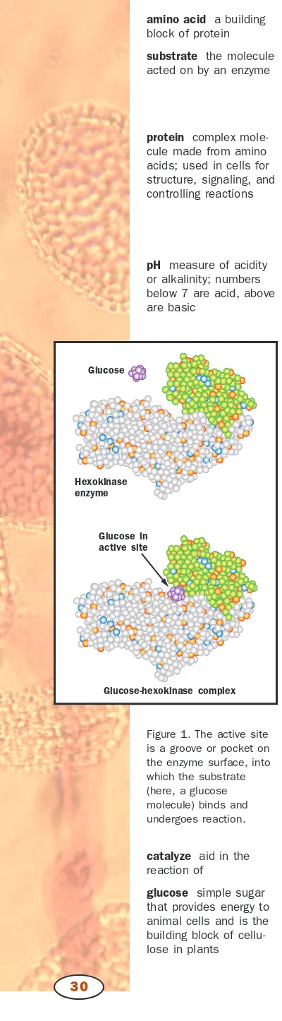
protein complex mole-cule made from amino acids; used in cells for structure, signaling, and controlling reactions
organelle membrane-bound cell compartment nanometer 10–9 meters; one-billionth of a meter
photographed or recorded with a digital camera. The SEM cannot see through a specimen as the TEM does, but can see only the surface where the metal coating is.
The SEM is capable of less resolution and useful magnification than the TEM. However, it produces dramatic three-dimensional images that can yield more information about surface topography than the flat images usu-ally produced by TEM.
Other Variations in Electron Microscopy
Both SEMs and TEMs can be equipped with a detector that monitors X rays given off by a specimen when it is bombarded by electrons. Other types of microscopes irradiate the specimen with ions or X rays and record ions,
Electron Microscopy
electrons, or X rays given off by the specimen. In both cases, the emitted particles and radiation yield information about the chemical composition of the specimen.
A scanning tunneling microscope measures the vertical movement of a tiny probe that is dragged over a specimen, producing a line representation of that movement. An atomic force microscope operates on a similar prin-ciple, but measures forces of attraction and repulsion between the specimen and the probe as the probe moves across the surface. In either case, multi-ple scan lines side by side produce images of the specimen surface, reveal-ing details as small as the “atomic terrain” of individual molecules. S E E A L S O
Light Microscopy; Microscopist
Sara E. Miller and Kenneth S. Saladin
Bibliography
Berger, Dee. Journeys in Microspace: The Art of the Scanning Electron Microscope.New York: Columbia University Press, 1995.
Gilmore, C. P. The Scanning Electron Microscope: World of the Infinitely Small. Green-wich, NY: Graphic Society, 1972.
Microworld Internet Guide to Microscopy. <mwrn.com/guide/electron_microscopy/ microscope.htm>. Includes lecture notes and guides to EM techniques and in-strumentation.
Slayter, Elizabeth M., and Henry S. Slayter. Light and Electron Microscopy.New York: Cambridge University Press, 1992.
WWW Virtual Library: Microscopy. <http://www.ou.edu/research/electron/mirror/>. Numerous links to other sites on all aspects of microscopy.
Electrophoresis
Electrophoresis is one of the most important techniques used by molecular biologists. To name only a few applications, deoxyribonucleic acid (DNA) electrophoresis is used to map the order of restriction fragmentswithin chromosomes, to analyze DNA variation within a population by restric-tion fragment length polymorphisms (RFLPs), and to determine the nu-cleotidesequence of a piece of DNA.
Electrophoresis refers to the migration of a charged molecule through a restrictive matrix, or gel, drawn by an electrical force. As the force drags the molecule through the gel, it encounters resistance from the strands of the gel, retarding its rate of migration. In gel electrophoresis, larger mole-cules migrate more slowly than smaller ones, and so the distance of migra-tion within a gel can be used to determine a molecule’s size.
Although it is possible to separate whole chromosomes using special-ized electrophoresis techniques, DNA that is to be analyzed by elec-trophoresis is usually cut into smaller pieces using restriction enzymes. Fragments of DNA prepared by treatment with restriction enzymes are commonly separated from one another, and their sizes determined, using a gel of agarose electrophoresis, a complex carbohydrate. DNA is negatively charged due to the phosphodiester bonds that join the individual nu-cleotide building blocks. DNA will therefore electrophorese toward the pos-itive electrode when placed in an electrical field. To visualize the results after electrophoresis, the gel is soaked in a solution that causes DNA to flu-oresce when exposed to ultraviolet light.
Electrophoresis
The rate of migration is
inversely proportional to the log-arithm of a molecule’s size.
restriction fragments block of RNA or DNA
usu-Treatment of the DNA sample with multiple restriction enzymes in var-ious combinations enables the researcher to generate a restriction map of the original DNA fragment, which identifies the sites at the DNA where the restriction enzymes are.
Many research questions require a detailed analysis of one specific DNA fragment in a complex mixture. In such cases, a radioactive DNA probe can be used to identify the fragment based on its nucleotide sequence. The method, known as hybridization, is based on the rules of complementary base pairing(A bonds to T, G bonds to C). A probe is designed whose sequence is complementary to the piece of DNA to be detected. The gel-separated DNA is first transferred to a nylon membrane using a tech-nique called a Southern blot.
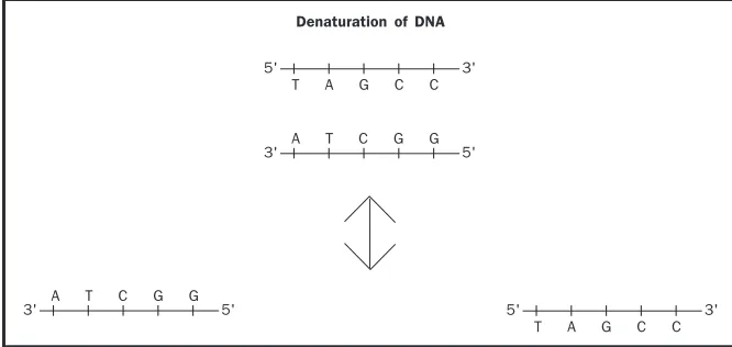
During the blotting procedure, the strands within the DNA double helix are separated from each other, or denatured, by treatment with a base. Because double-stranded DNA is more stable than single-stranded, during the hybridization the single-stranded probe will locate and bind to the single-stranded gel-separated fragment with complementary sequence.
Electrophoresis
An electrophoresis gel, which can be used to determine a molecule’s size.
complementary match-ing opposite
base pair two
nucleotides (either DNA or RNA) linked by weak bonds
Being fluorescent or radioactive, the position of the probe can be deter-mined using photographic methods. The target sequence can then be re-moved by cutting at the piece of the gel that contains it.
The most common technique for determining DNA sequence is the Sanger method, which generates fragments that differ in length by a single nucleotide. High-resolution polyacrylamide gel electrophoresis is then used to separate the fragments and to allow the sequence to be determined.
Electrophoresis of ribonucleic acid (RNA) is an integral procedure in many studies of gene expression. RNA is isolated, separated by elec-trophoresis, and then the gel-separated RNA fragments are transferred to a nylon membrane using a technique called a Northern blot. Hybridization with a single-stranded DNA probe is then used to determine the position of a specific RNA fragment.
DNA and RNA are relatively simple in terms of structure and compo-sition. Proteins, however, are composed of twenty different amino acids in various combinations, and proteins vary significantly in their three-dimensional structure. The composition of amino acids will affect the charge on the protein, which ultimately will affect its electrophoretic be-havior. The shape of a protein similarly will affect its rate of migration. As a result, a specialized technique, SDS-polyacrylamide gel electrophoresis (SDS-PAGE), is usually used to analyze proteins. In this method, protein samples are heated and then treated with the detergent sodium dodecyl sul-fate (SDS). Proteins treated in this way are unfolded, linear, and uniformly coated by negatively charged detergent molecules. The rate of migration of treated proteins is inversely proportional to the logarithm of molecular weight. Following electrophoresis, the protein in the gel can be stained to visualize all the proteins in a sample, or the proteins in the gel can be trans-ferred to a nylon membrane (Western blot) and specific ones detected with the use of enzyme-linked antibodies.
Regardless of the macromolecule being studied, gel electrophoresis is a crucial technique to the molecular biologist. Many scientific questions can be answered using electrophoresis, and as a result an active molecular biol-ogy research lab will have several benches that are devoted to the required specialized reagents and equipment. S E E A L S O DNA Sequencing
James E. Blankenship
Bibliography
Alberts, Bruce, et al. Molecular Biology of the Cell, 4th ed. New York: Garland Pub-lishing, 2000.
Stryer, Lubert. Biochemistry,4th ed. New York: W. H. Freeman and Company, 1995.
Emergency Medical Technician
An emergency medical technician (EMT) is a person who delivers the initial medical treatment to persons in crisis situations. Traditionally, EMTs are part of the medical team that travels by ambulance or helicopter to the site of the emergency situation. The most common medical crises to which EMTs are called include: injuries acquired during automobile accidents and roadway and home births; sudden myocardial infarctions (heart attacks); and wounds re-sulting from interpersonal violence (such as gun shots and stab wounds).
Emergency Medical Technician
S A N G E R , F R E D E R I C K ( 1 9 1 8 – )
English biochemist who received two Nobel Prizes in chemistry. The first came in 1958, for find-ing the amino sequence of insulin, the protein that helps regulate blood sugar levels, and the second, in 1980, for invent-ing a technique to sequence the nucleotides in a strand of deoxyribonucleic acid (DNA).
gene expression use of a gene to create the corresponding protein
protein complex mole-cule made from amino acids; used in cells for structure, signaling, and controlling reactions
amino acid a building block of protein
Emergency medical technicians must be trained and certified. There are five levels of EMT training, from First Responders, who are certified in ba-sic emergency medical care, to EMT-4 (paramedics), who are certified to administer drugs, read electrocardiograms, and use other advanced equip-ment in providing prehospital care. The training process is progressive, start-ing with EMT 1 (which includes First Responder trainstart-ing), requirstart-ing approximately 120 hours of training, through the paramedic level, requir-ing up to two years of trainrequir-ing. Hospitals, trauma centers, private ambu-lance companies, and fire and police departments employ emergency medical technicians. In fact, many firefighters are also certified EMTs.
In order to be well prepared for EMT training, a strong background in the sciences is important. High school courses such as biology, chemistry, mathematics, and physics are essential prerequisites for EMT training. A good driver’s education class is crucial as well, since many EMTs are also ambulance drivers who must negotiate challenging roadway situations in or-der to reach the crisis scene quickly and safely.
A career in emergency medicine can be very challenging. EMTs must maintain the difficult balance between compassion and emotional fortitude. Strong leadership and interpersonal skills are a must for an emergency med-ical technician. However, despite the challenges, it is very rewarding to help people and save lives daily. S E E A L S O Doctor, Family Practice; Doctor, Specialist; Medical Assistant, Nurse
Susan T. Rouse
Bibliography
New York Department of Labor. CareerZone. <http://www.explore.cornell.edu/ newcareerzone/profile.asp?onet=32508&cluster=4>.
U.S. Department of Labor. “Occupational Outlook Handbook.” Washington, DC: Government Printing Office, annually. <http://stats.bls.gov/oco/ocos101.htm>.
Endangered Species
Endangered species are species of plants or animals (or other life forms such as fungi) that are threatened with extinction. As well as being a biological term, “endangered” has a formal political meaning: nations, states, and other organizations evaluate the status of species and determine which are in the greatest danger of going extinct; these species are designated as endangered species. Other species that are declining rapidly in numbers, but are not yet believed to be on the brink of extinction, are designated as threatened species. In the United States, the Endangered Species Act protects such species.
Several factors can cause a species to become endangered. The most common cause is loss of habitat. Much of the world’s forests, grasslands, and wetlands are being transformed into agricultural and urban areas, and many species that lived in those habitats are unable to adapt to the new en-vironment. As a result, their numbers can drop greatly in a very short time. In some cases, human hunting or gathering of particular species can drive a species to the brink of extinction. This is the case of the rhinoceros, which has been killed in large numbers during the past century to meet market needs in certain areas of the world. The horn of the rhino is prized for dag-ger handles in the Middle East and for medicinal uses in parts of Asia. Tidag-gers
and sun bears in Asia have likewise been driven to the brink of extinction due to the huge market for animal parts that are believed by many to have potent medicinal powers.
Protection and Reestablishment
There are several ways that people can try to protect endangered species and to keep them from going extinct. One important way is to set up spe-cial protected areas around some of the last remaining populations of a species. China has created such reserves for the giant panda. However, for these reserves to be successful, they need to have the support of the resi-dent people that live around the reserve. In some cases, the reserves pro-vide the local people with jobs, and in other cases, some agricultural and even hunting activities are permitted within the reserve.
For some species, their habitat has essentially disappeared, or the species has declined to only a few individuals. In these instances, the only feasible way to try to preserve the species is to bring all the remaining individuals into captivity. One important function of zoos today is to house such en-dangered species. In some cases, captive breeding programs are initiated to
Endangered Species
increase the number of individuals of the endangered species. The ultimate goal of many of these captive breeding programs is to reintroduce the species back into the wild at some future date.
There are several ongoing reintroductions. In the 1980s, when the Cal-ifornia condor had declined almost to the point of extinction, the few re-maining individuals were captured and placed in captivity. A successful captive breeding program increased the numbers to several dozen individ-uals, and some have been released back into the wild. Reintroductions of endangered species are not always successful because the reintroduced ani-mals usually have lived only in captivity. Thus, it is often necessary to pre-pare these animals for their new life in the wild by teaching them how to catch their food and to avoid predators.
Probably the greatest success story of the recovery of an endangered species involves the national bird of the United States, the bald eagle. The bald eagle, like many other birds of prey, fell victim to the heavy use of pes-ticides by farmers in the 1950s, including DDT. Much of the DDT that was sprayed onto agricultural fields ran off into streams and rivers and lakes when it rained. Small aquatic life consumed some of this DDT, and it remained in their body tissue. When a small fish ate these small aquatic organisms, DDT accumulated in their bodies too and was passed on when a larger fish ate the smaller fish. This process has been referred to as bioac-cumulation, or biomagnification.
Thus, by the time the bald eagle ate the larger fish, it was eating cont-aminated food, and the eagles’ own tissues accumulated high concentrations of DDT. One unfortunate consequence of these high concentrations of DDT was the severe weakening of the eggshell laid by the eagle. They were so weak they would often break during the normal parental brooding of the eggs. As a result, the birth rates of the eagles plummeted at the same time the death rates from DDT poisoning rose.
In response to environmentalists like Rachel Carson, who saw how the use of these sorts of chemicals was harming wildlife, the United States banned further use of DDT and provided the bald eagle with special protection un-der its endangered species status. The eagle populations responded slowly, but in the 1990s the populations began to increase at a rapid rate. In the early twenty-first century, the bald eagle is seen commonly in many parts of the United States and Canada, and its numbers have increased substantially enough that it is no longer considered an endangered species. S E E A L S O
Bio-diversity; Carson, Rachel; Extinction; Pollution and Bioremediation
Mark A. Davis
Bibliography
Primack, Richard B. Essentials of Conservation Biology.Sunderland, MA: Sinauer As-sociates, 1998.
Endocrine System
The endocrine system is the interacting group of glands that secrete hor-mones, helping to control cells and organs throughout the body. How do cells and organs at different locations in the body communicate with each
Endocrine System
other to maintain the physiology of healthy living organisms? What hap-pens if organs do not communicate properly? These questions can be an-swered by understanding how organs of the nervous system and endocrine system function.
There are similarities and differences between how the human nervous system and endocrine system communicate with and control other organs. For example, the nervous system relies on electrical impulses and chemical neurotransmitters. Most endocrine organs do not transmit electrical in-formation but instead secrete hormones (from the Greek, meaning “to arouse or excite”), which are molecules that act as chemical messengers.
Endocrine System
Hypothalamus
Pineal Gland
Pituitary Gland
Parathyroids Thyroid
Thymus Adrenal
Cortex
Adrenal Medulla
Pancreas
Ovary (females)
Testis (males)
The endocrine organs in the human body.
Hormones are released into the bloodstream whereby they travel to organs they affect, known as target organs.
Endocrine organs are located throughout the body, and they have di-verse functions controlling events such as cell metabolism, blood sugar con-centration, digestion, the menstrual cycle in females, and the production of male and female gametes. Primary endocrine organs include the hypothal-amus, pituitary gland, pineal gland, thyroid and parathyroid glands, thymus, adrenal glands, pancreas, and male and female gonads, the testes and ovaries respectively. Other tissues serve endocrine functions through the hormones they produce. For example, the kidneys produce erythropoietin that stimu-lates formation of red blood cells, and the skin produces vitamin D, a steroid derivative required for calcium absorption by the small intestine.
Hormones
Hormones are “signaling” molecules because they influence the activity of other cells that may be far from where the hormone was produced. For a hormone to affect a target cell, it must attach to a receptor proteinon the target cell membrane or inside the cell. Hormone binding to a receptor trig-gers an intricate set of biochemical interactions that can affect the target cell in myriad ways. For example, hormones can influence cell metabolism, cell division, electrical activity, ribonucleic acid (RNA) and protein synthe-sis, or cell secretion.
There are several different types of hormones that vary in their chem-ical organization and functions. The majority of hormones are peptides. These consist of short sequences of amino acids; examples include insulin and growth hormone. The class of hormones called steroids are synthe-sized from cholesterol—examples include male sex steroids such as testos-terone and female sex steroids such as estrogen and progestestos-terone.
Hormone production by an endocrine organ is regulated by complex interactions, called feedback loops, between the endocrine organ and its target organs. Feedback loops are two-way modes of communication in which a target organ also releases molecules that regulate the endocrine or-gan. Feedback loops are designed to maintain hormone concentration within a normal range. Endocrine disorders in which hormone concentration be-comes abnormal can be difficult to diagnose and treat because of the com-plexity of feedback loops. One simple way to classify endocrine disorders is based on whether a condition is due to excess production (hypersecretion) or underproduction (hyposecretion) of hormone.
The Major Endocrine Glands
Located at the base of the brain, the pituitary gland produces many hor-mones that regulate other organs. Because of this, the pituitary is often re-ferred to as the “master” endocrine gland, although the term “central” endocrine gland is more correct because hormone release by the pituitary is primarily regulated by a brain structure called the hypothalamus, which acts to connect the nervous system to the endocrine system. The hypothal-amus produces hormones that stimulate or inhibit the release of pituitary hormones. The hypothalamus also produces antidiuretic hormone, which regulates water balance in the body by inhibiting urine formation by the
Endocrine System
kidneys, and a hormone called oxytocin, which stimulates uterine contrac-tions during childbirth and releases milk during breast-feeding.
Hormones released by the pituitary include growth hormone, which in-creases during childhood and stimulates the growth of muscle, bone, and other tissues. Sporadic bursts in growth hormone release often result in rapid growth “spurts” associated with adolescence. Hyposecretion of growth hormone can result in dwarfism, whereas hypersecretion of growth hormone can cause gigantism and other disorders. The pituitary also produces folli-cle-stimulating hormone and luteinizing hormone, which stimulate gamete production and sex steroid production in male and female reproductive or-gans, and prolactin, which stimulates milk formation in the mammary glands. Located adjacent to the larynx, the thyroid gland primarily produces thyroxine and triiodothyronine, collectively referred to as thyroid hormone. Thyroid hormone stimulates growth of muscles and bones, carbohydrate metabolism, and basal metabolic rate. Its production requires iodine; the lack of dietary iodine causes goiter, a thyroid gland that is overly enlarged in an effort to compensate for the thyroid hormone deficiency.
Effects of thyroid disorders in children and adults can differ widely. For example, hyposecretion of thyroid hormone in infants causes congen-italhypothyroidism, a disease characterized by mental retardation and poor body growth; hyposecretion in adults produces myxedema, with symptoms such as lethargy, weight gain, and dry skin. Conversely, hypersecretion of
Endocrine System
A frontal-view scintigram of a normal human thyroid. Part of the endocrine system, the thyroid controls basal metabolic rate.
larynx “voice box”; muscles at the top of the trachea that control pitch and loudness
congenital present at birth; inherited
thyroid hormone in adults causes Graves’ disease, a condition character-ized by weight loss, nervousness, and dramatic increases in body metabo-lism. The thyroid also produces calcitonin, a hormone that regulates blood calcium concentration.
The adrenal glands are small organs on the apex of each kidney. The outer layers of cells in the adrenal gland, called the adrenal cortex, produce several hormones that affect reproductive development; mineral balance; fat, protein, and carbohydrate balance; and adaptation to stress. The inner part, called the adrenal medulla, secretes epinephrine and norepinephrine, which activate the sympathetic nervous systemand stimulate the “fight-or-flight” response that helps the body cope with stressful situations, such as fear.
The pancreas produces insulin and glucagon, which function in oppos-ing fashion to regulate blood sugar (glucose) concentration. When blood glucose level rises—for example, after eating a sugar-rich meal—insulin lowers it by stimulating glucose storage in liver and muscle cells as long chains of glucose called glycogen. Conversely, between meals, blood glu-cose level decreases. In response, the pancreas releases glucagon, which stim-ulates glycogen breakdown and subsequent release of glucose into the bloodstream. One of the most well characterized endocrine disorders is di-abetes mellitus, resulting from hyposecretion of insulin or, more commonly, target cell insensitivity to it.
Endocrine functions of the gonads are addressed in articles on the male and female reproductive systems. The sex hormone testosterone regulates sperm production in males. Estrogen and progesterone influence egg mat-uration and release (ovulation) and control the uterine (menstrual) cycle in females.
Although the many hormones produced by human endocrine organs have a wide variety of actions, the common purpose of all hormones is to facili-tate organ-to-organ communication necessary for body physiology. S E E A L S O
Adrenal Gland; Anabolic Steroids; Blood Sugar Regulation; Female Reproductive System; Growth; Homeostasis; Hormones; Hypothala-mus; Nervous Systems; Pancreas; Pituitary Gland; Stress Response; Thyroid Gland
Michael A. Palladino
Bibliography
Hadley, Mac E. Endocrinology,5th ed. Upper Saddle River, NJ: Prentice Hall, 2000. Marieb, Elaine Nicpon. Human Anatomy and Physiology,5th ed. San Francisco:
Ben-jamin Cummings, 2000.
Endocytosis
The ability to internalize material from outside the cell is important for sev-eral cellular processes including the ingestion of essential nutrients, removal of dead or damaged cells from the body, and defense against microorgan-isms. Eukaryotic cellsinternalize fluid, large and small molecules, and even other cells from their surroundings by a process called endocytosis. During endocytosis, the plasma membrane of the cell forms a pocket around the material to be internalized. The pocket closes and then separates from the
Endocytosis
sympathetic nervous system branch of the nervous system that promotes heightened awareness, increased nutrient consumption, and other changes associated with “fight or flight”
glucose simple sugar that provides energy to animal cells and is the building block of cellu-lose in plants
glycogen complex car-bohydrate used as stor-age in animals and some other organisms
inside surface of the plasma membrane to form a membrane-enclosed bub-ble, or vesicle, containing the ingested material.
There are two main types of endocytosis that are distinguished by the size of the vesicle formed and the cellular machinery involved. Pinocytosis (cell drinking) describes the internalization of extracellular fluid and small macromoleculesby means of small vesicles. Phagocytosis(cell eating) de-scribes the ingestion of large particles such as cell debris and whole mi-croorganisms by means of large vesicles. While all eukaryotic cells are continually ingesting fluid and molecules by pinocytosis, only specialized phagocytic cells ingest large particles.
Specialized Phagocytic Cells Engulf Large Particles
Phagocytosis begins with the extension of large, handlike projections from the plasma membrane. The projections surround the particle and fuse to-gether so that the particle is completely engulfed in a large vesicle within the cell called a phagosome. Inside the cell, the phagosome fuses with an-other membranous organellecalled a lysosome, forming a single membra-nous organelle and mixing their contents in the process. Lysosomes, acting as the “stomach” of the cell, carry digestive enzymes that break down all types of biological molecules. Consequently, after a phagosome fuses with a lysosome, the digestive enzymes break down the ingested material into small molecules that are transported into the cytosoland made available for cell use. Many single-celled organisms like amoebasand ciliates use phago-cytosis as a means to acquire food. In multicellular animals, only specialized types of cells use phagocytosis. For example, in humans, specialized white blood cells called macrophages use phagocytosis to defend the body against infection by engulfing invading microorganisms and to remove cell debris from the body by ingesting damaged or old cells.
Endocytosis
A color-enhanced transmission electron micrograph of an amoeba engulfing green algal cell for food. In phagocytosis, a type of endocytosis, large vesicles ingest whole microorganisms.
macromolecules large molecules such as pro-teins, carbohydrates, and nucleic acids
phagocytosis engulfing of cells or large frag-ments by another cell, including immune system cells
organelle membrane-bound cell compartment
enzyme protein that con-trols a reaction in a cell
cytosol fluid portion of a cell, not including the organelles
All Eukaryotic Cells Constantly Ingest Fluid and
Molecules by Pinocytosis
In contrast to phagocytosis, pinocytosis begins with small, convex pits on the cell surface that collect material or fluid to be internalized. The convex pits expand into the interior of the cell forming small vesicles that pinch from the inside of the plasma membrane. All eukaryotic cells have a tinuous stream of vesicles budding from the plasma membrane. The con-stant removal of membrane from the plasma membrane would quickly deplete the plasma membrane if not for the balancing effects of another con-tinual process called exocytosis. Exocytosis is the process by which vesicles from inside the cell fuse with the plasma membrane to secrete material and fluid. So, pinocytosis brings fluid and material into the cell and removes membrane from the plasma membrane, while exocytosis expels fluid and mate-rial from the cell while adding membrane to the plasma membrane. Thus, the two processes work together to continuously recycle the plasma membrane.
The most thoroughly understood form of pinocytosis is receptor-mediated endocytosis. Receptor-receptor-mediated endocytosis selectively internalizes specific molecules that are found in low concentrations in the extracellular space, such as hormones, growth factors, antibodies, iron, enzymes, vita-mins, and cholesterol. The specific molecules to be internalized bind to pro-teins called receptors on the outside surface of the cell. Receptors are proteins that are embedded in the plasma membrane with portions of the protein extending outside the cell to form a binding site for a specific mol-ecule. Once molecules bind to their receptors, the receptors move within the plasma membrane and become concentrated in small depressions called clathrin-coated pits. Clathrin-coated pits are formed when many clathrin protein molecules interact with each other to form a convex, basketlike struc-ture on the inside of the plasma membrane that molds the membrane into a pit. The coated pits then progressively invaginate, or form inward, to form clathrin-coated vesicles that pinch off the plasma membrane into the cyto-plasm. Hence, the clathrin-coated vesicles carry the receptor proteins taken from the plasma membrane and their bound molecules taken from the ex-tracellular space.
Once a clathrin-coated vesicle pinches from the inner surface of the plasma membrane, the clathrin coat is removed. The “uncoated” vesicle, still carrying receptor proteins and their bound cargo molecules, fuses with another membranous organelle called an endosome. Endosomes function as “sorting stations.” In an endosome, molecules are sorted and packaged into new vesicles for transportation to various locations within the cell.
Receptors brought into the cell by receptor-mediated endocytosis have one of several fates after unloading their cargo and leaving the endosome: (1) they can be recycled back to the same area of plasma membrane from which they came; (2) they can be transported to another region of the plasma membrane; or (3) they can be transported to the lysosome where they are degraded. Thus, in contrast to phagocytosis, not all material brought into the cell by receptor-mediated endocytosis ends up in the lysosome for di-gestion.
Endocytosis
hormone molecule released by one cell to influence another
protein complex mole-cule made from amino acids; used in cells for structure, signaling, and controlling reactions
cytoplasm material in a cell, excluding the nucleus