Offspring Born From Chimeras Reconstructed
From Parthenogenetic and In Vitro Fertilized
Bovine Embryos
A. BOEDIONO,1,2*T. SUZUKI,1L.Y. LI,3ANDR.A. GODKE3
1United Graduate School of Veterinary Sciences, Yamaguchi University, Yamaguchi, Japan 2Faculty of Veterinary Medicine, Bogor Agricultural University, Bogor, Indonesia
3Department of Animal Science, Louisiana State University, Baton Rouge, Louisiana
ABSTRACT Chimeric embryos were produced by aggregation of parthenogenetic (Japanese Red breed) and in vitro fertilized (Holstein breed) bovine embryos at the Yamaguchi Research Station in Japan and by aggregation of parthenogenetic (Red Angus breed) and in vitro fertilized (Holstein breed) embryos at the St. Gabriel Research Station in Louisiana. After embryo reconstruction, live offspring were produced at each station from transplanting these embryos. The objective of this joint study was to evaluate the develop-mental capacity of reconstructed parthenogenetic and in vitro fertilized bovine embryos. In experiment I, chimeric embryos were constructed: by aggregation of four 8-cell (demi-embryo) parthenogenetic and four 8-cell stage (demi-embryo) IVF-derived blastomeres (method 1) and by aggregation of a whole parthenoge-netic embryo (8-cell stage) and a whole IVF-derived embryo (8-cell stage) (method 2). Similarly in experi-ment II, chimeric embryos were constructed by aggre-gating IVF-derived blastomeres with parthenogenetic blsatomeres. In this experiment, three categories of chimeric embryos with different parthenogenetic IVF-derived blastomere ratios (2:6; 4:4, and 6:2) were constructed from 8-cell stage bovine embryos. In experiment III, chimeric embryos composed of four 8-cell parthenogenetic and two 4-cell IVF-derived blasto-meres or eight 16-cell parthenogenetic and four 8-cell IVF-derived blastomeres were constructed. Parthenoge-netic demi-embryos were aggregated with sexed (male) IVF demi-embryos to produce chimeric blastocysts (experiment IV). In the blastocyst stage, hatching and hatched embryos were karyotyped. In experiment V, chimeric embryos that developed to blastocysts (zona-free) were cryopreserved in ethylene glycol (EG) plus trehalose (T) with different concentrations of polyvinyl-pyrrolidone (PVP; 5%, 7.5%, and 10%). In experiment I, the aggregation rate of the reconstructed demi-embryos cultured in vitro without agar embedding was significantly lower than with agar embedding (53% for 0% agar, 93% for 1% agar, and 95% for 1.2% agar, respectively). The aggregation was also lower when the aggregation resulted from a whole parthenogenetic and IVF-derived embryos cultured without agar than when cultured with agar (70% for 0% agar, 94% for 1%
agar, and 93% for 1.2% agar, respectively). The devel-opment rate to blastocysts, however, was not different among the treatments. In experiment II, the developmen-tal rates to the morula and blastocyst stages were 81%, 89%, and 28% for the chimeric embryos with parthenogenetic:IVF blastomere ratios of 2:6, 4:4, and 6:2, respectively. In experiment III, the developmental rate to the morula and blastocyst stages was 60% and 65% for the two 4-cell and four 8-cell chimeric embryos compared with 10% for intact 8-cell parthenogenetic embryos and 15% for intact 16-cell parthenogenetic embryos. To verify participation of parthenogenetic and the cells derived from the male IVF embryos in blasto-cyst formation, 51 embryos (hatching and hatched) were karyotyped, resulting in 27 embryos having both XX and XY chromosome plates in the same sample, 14 embryos with XY and 10 embryos with XX. The viability and the percentage of zona-free chimeric embryos at 24 hr following cryopreservation in EG plus T with 10% PVP were significantly greater than those cryopre-served without PVP (89% vs. 56%). Pregnancies were diagnosed in both stations after the transfer of chimeric blastocysts. Twin male (stillbirths) and single chimeric calves were delivered at the Yamaguchi station, with each having both XX and XY chromosomes detected. Three pregnancies resulted from the transferred 40 chimeric embryos at the Louisiana station. Two pregnan-cies were lost prior to 4 months and one phenotypically-chimeric viable male calf was born. We conclude that the IVF-derived blastomeres were able to stimulate the development of bovine parthenogenetic blastomeres and that the chimeric parthenogenetic bovine embryos were developmentally competent. Mol. Reprod. Dev. 53:159–170, 1999. r1999 Wiley-Liss, Inc.
Key Words:chimera; parthenogenetic; in vitro fertil-ization; cattle; offspring
Grant sponsor: Ministry of Education, Science and Culture, Japan; Grant number: 845160; Grant sponsor: Louisiana Agriculture Experi-mental Station, Louisiana State University Agriculture Center. *Correspondence to: Dr. A. Boediono, Faculty of Veterinary Medicine, Bogor Agricultural University, Jalan Taman Kencana 3, Bogor 16151, Indonesia.
Received 28 July 1998; Accepted 15 December 1998.
INTRODUCTION
Live offspring have occurred naturally in nonmamma-lian species from developing parthenogenetic embryos (White, 1978), and occasionally parthenogenetic em-bryos can be experimentally induced in invertebrates, and in some vertebrates (Nagy et al., 1979). Although parthenogenetically activated diploid mammalian em-bryos can develop in a normal pattern through the pre-implantation stage, these early conceptuses rarely reach the forelimb-bud stage (Kaufman et al., 1977). The diploid gynogenomes produced from fertilized mouse oocytes by suppression of the extrusion of the second polar body (Niemierko, 1975) or by subsequent removal of the male pronucleus (Modlinski, 1980; Bor-suk, 1982) develop to the 25-somite stage, as do the genetically similar diploid parthenogenomes (Surani and Barton, 1983). Barra and Renard (1988) have reported constructing diploid mouse embryos by fusing late 2-cell stage haploid parthenogenomes with late 2-cell stage haploid androgenones. These reconstructed embryos, however, developed into normal-appearing offspring after they were transferred to recipient mice. Pronuclear transfer experiments have shown that the parental genomes are not equivalent in supporting the embryogenesis in mice, and that both paternal and maternal genomes are required to support full term development of mouse embryos (Barton et al., 1984; McGrath and Solter, 1984; Surani et al., 1984). It has been shown that the paternal genome of the mouse was essential for the development of extra embryonic tis-sues and the maternal genome was essential for the embryogenesis (Barton et al., 1984; Surani et al., 1984). Genome imprinting during gametogenesis is thought to be responsible for the functional difference of parental genomes during embryogenesis (Surani et al., 1986, 1990).
Clear functional differences between the parental genomes of mammals have been revealed by demonstra-tion of lethality in embryos of parthenogenetic, gynoge-netic, and androgenetic uniparental genotypes in mice (Solter, 1988). Although mouse parthenogenomes are unable to complete embryogenesis in utero, diploid parthenogenomes are apparently capable of developing to chimeric adults when parthenogenetic cells contrib-ute to embryonic tissues after they were aggregated with in vivo-derived mouse embryos (Stevens et al., 1977; Surani et al., 1977), and have been found to produce viable germ cells of parthenogenetic origin (Stevens, 1978; Anderegg and Markert, 1986). The cellular interaction between parthenogenetic and in vivo-derived blastomeres in chimeric embryos has been shown to be critical to embryo development (Surani et al., 1987a,b; Thomson and Solter, 1988, 1989).
Various methods have been evaluated for the induc-tion of diploid parthenogenesis in bovine ova. The more
effective methods include exposure to Ca11ionophore
A23187 (Ware et al., 1989), ethanol, electric stimulation or a combination of ethanol and electric stimulation (Yang et al., 1994). The interest in bovine
parthenoge-netic development has risen during recent years for both scientific and economic purposes. Parthenogenetic activation of bovine oocytes has been induced by a number of methods with relatively high activation rates (Nagai, 1987; Aoyogi et al., 1994; Boediono and Suzuki, 1994; Goto et al., 1994; Presicce and Yang, 1994). In recent years, parthenogenetic bovine embryos have been transferred to recipient females but no offspring have been produced. In one study, estrus did not occur until 48 days after transfer of single partheno-genetic embryos (Fukui et al., 1992). In another study, the return to estrus was delayed until 67 days after transfer of aggregated parthenogenetic embryos, with pregnancy not being maintained after this period (Boediono and Suzuki, 1994), likely do to premature death of the conceptus (Boediono et al., 1995).
The primary objective of this joint study was to evaluate the developmental capacity of fresh and frozen-thawed reconstructed bovine parthenogenetic embryos with blastomeres of in vitro fertilized bovine embryos. In a series experiments conducted at two research stations, we investigated (1) the effect of agar embed-ding for protection of aggregated embryos from disaggre-gation during culture in vitro, (2) the contribution of parthenogenetic cells to the blastocyst formation after aggregation with an IVF-derived embryo, (3) the freez-ing media for cryopreservation of zona-free chimeric parthenogenetic blastocysts, and (4) the viability of chimeric blastocysts produced by aggregation of diploid parthenogenetic and IVF-derived embryos and their development to term following transfer to the recipient females.
MATERIALS AND METHODS The Yamaguchi Station
Parthenogenetic activation. The procedures for producing diploid parthenogenetic bovine embryos in experiments I, IV and V were previously reported (Boediono et al., 1995). In brief, oocytes were aspirated from cattle ovaries (Japanese Red breed) obtained from an abattoir and allowed to mature for 32 hr at 38.5°C
under 5% CO2in air. Maturation medium consisted of
Medium-199 (TCM-199; Gibco BRL, NY) supplemented with 5% (v/v) superovulated cow serum (SCS) (Boediono et al., 1994), 0.01 mg/ml follicle stimulating hormone (FSH, Denka, Kawasaki, Japan) and 50 µg/ml gentami-cin sulfate (Sigma, St. Louis, MO). To induce partheno-genetic activation, mature oocytes were suspended in culture medium contained 7% ethanol for 10 min and then they were treated with 5 µg/ml cytochalasin-D for 5 hr to suppress polar body extrusion and to produce diploid parthenogenomes. The embryos were then washed and cultured in vitro on feeder layers of bovine cumulus cells (Goto et al., 1988; Zhang et al., 1992) for further development during a 48- to 72-hr interval after activation.
(Boediono et al., 1994). Ovaries from dairy cows (Hol-stein) were collected from an abattoir one day later than the ovaries from cows where oocytes were to be used for parthenogenetic activation. Follicular oocytes were allowed to mature for 21 hr at 38.5°C under 5% CO2in air. Frozen-thawed sperm (Holstein) was washed twice with 2.5 mM caffeine in Brackett-Oliphant medium (B-O; Brackett and Oliphant, 1975) without bovine serum albumin. Sperm concentration was
ad-justed to 5 3106 spermatozoa per ml in B-O
supple-mented with 0.3% bovine serum albumin (BSA, Sigma) and 20 µg/ml heparin (Shimizu, Japan). A 100 µl aliquot of the sperm suspension was pre-incubated for 1 hr. In vitro-matured oocytes were transferred into fertiliza-tion droplets for inseminafertiliza-tion (20–25 oocytes/droplet). After 18 hr of sperm exposure, oocytes were washed and transferred to a polystyrene dish (4-well multidish; Nunclon, Roskilde, Denmark) containing TCM-199 supplemented with 5% SCS, 5 µg/ml insulin (Wako, Osaka, Japan) and 50 µg/ml gentamicin sulfate for further development.
Embryo reconstruction.Aggregation chimeras in experiments I, IV and V were produced by the following methods: (1) aggregation of four 8-cell parthenogenetic blastomeres and four 8-cell IVF-derived blastomeres, and 2) by aggregation of a whole parthenogenetic embryo (8-cell stage) and a whole IVF-derived embryos (8-cell stage). Embryo reconstruction was executed on day 2 following insemination (day 05day of insemina-tion). The procedure for opening the zona pellucida and insertion of blastomeres in method 1 was conducted as previously described by Tsunoda et al. (1986), with minor modifications. Embryo micromanipulation proce-dures were conducted under an inverted Nikon micro-scope with Narishige micromanipulation unit (Narish-ige Scientific Instrument, Tokyo, Japan). Briefly, a parthenogenetic 8-cell stage embryo was held by nega-tive pressure with a holding pipette and a microblade was used to make a rent in the zona pellucida. The injection pipette (30 µm in diameter) was inserted through the opening to remove four blastomeres (half of the embryo). The IVF-derived blastomeres (four of the 8-cell stage) were inserted into the parthenogenetic embryo (four of the 8-cell stage) (Fig. 1).
Aggregation of the embryos in method 2 was achieved by moving two zona-free embryos (8-cell stage) together until the blastomere aggregate was sufficiently stable (Fig. 2). Aggregated embryos produced by methods 1 and 2 were then cultured in culture medium (0.5 ml) in a polystyrene 4-well multidish (Nunclon) with a feeder cell layer of bovine cumulus cells covered with mineral oil (1 embryo/well). Aggregated embryos produced by method 1 were cultured in vitro within the zona pellu-cida and the aggregated embryos produced by method 2 were cultured without the zona pellucida, as previously described (Boediono et al., 1993).
All embryo aggregates were rechecked after 6 hr of incubation to ensure that the cells had not drifted apart. Morphological evaluations were made at 12-hr
intervals, with both unaggregated embryos and aggre-gated embryos with extruded blastomeres removed from the culture system. The chimeric embryos were then maintained in culture until day 9, and the origins of the different embryos that had developed into morpho-logically normal-appearing blastocysts were recorded.
The Louisiana Station
Parthenogenetic activation. The procedures for producing diploid parthenogenetic embryos in experi-ments II and III conducted at the St. Gabriel Research Station (Louisiana State University) were developed in this laboratory, and were similar to those reported by Boediono et al. (1994). Red Angus ovaries collected from a local abattoir were maintained in phosphate-buffered saline (PBS) with 5% fetal bovine serum (FBS) and 100 units/ml penicillin (Sigma) and 100 µg/ml streptomycin (Sigma) at room temperature and transferred to the laboratory within 6 hr. Cumulus-oocytes complexes were harvested from ovarian follicles 3–8 mm in diam-eter, using an 18-gauge needle. The oocytes were washed three times with 199 and then cultured in
TCM-199 with 10% FBS at 39°C in 5% CO2in an atmosphere
of humidified air for 22 hr. Matured bovine oocytes were then treated with 7% ethanol in PBS medium for 10 min, and washed thoroughly with fresh PBS medium immediately after the ethanol treatment. These oocytes were then cultured in TCM-199 with 10% FBS, 10 µg/ml cytochalsin B (Sigma) and 10 µg/ml cyclohexmide
(Sigma) at 39°C in 5% CO2 in an atmosphere of
humidified air for 16 hr. The oocytes were thoroughly washed at the end of the culture and then transferred into TCM-199 supplemented with 10% FBS on a granu-losa cell monolayer.
In vitro fertilization. In experiment II and III ovaries of dairy cows (Holstein) were collected from a local abattoir were maintained in PBS with 5% FBS and 100 units/ml penicillin (Sigma) and 100 µg/ml streptomycin (Sigma) at room temperature and trans-ferred to the laboratory within 6 hr. Cumulus-oocytes complexes were harvested with an 18-gauge needle from medium-size follicles (3–8 mm in diameter). The oocytes were washed three times with TCM-199 and then cultured in TCM-199 with 10% FBS at 39°C in 5%
CO2 in an atmosphere of humidified air for 22 hr.
Frozen sperm cells (from a fertile Holstein bull) were thawed, washed with Brackett-Oliphant medium (B-O medium) supplemented with 5 mM caffeine and then capacitated by the treatment of 0.1 mM calcium iono-phore A23187 for 1 min, as previously outlined by Zhang et al. (1992). Briefly, fertilization was conducted for 6 hr in droplets of 100 µl of B-O medium supple-mented with 5 mM caffeine and 1% bovine serum albumin (BSA, Fraction-IV, Sigma). The fertilized oo-cytes were washed twice with TCM-199 and then cultured in TCM-199 supplemented with 10% FBS at
39°C in 5% CO2in an atmosphere of humidified air.
during oocyte aspiration were washed with TCM-199 supplemented with 10% FBS, and recovered by centrifu-gation at 500gfor 5 min (Zhang et al., 1992). The cell pellet was suspended in TCM-199 with 10% FBS and then seeded on a 4-well culture plate (Nunc, France) in droplets of 50 µl TCM-199 supplemented with 10% FBS covered with equilibrated mineral oil. The granulosa cells were cultured at 39°C in 5% CO2in an atmosphere of humidified air. The culture medium was changed at 48-hr intervals, and the cells usually formed a confluent monolayer by day 2 of incubation.
Embryo reconstruction.In experiment II and III the zona pellucida of IVF-derived bovine embryos was removed by the treatment of acidic Tyrode’s solution and subsequent treatment of 0.25% pronase in PBS. The bovine embryos were then separated into single blastomeres by gently pipetting the zona-free embryos through a fine glass pipette (40 µm in diameter).
Micromanipulation was conducted in a microdroplet of PBS with 10% FBS and 10 µg/ml cytochalasin B with a pair of Leitz manipulator units under a Nikon inverted microscope. While a parthenogenetic embryo was held by a holding pipette (80 µm in diameter) via negative pressure, IVF-derived bovine blastomeres were then microinjected into the zona cavity of the parthenoge-netic embryo by an injection pipette (30 µm in diam-eter). Some of the parthenogenetic blastomeres were subsequently removed after the microinjection by using the same injection pipette. The reconstructed embryos were carefully washed and then cultured in TCM-199 supplemented with 10% FBS on a granulosa cell mono-layer for 4 days.
Experimental Design
Experiment I.Aggregated chimeras in experiment I were produced by the following methods: aggregation of
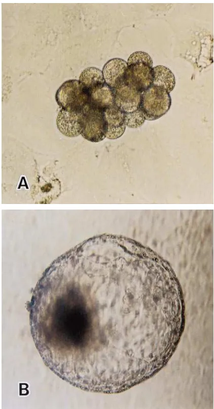
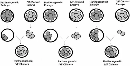
Fig. 1. Reconstructing an embryo produced by aggregation of four 8-cell parthenogenetic blastomeres and four 8-cell IVF-derived blasto-meres (A). In vitro development of a reconstructed embryo to a hatching blastocyst stage at day 7 post-insemination (B).
four 8-cell parthenogenetic blastomeres and four 8-cell IVF-derived blastomeres (method 1) and by the aggrega-tion of a whole parthenogenetic embryo (8-cell stage) and a whole IVF-derived embryo (8-cell stage) (method 2). This experiment was designed to evaluate two different methods of embryo reconstruction and three
concentrations of agar during embryo embedding (233
factorial arrangement) for the protection of aggregated embryos. Solutions of 0% (control), 1%, and 1.2% of agar (Difco) in 0.9% NaCl distilled water were prepared as the treatment groups in this experiment. The basic agar embedding procedure used in this study was that previously described by Willadsen (1979). While the respective agar solution was cooling, the aggregated embryos to be embedded were transferred to medium containing 5% SCS. When the agar had cooled from 39° to 37°C, 2 ml of the solution was poured into a petri dish (Falcon 1008, Becton Dickinson, Oxnard, CA) and then an aggregated embryo was immediately transferred to the agar solution with a fine bore Pasteur pipette. The agar containing the embryo was drawn and held in the tip of the pipette for ,30 sec and then expelled as a solid cylinder into the culture medium. This experi-ment was composed of five replicates that included 214 aggregated embryos.
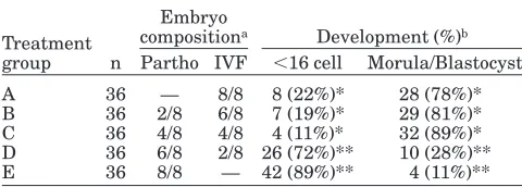
Experiment II.To evaluate the effect of blastomeric interaction on the development of bovine parthenoge-netic embryos, bovine IVF-derived blastomeres (Hol-stein breed) were microinjected into diploid bovine par-thenogenetic embryos (Red Angus breed) to produce parthenogenetic chimeric embryos. Three categories of
parthenogenetic/IVF chimeras were constructed: chi-meric embryos with two 8-cell parthenogenetic and six 8-cell IVF-derived blastomeres (treatment B), chimeric embryos with four 8-cell parthenogenetic and four 8-cell IVF-derived blastomeres (treatment C) and chi-meric embryos with six 8-cell parthenogenetic and two 8-cell IVF-derived blastomeres (treatment D) (Fig. 3). The intact 8-cell IVF-derived embryos and intact 8-cell parthenogenetic embryos prepared by the same micro-manipulation procedure were used as the IVF-derived embryo culture control (treatment A) and parthenoge-netic embryo culture control (treatment E). A total of 36 aggregated, parthenogenetic or intact embryos were allotted to respective treatments in three replications. Only viable appearing embryos (morphologically nor-mal) were used in this experiment.
Experiment III.To evaluate if stage of morphologi-cal development (number of cell cycles) between the parthenogenetic and the IVF-derived blastomeres was necessary to induce a stimulatory effect of IVF-derived blastomeres on the development of parthenogenetic blastomeres, parthenogenetic and IVF-derived blasto-meres at different developmental stages were combined to construct two combinations of aggregated embryos. The two aggregated combinations were chimeras com-posed of four 8-cell parthenogenetic and two 4-cell
IVF-derived blastomeres (treatment B) (n 5 40) and
chimeras composed of eight 16-cell parthenogenetic and four 8-cell IVF-derived blastomeres (treatment C) (n540). Intact 8-cell parthenogenetic embryos (n540)
Fig. 3. Reconstruction of parthenogenetic and in vitro–fertilized bovine embryos in experiment II. First, a chimeric embryo recon-structed from two 8-cell parthenogenetic blastomeres and six 8-cell IVF-derived blastomeres (first panel), followed by a chimeric embryo
and 16-cell parthenogenetic embryos (n 5 40) were used as the culture controls (treatments A and D).
Experiment IV. This experiment was designed to further evaluate the contribution of parthenogenetic cells to the blastocyst formation after aggregation with an IVF-derived embryo. Only the sexed (male) IVF embryos were aggregated with parthenogenetic em-bryos by one of two embryo reconstruction methods (method 1 or method 2, see experiment I) in an effort to produce aggregated chimeric embryos with long term developmental capabilities. The embryo sexing proce-dure used was that described previously by Itagaki et al. (1993). Briefly, to each of the tubes containing the samples (2–4 blastomeres from IVF-derived embryos), 100 ml of reaction mixture consisting of 10 mM
Tris-HCl (pH 8.9), 1.5 mM MgCl2, 80 mM KCl, 0.1% sodium
cholate, 0.1% Triton X-1000, 50 mM dNTPs, 0.2 mM DNA primer and 2 units of DNA polymerase were added, and then overlaid with 50 ml of mineral oil. The PCR amplification was performed with a DNA thermal cycler (TS-300, Iwaki Glass, Osaka, Japan) by 50 cycles of denaturation (94°C, 1 min), annealing (60°C, 1 min) and primer extension (72°C, 1 min). Ten microliter aliquots of amplified products were electrophoresed in 3% NuSieve 3:1 agarose (FMC BioProducts) gel in a Tris-borate-EDTA buffer.
After electrophoresis, the amplified fragments were visualized by ethidium bromide staining and ultravio-let illumination. To verify the participation of both the parthenogenetic and the IVF-derived embryonic cells in formation of the blastocyst, the aggregated embryos (hatching and hatched blastocysts) were karyotyped using the procedure described previously (Boediono et al., 1995). Briefly, the embryos were cultured with 0.04 µg/ml colcemid (Gibco) for 4 hr and suspended in 0.9% sodium citrate as a hypotonic solution for 20 min. Then, the embryos were fixed in a distilled water:acetic acid:methanol:sodium citrate solution (2:4:6:9) for 5 min followed by distilled water:acetic acid:methanol (1:2:3) for 1 to 2 min. The fixed embryos were placed on a glass slide and then a few drops of acetic acid were added. Chromosome preparations were stained in 5% Giemsa solution (Merck, Darmstadt) (pH 6.8) for 20 min and observed under phase contrast microscopy. The total number of cells/embryo were also evaluated using the same staining procedure.
Experiment V. This experiment evaluated several different freezing media for cryopreserving zona-free chimeric blastocysts. Blastocysts derived from aggrega-tion of a whole parthenogenetic and a whole IVF-derived embryos were randomly allotted to one of the following treatment groups: treatment A included 1.8 M ethylene glycol (EG)10.05 M trehalose (T), treatment
B 1.8 M EG10.05 M T15.0% PVP, treatment C 1.8 M
EG10.05 M T17.5% PVP and similarly treatment D
1.8 M EG10.05 M T but with 10.0% PVP. The freezing
procedures used in this experiment were similar to those previously described (Takagi et al., 1993), with minor modifications. To summarize, embryos were
ex-posed to the cryoprotectant for 5 min at room tempera-ture. Following this exposure, 2–5 embryos were loaded into 0.25 ml plastic straws. The loaded straws were then placed into a programmable freezer (ET-1, Fuji-hira, Japan) maintained at 0°C and held for 2 min. The embryos were cooled from 0° to –7°C at the rate of 1°C/min, seeded at –7°C with supercooled forceps, and then held at this temperature for 10 min. After seeding, the straws were cooled at rate of 0.3°C/min to –30°C, and then immediately plunged into liquid nitrogen. After 7–21 days of storage, embryos were thawed in a 30°C in water bath. The cryoprotectant was removed by a direct method (Suzuki et al., 1993), by washing the embryos several times in culture medium and cultured on feeder layers of bovine cumulus cells. Embryos were then evaluated microscopically at 24 hr post-thaw. A total of 113 zona-free aggregated blastocysts were allotted to respective treatments in four replications of this experiment.
Transfer of Reconstructed Embryos
At the Yamaguchi station, aggregated embryos pro-duced by either aggregation of four 8-cell parthenoge-netic and four 8-cell male IVF-derived blastomeres (n5
7) or the aggregation of a whole parthenogenetic with a
whole IVF-derived embryos (n53) were nonsurgically
transferred to the six recipient females (Holstein breed)
(1–2 embryos/female) on day 8 (estrus5day 0) of the
estrous cycle. One of the recipient females received a single frozen-thawed embryo cryopreserved using the 1.8 M EG plus 0.05 M T and 10% PVP treatment. Correspondingly, at the Louisiana station, a total of 40 co-cultured aggregated embryos from experiment III were nonsurgically transferred to 14 mixed breed beef recipient animals (2–4 embryos/female) either on day 7 or 8 of the estrous cycle. Pregnancy was diagnosed by ultrasonic scanning 40–45 days after embryo transfer and again by rectal palpation and/or ultrasound scan-ning at 60–65 days post-transfer.
Statistic Analysis
Combined effects of the aggregation methods and/or concentration of the agar embedding were evaluated using a 233 factorial arrangement (experiment I). The
percentage of the embryo developing to ,16 cells,
morula and blastocyst stages (experiment II and III) and the post-thaw re-expansion of embryos following cryopreservation (experiment V) were compared by Chi-square analysis. The mean number of cells per embryo (experiment IV) was evaluated across treat-ment groups using analysis of variance (ANOVA).
Dif-ferences at a probability value P of 0.05 or less was
regarded as significantly different.
RESULTS Experiment I
stage is shown in Table 1. The aggregation rate for embryos that were produced by method 1 and cultured
in vitro without agar embedding was less (P , 0.05)
than when similar embryos were agar embedded (19/ 36, 53% for 0% agar vs. 39/42, 93% for 1% agar, and 36/38, 95% for 1.2% agar, respectively). With recon-structed embryos produced by method 2 and cultured without agar there was less aggregation (P,0.05) than when embryos were cultured with agar (26/37, 70% for 0% agar vs.30/33, 94% for 1% agar, and 26/28, 93% for 1.2% agar, respectively). In contrast, the developmental rate to the blastocyst stage was not different among the treatments when evaluated using the number of aggre-gated embryos allotted per group.
Experiment II
After 96 hr of in vitro culture, the reconstructed embryos with more than 4 parthenogeneticly-derived blastomeres (treatments D and E) had significantly more embryos (P,0.05) developing prior to the 16-cell stage (72% and 89%, respectively) than those with 4 or less parthenogeneticly-derived blastomeres (22%, 19%, and 11%, respectively) (treatments A, B, and C) (Ta-ble 2).
In contrast, 81% of the parthenogenetic/IVF chime-ras composed of two 8-cell parthenogenetic and six 8-cell IVF-derived blastomeres in treatment B
devel-oped to morula and blastocyst stages, and 89% of the chimeric parthenogenetic/IVF chimeras composed of four 8-cell parthenogenetic and four 8-cell IVF-derived blastomeres in treatment C developed to morula and blastocyst stages, while only 28% of the parthenogenetic/ IVF chimeric embryos composed of six 8-cell partheno-genetic and two 8-cell IVF-derived blastomeres in treat-ment D the reached morula or blastocyst stage in vitro. Overall, the developmental rates to morula and blasto-cyst stages for aggregated embryos composed of 4 or more IVF-derived blastomeres (treatments B and C) were similar (78%) to that of the IVF-derived control embryos (treatment A). Correspondingly, a greater (P,
0.05) number of aggregated embryos with 4 or more IVF-derived blastomeres (treatments A, B, and C) developed to morulae and blastocysts during culture than did those of aggregated embryos with only 2 IVF-derived blastomeres (treatment D) (28%) and those of the parthenogenetic control group (treatment E) (11%).
Experiment III
Out of 40 of the chimeric embryos composed of four 8-cell parthenogenetic and two 4-cell IVF-derived blas-tomeres, 60% developed to the morula or blastocyst stage, while only 10% of the intact 8-cell parthenoge-netic embryos reached morula and blastocyst stages of development(P,0.05) (Table 3). Of 40 of the chimeric TABLE 3. In Vitro Development of Aggregated Bovine
Parthenogenetic/IVF-Derived Embryos After 4 Days in Co-Culture (Experiment III)
Treatment group n
Embryo
compositiona Development (%)b
Partho IVF ,16 cell Morula/Blastocyst
A 40 8/8 — 36 (90%)* 4 (10%)* B 40 4/8 2/4 16 (40%)** 24 (60%)** C 40 8/16 4/8 14 (35%)** 26 (65%)** D 40 16/16 — 34 (85%)* 6 (15%)*
aPartho 5 parthenogenetic blastomeres; IVF 5 in
vitro-fertilized blastomeres.
bValues within columns with different superscripts are
signifi-cantly different (P,0.05).
TABLE 1. Development of Agar Embedded and Nonembedded Aggregated Bovine Embryos Culture In Vitro (Experiment I)
Aggregation methodb n
Embryo compositiona
Agar %
No. aggregatedc
No. of blastocysts Partho IVF
Method 1 36 4/8 4/8 0% 19 (53%)* 16 (84%)
42 4/8 4/8 1.0% 39 (93%)** 36 (92%)
38 4/8 4/8 1.2% 36 (95%)** 32 (89%)
Method 2 37 8/8 8/8 0% 26 (70%)* 24 (92%)
33 8/8 8/8 1.0% 30 (94%)** 32 (97%)
28 8/8 8/8 1.2% 26 (93%)** 23 (88%)
aPartho5pathenogenetic blastomeres; IVF5in vitro-fertilized blastomeres.
bMethod 1: aggregation of four 8-cell parthenogenetic blastomeres and four 8-cell IVF-derived
blastomeres. Method 2: aggregation of a whole parthenogenetic embryo (8-cell stage) with a whole IVF-derived embryo (8-cell stage).
cValues within columns with different superscripts are significantly different (P,0.05).
TABLE 2. In Vitro Development of Aggregated Bovine Parthenogenetic/IVF-Derived Embryos After 4 Days
in Co-Culture (Experiment II)
Treatment group n
Embryo
compositiona Development (%)b
Partho IVF ,16 cell Morula/Blastocyst
A 36 — 8/8 8 (22%)* 28 (78%)* B 36 2/8 6/8 7 (19%)* 29 (81%)* C 36 4/8 4/8 4 (11%)* 32 (89%)* D 36 6/8 2/8 26 (72%)** 10 (28%)** E 36 8/8 — 42 (89%)** 4 (11%)**
aPartho 5 parthenogenetic blastomeres; IVF 5 in
vitro-fertilized blastomeres.
bValues within columns with different superscripts are
embryos composed of eight 16-cell parthenogenetic and four 8-cell IVF-derived blastomeres, 65% developed to morula or blastocyst stage, while only 15% of the intact 16-cell parthenogenetic embryos reached morula or blastocyst stage (P,0.05).
Experiment IV
Chimeric embryos produced either by aggregation of four 8-cell parthenogenetic and four 8-cell male
IVF-derived embryos (n528) or by aggregation of a whole
parthenogenetic and IVF-derived embryos (n 5 23)
were karyotyped. All embryos used in this experiment produced between four to 20 metaphase plates for evaluation. Karyotyping of aggregated embryos re-sulted in 27 embryos having XX and XY chromosome plates resulting from the same embryo, 14 embryos with XY and 10 embryos with XX chromosome plates (Table 4). The mean number of cells per blastocyst (6
SE) of the aggregated embryos produced by method 1
was less (P ,0.01) than those produced by method 2
(158665 vs. 202632).
Experiment V
The percentage of zona-free chimeric embryos that re-expanded 24 hr following cryopreservation in ethylene glycol and trehalose with 10% PVP was significantly
greater (P,0.01) than those similarly cryopreserved
without PVP (89% vs. 56%) (Table 5). Significantly
more aggregated embryos cryopreserved in ethylene
glycol and trehalose (P , 0.05) with 10% PVP
re-expanded 24 hr following cryopreservation than those
similarly cryopreserved in ethylene glycol and treha-lose with 5% PVP (89% vs. 67%).
Transfer of Chimeric
Parthenogenesis/IVF-Derived Embryos
At the Yamaguchi station, pregnancies were diag-nosed in two recipient dairy females after transfer of the chimeric embryos (one or two embryos/female) produced by aggregation of four 8-cell parthenogenetic and four 8-cell IVF-derived blastomeres (Table 6). After 60 days of gestation, one recipient female (receiving two aggregated day-7 blastocysts) lost her remaining fetus. Twin male calves (stillbirths) were delivered at 234 days of gestation from another recipient female receiv-ing two aggregated day-7 blastocysts. Both XX and XY chromosome plates (originating from white blood cells) were detected from each stillbirth calf (Fig. 4). One of two recipient females was diagnosed pregnant after transferring chimeric embryos (one or two embryos/ female) produced by aggregation of a whole parthenoge-netic and a whole IVF-derived embryos. A single male calf, with XX and XY chromosome plates (detected from white blood cells), was delivered at 261 days of gesta-tion. Skin pigment color and hair color pattern were both used as phenotypic confirmation of embryo chime-rism. The black and white color pattern on the abdomen originated from the Holstein breed (IVF-derived em-bryo), while the Red color pattern on the distal portion of right hind leg and the scapula region of the left front leg most likely originated from the Japanese Red breed (parthenogenetically-derived embryo) (Fig. 5).
At the Louisiana station, 40 aggregated parthenoge-netic/IVF morulae and blastocysts were transferred to 14 naturally synchronized recipient beef females (2–4 embryos/female) at the end of the culture interval. Three of the recipients were pregnant by ultrasonic examination on days 45–60 after transfer. Two of the pregnancies (with one fetus each) had a strong heart beat, and another had an irregular heart beat. Two of these pregnancies were lost after day 60 of gestation. These conceptuses were derived from chimeric embryos composed of four 8-cell parthenogenetic and four 8-cell IVF-derived blastomeres (experiment II). The remain-ing pregnancy was derived from chimeric embryos with four 8-cell parthenogenetic and two 4-cell IVF-derived blastomeres (experiment III). At 273 days of gestation, TABLE 4. Karyotyping of Reconstituted Embryos After Transferring Male
IVF-Derived Blastomeres to Parthenogenetic Bovine Embryos (Experiment IV)
Method of reconstructiona
No. of embryos
Sex chromosomes Total cell no./embryob
XX1XY XY XX
Method 1 28 15 (54%) 8 (29%) 5 (18%) 158665* Method 2 23 12 (52%) 6 (26%) 5 (22%) 202632** Total 51 27 (53%) 14 (27%) 10 (20%)
aMethod 1: aggregation of four 8-cell parthenogenetic blasromeres and four 8-cell
IVF-derived blastomeres. Method 2: aggregation of a whole parthenogenetic embryo (8-cell stage) with a whole IVF-derived embryo (8-cell stage).
bValues within columns with different superscripts are significantly different
(P,0.01).
TABLE 5. Development of Zona-Free Chimeric Blastocysts 24 hr Following Cryopreservation in 1.8 M
Ethylene Glycol Supplemented with 0.05 M Trehalose and Different Concentration of Polyvinylpyrrolidone
(PVP) (Experiment V)*
Concentration of PVP (%)
No. of embryos/ group
No. of viable embryos at 24 hr (%)
0% 32 18 (56%)a
5% 27 18 (67%)A
7.5% 27 20 (74%)
10% 27 24 (89%)bB
this pregnancy produced a live, viable male calf (Fig. 6). Similarly, this calf had a chimeric phenotypic coat color pattern with black and white from the Holstein IVF-derived embryo and red brindle hair coat pattern on the lower shoulder and the abdominal/flank regions indicat-ing parthenogenetic input from the Red Angus breed.
DISCUSSION
In an effort to protect the developing embryos, agar embedding procedures have been used for freezing of bovine demi-embryos (Rorie et al., 1987; Picard et al., 1988) and bovine nuclear transfer embryos (Wolfe and Kraemer, 1992). Whether agar embedding would be beneficial in enhancing pure parthenogenetic embryo development remains to be evaluated.
Results from the first experiment (experiment I) did show that the agar embedding method was useful for protection of aggregated embryos from disaggregation. The overall embryo developmental rate in this study however, was not affected by the presence of agar. A double-layer of agar embedding is usually used for in vivo culture of micromanipulated embryos, to protect them from disaggregation and from adhering to oviduc-tal tissue of the incubator female (Willadsen, 1979). Our results indicate that a single layer embedding method using a 1% concentration of agar would be sufficient for protection of aggregated bovine embryos from disaggregation during in vitro culture.
The in vitro development of pre-implantation parthe-nogenetic bovine embryos is generally low (Aoyagi et al., 1994; Goto et al., 1994; Presicce and Yang, 1994) and the exact cause of this markedly reduced in vitro development remains unclear. It has been proposed that genomic imprinting during gametogenesis may be responsible for the limited developmental capacity of the parthenogenetic mouse embryos (Barton et al., 1984; Surani et al., 1990). Although some parthenoge-netic mouse embryos are unable to develop to term (Kaufman et al., 1977), diploid parthenogenetic mouse
embryos have been shown to develop to adults as chimeras (Barton et al., 1984; McGrath and Solter, 1984; Surani et al., 1984), and to produce viable germ cells in chimeric adult mice (Stevens, 1978; Anderegg and Markert, 1986).
The results of experiments II and III in this study indicate that the bovine blastomeres of IVF-derived embryos were able to stimulate the development of parthenogenetic blastomeres in aggregated embryos, and this stimulatory effect could take place as early as the 4-cell stage during pre-implantation embryo devel-opment. Furthermore, the number of IVF-derived blas-tomeres in the aggregated embryos appeared to be closely related to the developmental capacity of the reconstructed bovine embryos. Four or more, IVF-de-rived bovine blastomeres were apparently needed to enhance the in vitro developmental capacity of the parthenogenetic bovine embryos to the level of the IVF-derived embryos. It was also noted that as few as two IVF-derived blastomeres per embryo were capable of partially enhancing the development of the chimeric parthenogenetic embryos. Pregnancies from the trans-ferred chimeric parthenogenetic embryos imply that these embryos were developmentally competent at rela-tively early stages of in vitro development.
It is presently unknown how the stimulatory effect of the IVF-derived bovine blastomeres is mediated in these reconstructed embryos. The growth factors pro-duced by the IVF-derived bovine blastomeres (Watson et al., 1992) might have played a role in the enhanced development of the aggregated parthenogenetic bovine embryos in the present study. It should not be over-looked that developing intercellular junctions between blastomeres of different origins might play a role in communication and subsequently enhanced develop-ment of the chimeric parthenogenetic embryos (Duci-bella and Anderson, 1975; Duci(Duci-bella et al., 1975).
The results of the present study indicate that parthe-nogenetic bovine embryos were stimulated by the IVF-TABLE 6. Summary of Transfers of Reconstructed Embryos Produced by Aggregation Between
Parthenogenetic and IVF-Derived Embryos
Embryo recipienta
No. of embryos transferred
Morphological stage
of the embryo Diagnosis
Sex of offspring
Chromosome plates
Method 1
A 2 Blastocyst (day 7) Pregnantb — —
Hatching (day 7)
B 2 Blastocyst (day 7) Pregnant Male XX1XY
Hatching (day 7) Male XX1XY
C 1 Hatched (day 9) Not pregnant — —
D 2 Hatched (day 8)c Not pregnant — —
Hatched (day 8)c
Method 2
E 2 Hatching (day 8) Not pregnant — —
Hatched (day 8)
F 1 Hatched (day 8)c Pregnant Male XX1XY
aMethod 1: aggregation of an IVF-derived demi-embryo (8-cell stage) with parthenogenetic demi-embryo
(8-cell stage). Method 2: aggregation of a whole IVF-derived embryo (8-cell stage) with a whole parthenoge-netic embryo (8-cell-stage).
derived bovine blastomeres over a range of developmen-tal stages in pre-hatched and post-hatched embryos. Indications are that the less advanced IVF-derived bovine blastomeres were capable of enhancing develop-ment of the more advanced parthenogenetic bovine blastomeres.
In the mouse, fetal development of parthenogenetic conceptuses is most often restricted by the lack of development beyond the implantation stage (Kaufman, 1983). The life span of parthenogenetically activated cells has been reported to be extended when chimeras were made from parthenogenetic and in vivo-fertilized embryos (Stevens et al., 1977; Stevens, 1978). However, the contribution of parthenogenetic cells to chimeras is generally considered low, representing not more than 20% of the total population of cells in the chimeric mouse (Surani et al., 1977). The survival and
integra-tion of parthenogenetic cells, in such chimeras, are probably influenced to a considerable extend by environ-mental conditions that are mediated by the cells from the IVF-derived embryo. There is evidence for meta-bolic cooperation between genetically diverse cell types through permeable cell junctions that enable metaboli-cally deficient cells to function in a normal manner (Pitts and Burk, 1976). Similar interactions between parthenogenetic and IVF-derived blastomeres might enhance parthenogenetic cells in the reconstructed embryo.
To verify the participation of both parthenogenetic and IVF-derived embryonic cells in blastocyst forma-tion, aggregated embryos (hatching and hatched blasto-cysts) were karyotyped. The karyotyped embryos in experiment IV resulted in 53% of the embryos having
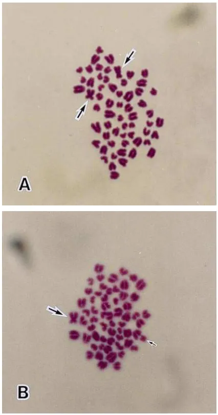
Fig. 4. Metaphase spreads of an embryo transfer chimeric calf (male) produced by aggregation of a whole parthenogenetic embryo (8-cell) and a whole IVF-derived embryo (8-cell): (A) 60, XX and (B) 60, XY. Long arrow5X chromosome, short arrow5Y chromosome.
Fig. 5. A chimeric male calf resulting from the transfer of a frozen-thawed embryo originally produced by aggregation of a whole 8-cell parthenogenetic embryo (Japanese Red) and a whole IVF-derived 8-cell embryos (Holstein). Arrow 5 a red color pattern originating from a parthenogenetic embryo.
XX and XY chromosome plates in the same embryo sample, 27% of the embryos had XY and 20% of the embryos had XX chromosome plates. These findings indicate that the XX chromosomes originated from parthenogenetic blastomeres and the XY chromosomes originated from the IVF-derived blastomeres. These results further indicate that both parthenogenetic and IVF-derived cells can contribute to embryo and concep-tus development, although the distribution rate of these different cell types could not be analyzed using this procedure.
In the earliest reports of chimeric mice births, it was evident that the sex ratio was heavily biased in favor of the male, with intersexes relatively uncommon. This may be due to the gene for H-Y antigen production located on the Y chromosome, which could influence the development of the undifferentiated gonad. In sheep, Tucker et al. (1974) reported that two XX-XY chimeras’ offspring were male phenotypes at birth. In a subse-quent report (Fehilly et al., 1984), 26 of the 36 recon-structed-embryo lambs born had a similar visible male pattern. In the present study, all calves born were phenotypically males with XX-XY chromosome plates. The results further suggest that when multiple-sex are used to make chimeric embryos, the male component exerts a marked effect on sexual differentiation, and consequently most chimeric embryos will develop as phenotypic males.
Embryos frozen in ethylene glycol can be rehydrated directly in a holding medium without step-wise dilution of cryoprotectant (Suzuki et al., 1993). In a subsequent report (Suzuki et al., 1995), a low concentration of trehalose was found to be the most beneficial for the cryopreservation of zona-free blastocysts. The presence of trehalose is thought to reduce osmotic shock while the zona-free blastocysts are suspended in the holding medium for rehydration. Also, Leibo and Oda (1993) have reported that PVP was effective in protecting mouse embryos during the freezing procedure. In experi-ment V, along with ethylene glycol (cryoprotectant) and trehalose (sugar, as a nonpermeating agent), a macro molecular component (PVP) was evaluated. Since the mechanism of protection of the large polymer, PVP (m.w. 530,000) is not clear, it is suggested that this substance coats the cells immediately following thaw-ing, giving them physical protection against osmotic stressors. Optimum viability of cryopreservation of zona-free chimeric blastocysts in the present study was obtained when the embryos were cryopreserved in 1.8 M ethylene glycol and 0.05 M trehalose with 10% PVP. To our knowledge, this is the first report describing parthenogenetic cells derived from cattle developing to term in utero, and their contribution to the production of live chimeric offspring. The results of this study indicate that chimeric blastocysts can be produced by aggregation of blastomeres, demi-embryos or whole embryos obtained from both parthenogenetic and IVF-derived embryos.
In an attempt to increase the participation of parthe-nogenetic cells in the aggregated embryo, inserting
blastomeres obtained from parthenogenetic embryos at an advanced stage (16-cell stage) into IVF-derived embryos at a less advanced stage (4-cell stage) is continuing. The idea is that the more advanced blasto-meres (from the parthenogenetic embryo) may contrib-ute to the inner cell mass (ICM) and the less advanced blastomeres (from the IVF-derived embryo) would de-velop into the trophectoderm (see reviews by Kelly et al., 1978; Godke and Rorie, 1993). As a consequence, this approach could produce live viable transplant calves from parthenogenetic cells.
From the results of the present study, we conclude that the IVF-derived bovine blastomeres were able to enhance the development of the parthenogenetic bovine blastomeres in chimeric embryos, and that the chimeric parthenogenetic bovine embryos can be developmen-tally competent.
ACKNOWLEDGMENTS
A portion of this joint research is a part of the Western Federal Regional Project W-171. Approved by the Director of the Louisiana Experimental Station as manuscript no. 97-11-0292.
REFERENCES
Anderegg C, Markert CL. 1986. Successful rescue of microsurgically produced homozygous uniparental embryos via production of aggre-gation chimeras. Proc Natl Acad Sci 83:6509–6513.
Aoyagi Y, Konishi M, Wada T, Takedomi T. 1994. Unaged bovine oocytes successfully develop to blastocysts after parthenogenetic activation or nuclear transfer. Theriogenology 41:157.
Barra J, Renard JP. 1988. Diploid mouse embryos constructed at the late 2-cell stage from haploid parthenotes and androgenotes can develop to term. Development 102:773–779.
Barton SC, Surani MAH, Norris ML. 1984. Role of paternal and maternal genomes in mouse development. Nature 311:374–376. Boediono A, Ooe M, Yamamoto M, Takagi M, Saha S, Suzuki T. 1993.
Production of chimeric calves by aggregation of in vitro fertilized bovine embryos without zonae pellucidae. Theriogenology 40:1221– 1230.
Boediono A, Takagi M, Saha S, Suzuki T. 1994. The influence of day 0 and day 7 superovulated cow serum during in vitro development of bovine oocytes. Reprod Fertil Dev 6:261–264.
Boediono A, Saha S, Sumantri C, Suzuki T. 1995. Development in vitro and in vivo of aggregated parthenogenetic bovine embryos. Reprod Fertil Dev 7:1073–1079.
Boediono A, Suzuki T. 1994. Pregnancies after transfer of aggregated parthenogenetic bovine activated oocytes. Theriogenology 41:166. Borsuk E. 1982. Pre-implantation development of gynogenetic diploid
mouse embryos. J Embryol Exp Morphol 19:215–222.
Brackett BG, Oliphant G. 1975. Capacitation of rabbit spermatozoa in vitro. Biol Reprod 12:260–274.
Ducibella T, Albertini DF, Anderson E, Briggers J. 1975. The pre-implantation mammalian embryo: Characterization of intracellular junctions and their appearance during development. Dev Biol 45:231–250.
Ducibella T, Anderson E. 1975. Cell shape and membrane changes in the eight-cell mouse embryo, prerequisites for morphogenesis of the blastocyst. Dev Biol 47:45–58.
Fehilly CB, Willadsen SM, Tucker EM. 1984. Experimental chimerism in sheep. J Reprod Fertil 70:347–351.
Fukui Y, Sawai K, Furudate M, Sato N, Iwazumi Y, Ohzaki K. 1992. Parthenogenetic development of bovine oocytes treated with ethanol and cytochalasin B after in vitro maturation. Mol Reprod Dev 33:357–362.
Goto K, Kajihara Y, Kosaka S, Koba M, Nakanishi Y, Ogawa K. 1988. Pregnancies after co-culture of cumulus cells with bovine embryos derived from in vitro fertilization of in vitro matured follicular oocytes. J Reprod Fertil 83:753–758.
Goto K, Ishida M, Ookutsu S, Nakanishi Y. 1994. Activation of unaged bovine oocytes by various parthenogenetic stimuli. Theriogenology 41:207.
Itagaki Y, Sato S, Shitanaka Y, Kudo T, Yamaguchi Y, Sutou S. 1993. Sexing of bovine embryos with male-specific repetitive DNA by polymerase chain reaction: sexing of bovine embryos and production of calves with predicted sex. J Reprod Dev 39:65–72.
Kaufman MH. 1983. Early mammalian development: parthenogenetic studies. Cambridge: Cambridge University Press.
Kaufman MH, Barton SC, Surani MAH. 1977. Normal postimplanta-tion development of mouse parthenogenetic embryos to the forelimb bud stage. Nature 265:53–55.
Kelly SJ, Mulnard JG, Graham CF. 1978. Cell division and cell allocation in early mouse development. J Embryol Exp Morphol 48:37–51.
Leibo SP, Oda K. 1993. High survival of mouse oocytes and embryos cooled rapidly or slowly in ethylene glycol plus polyvinylpyrrolidone. Cryo-Letters 14:133–144.
McGrath J, Solter D. 1984. Completion of mouse embryogenesis requires both the maternal and paternal genomes. Cell 37:179–183. Modlinski JA. 1980. Pre-implantation development of microsurgically obtained haploid and homozygous diploid mouse embryos and effects of pretreatment with cytochalasin B on enucleated eggs. J Embryol Exp Morphol 60:153–161.
Nagai T. 1987. Parthenogenetic activation of cattle follicular oocytes in vitro with ethanol. Gamete Res 16:243–249.
Nagy A, Rajki K, Bakos J, Csanyi V. 1979. Genetic analysis in carp (Cyprinus carpio) using gynogenesis. Heredity 43:35–40.
Niemierko A. 1975. Induction of triploidy in the mouse by cytochalsin B. J Embryol Exp Morphol 34:279–289.
Picard L, Schneider U, Betteridge KJ, King, WA. 1988. Effects of zona pellucida, agar embedding, and culture on the survival of microma-nipulated bovine embryos after freezing and thawing. J In Vitro Fertil Embryo Transf 5:268–274.
Pitts JD, Burk RR. 1976. Specificity of junctional communication between animals cells. Nature 264:762–764.
Presicce GA, Yang X. 1994. Development of 24 hour in vitro matured bovine oocytes following parthenogenetic activation by ethanol and cycloheximide treatment. Theriogenology 41:277.
Rorie RW, Pendelton RJ, Pool SH, Youngs CR, Godke RA. 1987. The viability of bovine ‘‘half ’’ embryos produced before or after liquid nitrogen freezing. In: Feichtinger W, Kemeter P, editors. Future aspects in human in vitro fertilization. Berlin: Springer-Verlag. p 26–35.
Solter D. 1988. Differential imprinting and expression of maternal and paternal genomes. Annu Rev Genet 22:127–46.
Stevens LC. 1978. Totipotent cells of parthenogenetic origin in a chimaeric mouse. Nature 276:266–267.
Stevens LC, Varnum DS, Eicher EM. 1977. Viable chimaeras produced from normal and parthenogenetic mouse embryos. Nature 269:515– 517.
Surani MAH, Barton SC, Kaufman MH. 1977. Development to term of chimaeras between diploid parthenogenetic and fertilized embryos. Nature 270:601–603.
Surani MA, Barton SC. 1983. Development of gynogenetic eggs in the mouse: implications for parthenogenetic embryos. Science 341:1034– 1036.
Surani MAH, Barton SC, Norris ML. 1984. Development of reconsti-tuted mouse eggs suggests imprinting of the genome during gameto-genesis. Nature 308:548–550.
Surani MAH, Barton SC, Norris ML. 1986. Nuclear transplantation in the mouse: Heritable differences between parental genomes after activation of the embryonic genome. Cell 45:127–136.
Surani MAH, Barton SC, Norris ML. 1987a. Experimental reconstruc-tion of mouse eggs and embryos: An analysis of mammalian development. Biol Reprod 36:1–16.
Surani MAH, Barton SC, Norris ML. 1987b. Parental chromosomes confer spatial specificity in androgenetic-parthenogenetic mouse chimaeras. Nature 320:1329–1332.
Surani MA, Kothary R, Allan ND, Singh PB, Fundele R, Ferguson-Smith AC, Barton SC. 1990. Genome imprinting and development in the mouse. Development (suppl):89–98.
Suzuki T, Takagi M, Yamamoto M, Boediono A, Saha S, Sakakibara H, Oe M. 1993. Pregnancy rate and survival in culture in vitro fertilized bovine embryos frozen in various cryoprotectants and thawed using a one-step system. Theriogenology 40:651–659. Suzuki T, Saha S, Sumantri C, Takagi M, Boediono A. 1995. The
influence of polyvinylpyrrolidone on freezing of bovine IVF blasto-cysts following biopsy. Cryobiology 32:505–510
Takagi M, Boediono A, Saha S, Suzuki T. 1993. Survival rate of frozen-thawed bovine IVF embryos in relation to exposure time using various cryoprotectants. Cryobiology 30:306–312.
Thomson JA, Solter D. 1988. The developmental fate of androgenetic, parthenogenetic and gynogenetic cells in chimeric gastrulating mouse embryos. Genes Dev 2:1344–1351.
Thomson JA, Solter D. 1989. Chimeras between parthenogenetic or androgenetic blastomeres and normal embryos: allocation to the inner cell mass and trophectoderm. Dev Biol 131:580–583. Tsunoda Y, Yasui T, Nakamura K, Uchida T, Sugie T. 1986. Effect of
cutting the zona pellucida on the pronuclear transplantation in the mouse. J Exp Zool 240:119–125.
Tucker EM, Moor RM, Rowson LEA. 1974. Tetraparental sheep chimeras induced by blastomere transplantation. Immunology 26: 613–621.
Ware CB, Barnes FL, Maiki-Laurila M, First NL. 1989. Age depen-dence of bovine oocyte activation. Gamete Res 22:265–275. Watson AJ, Hogan A, Hahnel A, Wiemer KE, Schutlz GA. 1992.
Expression of growth factor ligand and receptor genes in the pre-implantation bovine embryos. Mol Reprod Dev 31:87–95. White WK. 1978. Modes of speciation. San Francisco: WH Freeman. Willadsen SM. 1979. A method for culture of micromanipulated sheep
embryos and its use to produce monozygotic twins. Nature 277:298– 300.
Wolfe BA, Kraemer DC. 1992. Methods in bovine nuclear transfer. Theriogenology 37:5–15.
Yang X, Presicce GA, Moraghan L, Jiang S, Foote RH. 1994. Synergis-tic effect of ethanol and cyclohexamide on activation of freshly matured bovine oocytes. Theriogenology 41:395–403.