Original article
Inadvertent BSA-induced elution of IgE in the BSA-RAST
Background: Bovine serum albumin (BSA) is widely used to block nonspecific binding in immunochemical assays. Whereas a previous study had indicated that soluble allergen present during the incubation with anti-IgE in the RAST did not affect bound IgE, we reinvestigated this in the current study, using IgE elution from BSA by soluble BSA as a test system.
Methods: Sepharose-coupled BSA (0.08, 0.4, 2, or 10mg BSA/test) was incubated overnight with serum and washed. The Sepharose was then incubated with different concentrations of soluble BSA (0, 12, 60, 300, or 1500mg/test), washed again, and incubated with radioactive anti-IgE. The effect on IgE binding was investigated for various incubation periods (t=0, 1, 2, 4, and 20 h). Results: Incubation in buffer without BSA did not change IgE binding. Soluble BSA eluted IgE antibodies from immobilized BSA by up to 85%. If the BSA density on the solid phase wasi2 mg/test, the elution efficiency was dependent on the levels of both immobilized BSA and soluble BSA. At lower densities, the dissociation was dependent only on the concentration of soluble BSA. The time needed to obtain 50% IgE elution (tK) was less if the density of immobilized BSA decreased. Below the critical density (0.8mg BSA/mg solid phase),tKwas independent of the coating density (45 min). Probably all IgE antibodies are monovalently bound below this density.
Conclusions: Dissociation of IgE from immobilized protein in the presence of soluble protein should be taken into account, particularly when IgE to mammalian serum albumin is involved (milk, meat, or animal dander).
I. Kleine Budde1, P. G. de Heer1, S. O. Stapel1, J. S. van der Zee2, R. C. Aalberse1
1Department of Immunopathology, CLB and Laboratory
of Experimental and Clinical Immunology, University of Amsterdam, Amsterdam;2Department of Pulmonology,
Academic Medical Center, University of Amsterdam, Amsterdam, The Netherlands
Key words: allergy; BSA; IgE; IgE elution; RAST.
Rob C. Aalberse, PhD, CLB Plesmanlaan 125 PO Box 9190 1066 CX Amsterdam The Netherlands
Accepted for publication 9 July 2001
The radioallergosorbent test (RAST) (1) is a well-established method for allergy diagnosis. The principle is based on the detection of IgE binding to allergen on a solid phase by labeled antihuman IgE. The latter reagent is often diluted in a buffer containing bovine, sheep, or horse serum. If the solid phase contains some immunoglobulin G (IgG), IgG in heterologous serum must be present to prevent binding of labeled antihuman IgE via bivalent human antibodies directed to heterologous IgG (2). Bovine serum albumin (BSA) is a widely used blocker in, for example, ELISAs and immunoblotting experiments.
In the current study, we analyzed IgE binding to BSA. IgE antibodies to albumin have been shown to be in large part responsible for allergy to, for instance, bovine and ovine meat (3, 4), cow’s dander (5), and cow’s milk (5). In a BSA-RAST, the incubation with labeled anti-IgE is also performed in the presence of
heterologous serum and soluble BSA. No effect by this BSA on IgE binding was expected, according to a study by van Milligen et al. (6), in which IgE binding to the major cat (Felis domesticus) allergen Fel d 1, and Fel d 1-peptides was investigated. Fel d 1-bound Sepharose (0.25–0.5mg/test) was incubated overnight with serum, and after extensive washing, it was shown that IgE was not eluted by incubation with soluble Fel d 1 (2.4mg) for 16 h. Only IgE bound to low-affinity Fel d 1-peptides (8mg/test) was eluted by Fel d 1. In RAST-inhibition experiments, in which serum is preincubated with the allergen before addition to Sepharose, less protein is needed to inhibit binding. In such a preincubation protocol, IgE binding to Fel d 1 was inhibited by >95% by 2.4 mg of Fel d 1.
In this paper, we present data showing that, unexpectedly, soluble BSA added after incubation of BSA-allergosorbent with serum eluted IgE bound to immobilized BSA. We determined the levels of soluble and immobilized BSA at which this elution became significant, and how long it took for 50% of IgE antibodies to be eluted.
Abbreviations.BSA: bovine serum albumin; FCS: fetal calf serum; HSA: human serum albumin; Ig: immunoglobulin; RAST: radioallergosorbent test.
Material and methods
Materials
The materials included bovine albumin fraction V (Boehringer Mannheim, Heidelberg, Germany), human serum albumin (HSA) (CLB, Amsterdam, The Netherlands), fetal calf serum (FCS) (Bodinco, Alkmaar, The Netherlands), Tween-20 (Merck, Darmstadt, Germany), EDTA (Siegfried Synopharm, Switzerland), NaN3 (Merck, Darmstadt, Germany), and CNBr-activated Sepharose (Pharmacia Fine Chemicals, Uppsala, Sweden).
Buffers
BSA-sheep.The contents were PBS with 3 mg/ml BSA, 0.5% (v/v) sheep serum, 4.5% (v/v) bovine serum, 0.2% Tween-20, and 10 mM EDTA, 0.05% NaN3.
HSA-sheep Ig. The contents were PBS with 100mg/ml HSA, 20 mg/ml sheep Ig, 0.1% Tween-20, and 0.06% NaN3. Sheep Ig was purified with caprylic acid as described by McKinney & Parkinson (7).
HSA-PBS. The contents were PBS with 3 mg/ml HSA, 0.2% Tween-20, 10 mM EDTA, and 0.05% NaN3.
Plasma and serum samples
Plasma samples were derived (Haemonetics Plasma Collection System, Haemonetics Corporation, Braintree, MA, USA) from volunteers, after informed consent. Plasma samples nos. 163 and 151 were defibrinated by recalcification and dialyzed. Plasma no. 163 (total IgE: 2500 IU/ml) was positive for IgE to common inhalant allergens (house-dust mite, grass pollen, birch pollen, cat dander, dog dander, and Alternaria), and to BSA (17 IU specific IgE/ml, determined in a BSA-free system). Plasma sample no. 151 (total IgE: 1600 IU/ml) was used as negative control in the BSA-RAST. This plasma was positive for IgE to house-dust mite, grass pollen, birch pollen, cat dander, dog dander,Alternaria, and Aspergillus. Serum samples s2910 and s3123 contained totals of 666 and 175 IgE/ ml, respectively.
Labeling
Affinity-purified sheep antihuman IgE (M1294, CLB, Amsterdam) was labeled by the125I chloramine-T method (8), except that after iodination and reduction HSA was used instead of BSA as carrier protein. Labeled anti-IgE was diluted in PBS/0.3% HSA/0.01 M EDTA/0.05% NaN3and stored atx20uC.
FCS-RAST
FCS (1.7 mg protein) was coupled to 100 mg CNBr-activated Sepharose (8), after which the Sepharose was suspended at 4 mg/ml in HSA-PBS. The RAST was performed as described previously (8, 9). Human serum (50ml) was incubated overnight with FCS-Sepharose (250ml). After washing five times, 125I-labeled sheep antihuman IgE was added in the presence of 500ml BSA-sheep Ig, HSA-sheep Ig, or HSA-PBS, and incubated overnight. After washing four times, bound radioactivity was determined, and expressed as percentage of added counts. The experiments were performed in duplicate.
BSA-RAST
BSA was coupled to CNBr-activated Sepharose (8) in different densities: 16, 80, 400, and 2000mg BSA per 100 mg Sepharose.
Sepharose was suspended at 2 mg/ml in HSA-PBS. BSA-Sepharose was incubated with human serum (50ml per 0.5 mg Sepharose) while rotating overnight. After washing five times, 500ml of HSA-sheep Ig containing 0, 24, 120, 600, or 3000mg/ml BSA (final volume 700ml) was added to the Sepharose, which was incubated for different time periods (0, 1, 2, 4, or 20 h). Next, Sepharose was washed five times, and incubated overnight with radioactive anti-IgE in 500ml HSA-sheep Ig. Tests without addition of HSA-HSA-sheep Ig (t=0) were performed in 10-fold, and the other tests in duplicate. Bound radio-activity was converted to international units (IU) IgE per ml serum, using as calibration system mouse/human chimeric antibodies direc-ted to Der p 2 in combination with Sepharose-coupled mite extract (10).
Calculation of BSA density on Sepharose
CNBr-activated Sepharose (1 ml packed gel=300 mg Sepharose) consists of beads with an average diameter of 105mm (range 45–165mm). Assuming that Sepharose beads are smooth spheres, the surface of one bead is 34 600mm2. Per test, 0.5 mg Sepharose (3000 beads) was used, corresponding to a surface area of 104 mm2. Thus, 0.08, 0.4, 2, or 10mg BSA per 0.5 mg Sepharose corresponds to 0.8, 3.8, 19.2, and 96.2 ng per mm2, respectively. Obviously, these values are overestimated, because the surface area is larger than that of smooth spheres. Moreover, no allowance has been made for incom-plete coupling.
Results
BSA and HSA in the RAST
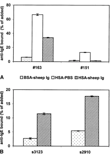
As one of the control tests for experiments described elsewhere (11), we performed RAST experiments in which IgE antibodies to FCS were analyzed. We discovered that soluble BSA, a main component of FCS, present during the incubation with radioactive anti-IgE, affected the IgE binding. Fig. 1A shows that IgE binding decreased after incubation of radioactive anti-IgE in BSA-containing buffer compared to buffer with HSA. To avoid binding of human IgG antibodies to bovine IgG on the Sepharose, sheep and/or bovine immunoglobulin must be added to the buffer. This is illustrated by the results with negative control plasma no. 151. When this plasma was incubated with anti-IgE in HSA-PBS in the absence of sheep and/or bovine immunoglobulin, significant binding of 125I-labeled sheep anti-IgE to FCS-Sepharose was observed. This binding was abolished by the addition of sheep immunoglobulin.
Fig. 1B shows that the elution of IgE from immo-bilized BSA by soluble BSA is not a unique property of plasma no. 163. With two other sera containing specific IgE, IgE elution was also found in the BSA-RAST: respectively, 3.2 and 5.8 IU IgE/ml serum were bound in the absence of soluble BSA, whereas these values were 0.5 and 1.3 IU IgE/ml, respectively, if soluble BSA was present.
respectively, in the absence of BSA, and 53.7% and 32.5%, respectively, when BSA was present. In contrast, the RAST of plasma no. 163 with BSA-Sepharose resulted in 42.5% of radioactive anti-IgE being bound if the last incubation was performed in HSA sheep-Ig buffer, while the result was 3.6% if the incubation was performed in a mixture of BSA and sheep-Ig.
Dose dependency of IgE elution
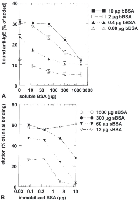
The elution of BSA-specific IgE antibodies by soluble BSA was studied in more detail in the BSA-RAST. After incubation of plasma no. 163 with different densities of BSA on the Sepharose, different concentra-tions of soluble BSA were added. The incubation was performed for 20 h before radiolabeled anti-IgE was added. The results are shown in Fig. 2A. When 2 or 10mg of immobilized BSA per test was used, the elution was dependent on both the density of immobilized BSA and the concentration of soluble BSA. However, with lower densities of immobilized BSA (j0.4mg), the dissociation was dependent only on the concentration of soluble BSA (Fig. 2B). The amounts of IgE antibodies eluted from 0.08, 0.4, 2, and 10mg immobilized BSA per test by 1.5 mg soluble BSA, expressed as percentage of initially IU/ml bound IgE, were 76%, 79%, 83%, and 85%, respectively.
Kinetics of dissociation
The elution of IgE antibodies from BSA coupled to Sepharose at four densities by different concentrations of soluble BSA was determined as a function of time. Short incubation times were effective only with lower densities of immobilized BSA. IgE antibodies were not significantly eluted by buffer without BSA: the concentrations of bound IgE to 0.08, 0.4, 2, and 10mg coupled BSA per test were at t=0 and t=20, respectively, 2.9 and 2.5 IU/ml serum, 8.3 and 8.2 IU/ml serum, 13.9 and 14.9 IU/ml serum, and 16.9 and 15.7 IU/ml serum.
The kinetics of the dissociation of IgE from Sepharose-bound BSA by soluble BSA was investigated in more detail. Fig. 3A shows the exponential phase of the amount of IgE antibodies eluted from immobilized BSA. The time needed to elute 50% of IgE antibodies (tK) in this first phase was plotted against the density of
immobilized BSA (Fig. 3B). The critical density, below whichtKis independent of the density of immobilized
BSA, was estimated to be approximately 0.8mg BSA per mg Sepharose.
Discussion
In the current study, we unexpectedly found that soluble allergen, added after incubation of allergen-specific IgE with allergosorbent, elutes IgE antibodies. The influence of soluble allergen on IgE binding in the RAST is, of course, well-established when allergen is
preincubatedwith serum. RAST-inhibition experiments with antigen are based on this principle (12). van Milligen et al. (6) showed that concentrations of the allergen Fel d 1 able to inhibit IgE binding in an inhibition RAST did not influence IgE binding when added after the binding of IgE to immobilized Fel d 1. IgE antibodies did not elute from Fel d 1 on solid phase (0.25–0.5mg/0.5 mg Sepharose) in the presence of 2.4mg soluble Fel d 1. However, according to the results of the current study, IgE elution is to be expected only at higher levels of soluble Fel d 1. We found that 12mg soluble BSA is required to decrease IgE binding to 0.4mg immobilized BSA with 25%.
It was confirmed in the current report that sheep IgG should be present during incubation with radioactive sheep anti-IgE in a FCS-RAST. This has been reported previously for RAST experiments with allergen extracts containing sheep or cow IgG as in meat, milk, or dander (2). If bivalent antibodies to bovine or ovine IgG are present in human serum, false-positive results may be obtained by binding of these human antibodies with one antigen-binding site to IgG on Sepharose, and with the other site to radioactive sheep antihuman IgE. The results of our FCS-RAST were surprising, since IgG was expected to be low in fetal serum. According to the manufacturer, the concentration of gamma globulin in
FCS is less than 0.5% of total protein, which corresponds to <85 ng IgG per test. The results of this paper (Fig. 2A) indicate that as little as 80 ng allergen per test suffices to give a strong response (>10% binding). Thus, the effect of sheep IgG in the FCS-RAST is not incompatible with a low amount of IgG in FCS.
We showed that binding of IgE to immobilized BSA was not significantly influenced (<1.5 IU IgE/ml serum) by incubation in buffer without BSA for 20 h. IgE antibodies might be monovalently or bivalently bound to immobilized BSA. If IgE binding was bivalent in our tests, full dissociation was not easily accom-plished in buffer because the IgE antibodies that dissociate with one of the antigen-binding sites from BSA are likely to rebind before the second site dissociates. In the presence of soluble BSA, reassocia-tion of the first site is prevented, and after dissociareassocia-tion of the second antigen-binding site, the IgE antibody will be released from the Sepharose. The binding of IgE to BSA might also be monovalent. However, this may
seem incompatible with the requirement for soluble BSA to dissociate the complex. IgE antibodies elute in the presence of soluble BSA, a fact which indicates that IgE occasionally dissociates from immobilized BSA. Thus, for monovalent binding, elution was expected even in the absence of BSA. However, IgG4 antibodies, which are functionally monovalent (8, 13), also require soluble BSA for dissociation (data not shown). The lack of dissociation in buffer may be explained by assuming the presence of an ‘‘unstirred layer’’. An antibody is assumed to have three possible states:
1) bound
2) unbound at a negligible distance from the antigen 3) freely diffusing in the medium.
In the second state, rebinding of IgE antibodies is more likely than diffusion from the ‘‘unstirred layer’’ into the medium. However, soluble BSA prevents reassociation.
The dissociation of IgE described in the current study
Figure 2. Dissociation of IgE from immobilized BSA by different concentrations of soluble BSA. Incubation with soluble BSA was performed for 20 h. A) IgE antibodies of plasma no. 163 fixed to different densities of BSA coupled to Sepharose were eluted by various concentrations of soluble BSA. B) Elution of IgE antibodies by soluble BSA expressed as percentage of IgE binding in absence of soluble BSA. bBSA: bound BSA; sBSA: soluble BSA.
Figure 3. A) Logarithmic transformation of dissociation of IgE antibodies bound to immobilized BSA by soluble BSA. After overnight incubation of plasma no. 163 with BSA-Sepharose, IgE was eluted by addition of 1500mg soluble BSA. Coupled BSA per
test: 0.08mg (open triangles), 0.4mg (solid triangles), 2mg (open
squares), 10mg (solid squares). B) Time needed to reduce IgE
cannot be completely explained by assuming bivalent binding of IgE antibodies, since the dissociation time (tK) did not change below 0.8mg BSA per mg
Sepharose. These results suggest that all IgE binding is monovalent below this critical density. Thus, in the case of a high concentration of immobilized BSA, most IgE is bound bivalently and dissociates slowly, whereas at a low concentration of immobilized BSA, most IgE is bound monovalently and dissociates rapidly in the presence of soluble BSA. The critical density of 0.8mg/ mg Sepharose is equivalent to one BSA molecule per 29 nm2. Since the distribution of molecules on a sphere is hexagonal, the mean distance between molecules on a bead surface of 104 mm2 is 4.4 nm. The maximum distance between two antigen-binding sites of an antibody, which is 150 A˚ (15 nm), is approximately 3.5 times this value. However, the surface of a Sepharose bead is rough rather than smooth. Moreover, not all coupled proteins are available for antibody binding, because some proteins are bound in antibody-inaccessible pores of the Sepharose (data not shown). Assuming that the surface of a bead is three times larger than indicated and that only 25% of coupled proteins are available for antibody binding, the distance between two BSA molecules is around the critical density for bivalent or monovalent binding (15 nm).
Posner et al. (14) also found a rapid and a slow phase in IgE dissociation. They monitored the dissociation of bivalent ligand-bivalent receptor aggregates in solution. The dissociation of a bivalent ligand with two identical 2,4,-dinitrophenyl (DNP) groups from fluorescein-labeled, monoclonal, DNP-specific murine IgE was promoted by a monovalent ligand. In this system, two types of IgE-ligand aggregates were formed, linear chains and closed chains, consisting of at least trimers of IgE. One rapid and one slow dissociation compo-nents were found. It was postulated that the fast phase was induced by dissociation of monovalently bound IgE.
It may not be possible to elute all IgE antibodies with allergen. Approximately 15% of IgE antibodies were not eluted in the presence of 1.5 mg soluble BSA. Why some IgE antibodies were bound in a reversible way, while others were apparently irreversibly bound, is not clear. Possibly, secondary changes in the antibody-allergen complex lead to a tighter interaction. This hypothesis might be verified by varying the incubation time of IgE with immobilized BSA.
In the experiments described in the current study, soluble BSA was removed by washing before incuba-tion with radioactive anti-IgE. In a ‘‘standard’’ RAST, soluble BSA is present during this anti-IgE incubation. Since anti-IgE will also bind eluted IgE, the RAST results might even be more disturbed in these experi-ments. Thus, two factors decrease the binding of label to the solid phase:
1) decreasing quantities of IgE on the solid phase 2) decreasing availability of labeled anti-IgE due to
neutralization with eluted IgE.
In this paper, we showed that soluble BSA dissociates BSA-specific IgE antibodies from immobilized BSA in the RAST. Presumably, a similar effect occurs in immunoblot experiments. Thus, the effect of soluble BSA should be taken into account to avoid under-estimation of the IgE antibody levels to BSA and cow’s milk, meat, or dander. Moreover, since BSA cross-reacts with serum albumin from other animals (such as sheep, cat, dog, and horse), soluble BSA may also influence RAST results for these components. We showed that this inadvertent elution of IgE by BSA may be avoided by using HSA instead of BSA in buffers.
Acknowledgment
This project was funded by The Netherlands Asthma Foundation (project no. 32.95.13).
References
1. WIDEL, BENNICHH, JOHANSSONSG. Diagnosis of allergy by anin vitrotest for allergen antibodies. Lancet 1967;2: 1105–1107.
2. FOUCARDT, BENNICHH, JOHANSSONSG, LUNDKVISTU. Human antibodies to bovine alpha-globulin. Int Arch Allergy Appl Immunol 1975;48:812–823. 3. FIOCCHIA, RESTANIP, RIVAE, et al. I.
Specific IgE to BSA and OSA in atopic, beef sensitive children. J Am Coll Nutr 1995;14:239–244.
4. FIOCCHIA, RESTANIP, RIVAE, et al. II. Effects of food processing and enzymatic digestion on the allergenicity of bovine and ovine meats. J Am Coll Nutr 1995;14:245–250.
5. SZEPFALUSIZ, EBNERC, URBANEKR, et al. Detection of IgE antibodies specific for allergens in cow milk and cow dander. Int Arch Allergy Immunol 1993;102: 288–294.
6. vANMILLIGENFJ, vAN’THOFW, vAN DEN BERGM, AALBERSERC. IgE epitopes on the cat (Felis domesticus) major allergen Fel d I: a study with overlapping synthetic peptides. J Allergy Clin Immunol 1994;93:34–43.
7. MCKINNEYMM, PARKINSONA. A simple, non-chromatographic procedure to purify immunoglobulins from serum and ascites fluid. J Immunol Methods 1987;96:271–278.
9. vAN DERZEEJS,DEGROOTH, vAN SWIETENP, JANSENHM, AALBERSERC. Discrepancies between the skin test and IgE antibody assays: study of histamine release, complement activationin vitro, and occurrence of allergen-specific IgG. J Allergy Clin Immunol 1988;82: 270–281.
10. SCHUURMANJ, PERDOKGJ, LOURENSTE, PARRENPW, CHAPMANMD, AALBERSE RC. Production of a mouse/human chimeric IgE monoclonal antibody to the house dust mite allergen Der p 2 and its use for the absolute quantification of allergen-specific IgE. J Allergy Clin Immunol 1997;99:545–550.
11. KLEINEBUDDEI, AALBERSM, AALBERSE RC, vAN DERZEEJS, KNOLEF. Reactivity to IgE-dependent histamine-releasing factor is due to monomeric IgE. Allergy 2000;55:653–657.
12. GLEICHGJ, LARSONJB, JONESRT, BAER H. Measurement of the potency of allergy extracts by their inhibitory capacities in the radioallergosorbent test. J Allergy Clin Immunol 1974;
53:158–169.
13. vAN DERZEEJS, vANSWIETENP, AALBERSE RC. Serologic aspects of IgG4 antibodies. II. IgG4 antibodies form small, nonprecipitating immune complexes due to functional monovalency. J Immunol 1986;137: 3566–3571.