ContentslistsavailableatScienceDirect
Colloids
and
Surfaces
A:
Physicochemical
and
Engineering
Aspects
j o u r n al ho me p a g e :w w w . e l s e v i e r . c o m / l o c a t e / c o l s u r f a
Bovine
serum
albumin
(BSA)
and
cleaved-BSA
conjugated
ultrasmall
Gd
2
O
3
nanoparticles:
Synthesis,
characterization,
and
application
to
MRI
contrast
agents
Md.
Wasi
Ahmad
a,
Cho
Rong
Kim
a,
Jong
Su
Baeck
b,
Yongmin
Chang
b,d,∗,
Tae
Jeong
Kim
c,d,
Ji
Eun
Bae
d,
Kwon
Seok
Chae
d,e,
Gang
Ho
Lee
a,d,∗aDepartmentofChemistry,CollegeofNaturalSciences,KyungpookNationalUniversity(KNU),Taegu702-701,SouthKorea
bDepartmentofMolecularMedicineandMedical&BiologicalEngineering,SchoolofMedicine,KNU,Taegu702-701,SouthKorea
cDepartmentofAppliedChemistry,CollegeofEngineering,KNU,Taegu702-701,SouthKorea
dDepartmentofNanoscienceandNanotechnology,KNU,Taegu702-701,SouthKorea
eDepartmentofBiologyEducation,Teacher’sCollege,KNU,Taegu702-701,SouthKorea
h
i
g
h
l
i
g
h
t
s
•BSAandC-BSAconjugatedultrasmall
Gd2O3 nanoparticles were
synthe-sized.
•BSAcanbindmanyultrasmallGd2O3
nanoparticleswhereasC-BSAcannot.
•Largewaterprotonrelaxivitieswere
observed.
•HighcontrastMRimagesinamouse
liverafterintravenousinjectionwere
observed.
•These nanoparticles are potential
MRIcontrastagents.
g
r
a
p
h
i
c
a
l
a
b
s
t
r
a
c
t
a
r
t
i
c
l
e
i
n
f
o
Articlehistory:
Received15November2013
Receivedinrevisedform23February2014 Accepted1March2014
Availableonline12March2014
Keywords:
BSA C-BSA
UltrasmallGd2O3nanoparticle
Nanoparticlecarrier MRI
Contrastagent
a
b
s
t
r
a
c
t
Bovineserumalbumin(BSA)(Mn=66.5kD,size=14×4×4nm)isanattractivebiologicalmoleculefor
biomedicalapplicationsbecauseofitswater-solubilityandbio-compatibility.Itcanalsobindmany
ultrasmallnanoparticles(NPs)asconfirmedinthisstudy.Wesynthesizedpolyethyleneglycoldiacid
(PEGD)coatedultrasmallGd2O3nanoparticles(PEGD-GNPs,thecoredavg=2.0nm),whichwerethen
con-jugatedtoBSAandcleaved-BSA(C-BSA)(i.e.BSA-PEGD-GNPsandC-BSA-PEGD-GNPs)throughamide
bonding.Largerelaxivities wereobservedinbothaqueous samplesolutions(r1=6.0s−1mM−1 and
r2=28.0s−1mM−1 forBSA-PEGD-GNPsandr1=7.6s−1mM−1 andr2=22.0s−1mM−1 for
C-BSA-PEGD-GNPs).ThreeteslaT2magneticresonanceimaging(MRI)inamouseaftertheinjectionofanaqueous
samplesolutionofBSA-PEGD-GNPsintoamousetailveinrevealedsignificantnegativecontrast
enhance-ments.LargerelaxivitiesandinvivoMRimagesprovethatBSA-PEGD-GNPsandC-BSA-PEGD-GNPsare
potentialMRIcontrastagents.
©2014ElsevierB.V.Allrightsreserved.
Abbreviations:BSA,bovineserumalbumin;C-BSA,cleavedBSA;PEGD,polyethyleneglycoldiacid;NP,nanoparticle;GNP,Gd2O3NP;BRB,Britton–Robinsonbuffer;PBS,
phosphatebuffersolution;MRI,magneticresonanceimaging;NCT,neutroncapturetherapy;CT,X-raycomputedtomography;DTPA,diethylenetriaminepentaaceticacid;PBS, phosphatebuffersolution;NHS,N-hydroxysuccinimide;EDC,1-ethyl-3-(3-dimethylaminopropyl)carbodiimide;GPC,gelpermeationchromatograph;HVEM,highvoltage electronmicroscope;XRD,X-raydiffraction;ICPAES,inductivelycoupledplasmaatomicemissionspectrometer;FT-IR,Fouriertransform-infrared;TGA,thermo-gravimetric analyzer;DU145,humanprostatecancercell;NCTC1469,normalmousehepatocytecell.
∗Correspondingauthors.Tel.:+82539505340;fax:+82539506330.
E-mailaddresses:[email protected](Y.Chang),[email protected](G.H.Lee).
http://dx.doi.org/10.1016/j.colsurfa.2014.03.011
68 Md.W.Ahmadetal./ColloidsandSurfacesA:Physicochem.Eng.Aspects450(2014)67–75
1. Introduction
Biologicalmoleculeshave attracted considerableinterest for applicationtonanomedicinebecausetheyarefullybiocompatible
andwater-soluble.Bovineserumalbumin(BSA)isanimportant
carrierproteininbloodplasmaforseveralionsandmolecules.Ithas alargedimension(14×4×4nm)andheavymass(Mn=∼66.5kD).
Thus,itmayalsocarryseveralultrasmallnanoparticles(NPs)with asmallersizeandmassthanBSA.Biologicalmoleculeshave sev-eraladvantagesoversmallmoleculesandpolymersforbiomedical applications.First, the water-solubility of surface-modifiedNPs generallyincreaseswithincreasingmassoftheligands[1,2]and thus,biologicalmoleculeswillprovideenhancedwater-solubility forNPs.Second,theNPsconjugatedtobiologicalmoleculescan
remain in the blood for a longer duration than free NPs and
Gd-chelates,allowinglongerimagingtimes(socalledblood-pool imagingagents)andahigherlikelihoodofdeliveringNPstothe targetedareasinabody[3–6].
ThisstudymakesuseofBSAandultrasmallGd2O3NPs(GNPs)
for magnetic resonance imaging (MRI). Ultrasmall GNPs have
shownlongitudinal(r1)andtransverse(r2)waterproton
relax-ivities larger than those of Gd-chelates because of the dense
populationofGd(III)inNPs[7,8].Therefore,BSAwhichcouldbind severalultrasmallGNPs maybeusefulfor MRI.Here, ultrasmall
GNPs can bealso appliedto X-raycomputed tomography (CT)
asCTcontrastagentsandneutroncapturetherapy(NCT)asNCT agentsbecauseGdhasalargeX-rayattenuationpower(∼2.5times
strongerthancommercialiodineCTcontrastagents)[9–12]anda verylargeneutroncapturecrosssection(∼254,000barns)[13–16].
Thisimplies thatBSAconjugatedultrasmallGNPs couldbealso usefulforCTandNCT.
PreviousstudiesonMRIusingBSAincludeBSA[Gd-chelates]n
andBSA-GNPs(d=20–40nm)[3–6,10,17].Enhancedwaterproton relaxivities have beenobserved in both systems after conjuga-tiontoBSA.Inaddition,theinvivoapplicationofBSA[Gd-DTPA]n
(DTPA=diethylenetriaminepentaaceticacid)showedalonger
cir-culationtime intheblood than Gd-DTPA, providing longerMR
imagingtimesinbraintumorsandbloodvesselsinrats[3,4]. ThisstudyexaminedBSAandcleavedBSA(C-BSA)(<7.0kD) con-jugatedultrasmallGNPsforMRIcontrastagents.Here,ultrasmall nanoparticles(d<3nm)areclinicallyimportantbecausetheycan beexcretedthroughrenalsystem[18]andtherefore,haveahigh potentialforbiomedical applications.ToconjugateGNPstoBSA andC-BSA,theGNPsweresurfacemodifiedwith polyethylenegly-coldiacid(PEGD)(Mn=600)andthenconjugatedtoBSAandC-BSA viaamidebonding.Theparticlesizes,surfacemodifications, num-berofultrasmallGNPsconjugatedtoBSAandC-BSA,waterproton relaxivities,cellulartoxicities,andinvivoMRimagesusingamouse werecharacterized.TheBSAconjugatedGNPswerefurtherapplied toMRIinamouseandnegativecontrastenhancementsin3TT2MR imageswereclearlyobserved.
2. Experimentals
2.1. Chemicals
AllchemicalssuchasGdCl3·xH2O(99.9%),NaOH(>99.9%), tri-ethyleneglycol(99%),polyethyleneglycoldiacid(PEGD)(Mn=600), BSA (Mn=∼66.5kD), phosphate buffersolution (PBS) (pH=7.2),
HCl (>99%), N-hydroxysuccinimide (NHS) (98%),
1-ethyl-3-(3-dimethylaminopropyl) carbodiimide (EDC) (>97%), boric acid
(>98%),phosphoricacid(>97%),andacetic acid(98%)were pur-chasedfromSigma-Aldrichandusedasreceived.Triplydistilled waterwasusedforbothwashingtheproductsandpreparingthe MRIsamplesolutions.
2.2. SynthesisofPEGDcoatedultrasmallGNPs(PEGD-GNPs)
PEGD-coatedultrasmallGNPsweresynthesizedusingone-pot
process(Scheme1a).Twoseparatesolutionswereprepared.One isaprecursorsolutionmadefrom5mmolofGdCl3·xH2Oin25mL oftriethyleneglycol,andtheotherisaNaOHsolutionmadefrom 15mmolofNaOHin10mLoftriethyleneglycol.Theprecursor solu-tionwasheatedto60◦Cwithmagneticstirringunderatmospheric conditionsuntiltheprecursorwasdissolvedcompletelyin triethy-leneglycol.ANaOHsolutionwasaddedtotheprecursorsolution. Themixedsolutionwasstirredmagneticallyat180◦Cfor4h.The solutiontemperaturewasreducedto80◦Cand8mmolofPEGDwas addedtothesolutionforsurfacecoating.AnexcessofPEGDwas
usedtoensurethatonlyone–COOHgroupamongthetwo–COOH
groupsinPEGDcouldbeusedforconjugationtoultrasmallGNPs becausetheother–COOHgroupshouldbefreeforconjugationto BSA(orC-BSA).Thesolutiontemperaturewasagainincreasedto 180◦Candstirredforanadditional4h.Thesolutionwascooled toroomtemperatureandtransferredtoa1Lbeakercontaining 500mLoftriplydistilledwater.Thiswasstirredmagneticallyfor 10minandstoredforaweekorsountilthePEGD-coatedultrasmall
GNPssettledinabeakerbottom.Thesupernatantwasdecanted
andtheremainingsamplesolutionwaswashedagainwithtriply distilledwater.Thisprocedurewasrepeatedthreetimes.
2.3. SynthesisofC-BSA
To prepare C-BSA, 10mmol of l-serine and 10mmol of l
-histidineweredissolvedin 300mLofaBritton–Robinsonbuffer
(BRB) (40mM phosphate, 40mM acetate, and 40mM borate)
(pH=5–6)at60◦Candunderatmosphericconditions(Scheme1b)
[19].Here,apHof5–6ofthebuffersolutionwasachievedbyadding NaOHslowlytotheoriginalbuffersolution.Aftermagneticstirring for30min,156mgofBSAwasaddedtothesolution,andthe solu-tionwasstirredmagneticallyfor36hat60◦C.Afterthereaction wascomplete,thewaterwasevaporated.Thiscleavagereaction
wasrepeated10timestoobtainenoughC-BSA.Themassesof
C-BSAswerecharacterizedbygelpermeationchromatography(GPC) andtheresultissummarizedinSupportingInformation.Twomajor
C-BSAswithmassesof6.67and2.01kDwereobservedfromGPC
analysis.TheC-BSAwasusedwithoutfurtherpurificationbecause thewater-solubleC-BSAcouldnotbeseparatedfromother water-solublereagentsusedinthereaction.Ontheotherhand,thewater solublereagentswerelaterremovedafterconjugationofC-BSAsto
PEGD-GNPsthroughamidebonding.
2.4. SynthesisofBSA-PEGD-GNPsandC-BSA-PEGD-GNPs
BSA-PEGD-GNPs and C-BSA-PEGD-GNPs were synthesized
usinganEDC/NHScouplingmethod(Scheme1c)[20,21].Inthis
reactionamidebondswereformedbetween–COOHofPEGD-GNPs
and–NH2ofBSA(orC-BSA).Thereactionwascarriedoutatroom
temperatureandunderatmosphericconditions.SolutionpHwas
fixedat6.0byadding1mMHCltotheoriginalPBSwithpHof7.2.
5mmolofEDCand5mmolofNHSwereaddedto30mLofPBS
Scheme1.Synthesesof(a)theultrasmallGNPsandPEGD-GNPs,(b)theC-BSA,and(c)theBSA-PEGD-GNPsandC-BSA-PEGD-GNPs.
otherreagentsusedinsynthesisofC-BSA,wereremovedfromthe
products.Portionsofthesampleswereevaporatedtoapowder
formin airandtheremainingportionsweredilutedwithtriply distilledwatertoprepareaqueoussamplesolutionsfortheMRI experiments.
2.5. Generalcharacterization
TheparticlediametersoftheultrasmallGNPsweremeasured
with a high voltage electron microscope (HVEM) (JEOL
JEM-ARM1300S,1.2MeVaccelerationvoltage).Acoppergrid(PELCO
No. 160, TED PELLA,INC.) covered with an amorphous carbon
membrane was placed onto a filter paper and a sample
solu-tiondilutedintriplydistilledwaterwasdroppedontothecopper gridusingamicropipette (Eppendorf,2–20L).Thecoppergrid
wasdried inair for onehour toremove thesolvent.The
crys-tal structure of ultrasmall GNPs was examinedusing an X-ray
diffraction(XRD)spectrometer(Philips,X-PERTPROMRD)with
unfilteredCu-K␣radiationof1.54184 ˚A,scanningstepof0.033◦,
andscanrangeof2=15–100◦.TheconcentrationofGdinthe sam-plesolutionwasdeterminedusinganinductivelycoupledplasma atomicemission spectrometer(ICPAES) (ThermoJarrell AshCo.,
IRIS/AP).The surfacecoatingof theultrasmallGNPs withPEGD
andconjugationofPEGD-GNPstoBSA(orC-BSA)wereinvestigated usingaFouriertransform-infrared(FT-IR)absorption spectrome-ter(MattsonInstruments,Inc.,Galaxy7020A).Thepowdersamples
were dried ona hot plate at 50◦C in a hood for one week to
minimizethewatercontent.TorecordtheFT-IRabsorption spec-tra(400–4000cm−1),pelletsofthepowdersamplesinKBrwere
prepared.The amountof surface coatingwasestimated witha
thermo-gravimetricanalyzer(TGA)(TAInstruments,SDT-Q600).
TheTGAcurvesofthepowdersampleswererecordedbetween
roomtemperatureand700◦Cwhile airflowed.Theamountsof
PEGD,BSA,andC-BSAperGNPwereestimatedbyrecordingtheTGA curves.Waterdesorptionbetweenroomtemperatureand∼110◦C
70 Md.W.Ahmadetal./ColloidsandSurfacesA:Physicochem.Eng.Aspects450(2014)67–75
2.6. Relaxivityandmapimagemeasurement
Boththelongitudinal(T1)andtransverse(T2)relaxationtimes and longitudinal (R1) and transverse (R2) map images of the
aqueoussamplesolutions of BSA-PEGD-GNPsand
C-BSA-PEGD-GNPsweremeasuredusing a1.5Tmagneticresonance imaging
(MRI)instrument(GE1.5TSignaAdvantage,GEmedicalsystem) equippedwithaKneecoil(EXTREM).Aqueoussamplesolutionsat differentGdconcentrationswerepreparedbydilutingthe origi-nalsamplesolutionswithtriplydistilledwater.Bothmapimages andrelaxationtimesweremeasuredusingthesesolutions.The lon-gitudinal(r1)and transverse(r2)waterprotonrelaxivitieswere estimatedfrom theslopesin the plots of1/T1 and 1/T2 versus theGdconcentration,respectively.Thetypicalparametersusedto
measuretherelaxation timesandmapimageswereasfollows:
theexternal MRfield (H)=1.5T,thetemperature(T)=22◦C,the numberofacquisition(NEX)=1,thefield ofview(FOV)=16cm, the phase FOV=1, the matrix size=512×512, the slice
thick-ness=5mm,thespacinggap=0,thepixelbandwidth=61.0547,the repetitiontime(TR)=2009ms,andtheechotime(TE)=9ms.
2.7. Invitrocytotoxicitymeasurement
Thein vitro cytotoxicityof theaqueoussample solutions of
BSA-PEGD-GNPsandC-BSA-PEGD-GNPswasmeasuredusingboth
humanprostate cancer(DU145)and normal mousehepatocyte
(NCTC1469)cells.ACellTiter-GloLuminescentCellViabilityAssay (Promega,WI,USA)wasusedtomeasurethecytotoxicity.Inthis
assay,theintracellularATPwasquantifiedusingaluminometer (Victor3,PerkinElmer).Thecellswereseededontoa24-wellcell cultureplateandincubatedfor24h(5×10⁴celldensity,500L
cellsperwell,5%CO2,37◦C).Aseriesoftestsamplesolutions(0, 10,100,and200M)werepreparedbydilutingtheoriginalsample
solutionswithasterilephosphatebuffersalinesolution.∼2mLof
eachtestsolutionwasaddedtothecellculturemedia.Thetreated cellculturemediawerethenincubatedfor48h.Thecellviabilityof eachcellwasdeterminedandnormalizedwithrespecttothatofthe controlcellwith0.0MGdconcentration.Themeasurementsforall testcellswererepeatedtwicetoobtaintheaveragecellviabilities.
2.8. Invivo3TT2MRimagemeasurement
A3TMRIinstrument(SIEMENS3.0TMAGNETOMTrioaTim)
wasusedtomeasuretheT2 spinecho(SE)imagesof amouse.
Theanimalexperiment inthis studywascarriedoutunderthe
permissionandguidanceoftheKNUanimalcommittee.AnICR
female mouse (ICR—Institute of Cancer Research, USA) with a
weight of ∼100g was used for the MR image measurements.
Themouse wasanesthetized by1.5% isofluranein oxygen. The
measurementsweremadebeforeandafterinjectingthesample
solutionintoamousetailvein.Theinjectiondosewas∼250L
(∼0.1mmol Gdkg−1). After the measurement, the mouse was
revivedfromanesthesia,placedintoacage,andgivenafreeaccess
toboth food and water. During themeasurement, each mouse
wasmaintainedat∼37◦Cusingawarmwaterblanket.The
typ-ical measurement parameters were as follows: the H=3T, the
T=37◦C,theNEX=3–4,theFOV=60mm,thephaseFOV=30mm, thematrixsize=128×256,theslicethickness=1mm,thespacing
gap=0.1mm,theTR=2690ms,andtheTE=37ms.
3. Resultsanddiscussion
3.1. Particlediameter(d)andcrystalstructureofultrasmallGNPs
Fig. 1a–d shows HVEM images of the as-prepared
ultra-smallGNPs,BSA-PEGD-GNPs,andC-BSA-PEGD-GNPs.Theparticle
diametersoftheultrasmallGNPsrangedfrom1to3nmwiththe
davgof2.0nm(Fig.1aandb).TheHVEMimageofBSA-PEGD-GNPs
indicatedthatmanyultrasmallPEGD-GNPswereconjugatedtoa
BSA(Fig.1c).Ontheotherhand,theHVEMimageof C-BSA-PEGD-GNPsshowedthatmanyC-BSAswereconjugatedtoeachultrasmall PEGD-GNP(Fig.1d).Aswillbediscussedlater,thisisconsistent
withthenumbersof PEGD-GNPs conjugatedtoBSA andC-BSA
estimatedfromTGAanalyses.
TheXRDpatternsofpowdersamplesofbothBSA-PEGD-GNPs
andC-BSA-PEGD-GNPsweremeasuredbeforeandafterTGAand
areprovidedinFig.2.TheXRDpatternsoftheas-prepared pow-dersampleswerebroad,duelikelytoultrasmallparticlediameters
[22].Ontheotherhand,theXRDpatternsoftheTGA-treated
pow-dersamplesrevealedsharp peaks,correspondingtomonoclinic
GdPO4.AllpeaksafterTGAanalysiscouldbeassignedtomonoclinic GdPO4andthepeakpositionswithsufficientintensitiesaremarked with‘*’inFig.2andtheMillerindex(hkl)assignmentsofthese peaksareprovidedinSupportinginformation.Theformationof GdPO4afterTGAanalysisisbecausetheEDC/NHScouplingreaction wascarriedoutinPBS.Thatis,thePO43−ionswerelikelyattached
to amine groups of BSA-PEGD-GNPs and C-BSA-PEGD-GNPs
20 40 60
* : GdPO4
* * * * * * * * * * * * * * * * * * *
2
In te nsit y (Ar b . U ni ts )as prepared after TGA
*
(a)
20 40 60
* * *
*
* : GdPO4
* * * * * * * * * * * * * *
* after TGA
as prepared
2
Inte n sity (Arb. Uni ts ) (b) *Fig.2. XRDpatternsofpowdersamplesof(a)BSA-PEGD-GNPsand(b) C-BSA-PEGD-GNPsbefore(i.e.as-prepared)andafterTGAanalysis.AllpeaksafterTGAanalysis couldbeassignedtomonoclinicGdPO4andonlythepeakpositionswith
suffi-cientintensitiesaremarkedwith‘*’(Millerindex(hkl)assignmentsareprovided inSupportinginformation).
throughhydrogenbonding[23–25].Theestimatedcellconstants area=6.644 ˚A,b=6.841 ˚A,c=6.328 ˚A,andˇ=103.976◦ for TGA-treatedBSA-PEGD-GNPsanda=6.643 ˚A,b=6.839 ˚A,c=6.326 ˚A,and ˇ=104.001◦forTGA-treatedC-BSA-PEGD-GNPswhichare consis-tentwiththevaluesreportedinPCPDFWIN[26].
3.2. SurfacemodificationofultrasmallGNPs
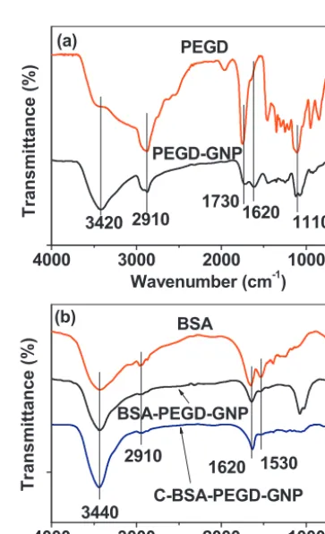
ThesurfacecoatingofultrasmallGNPswithPEGDfollowedby conjugationtoBSAorC-BSAwasinvestigatedbyFT-IRabsorption spectroscopy.Asmentionedpreviously,onegroupamongthetwo
–COOHgroupsineachPEGDwasconjugatedtoanultrasmallGNP
andtheotherwasleftfree foramidebondingtoBSA orC-BSA.
These were confirmed from the two different C=O stretching
vibrations in theFT-IR absorption spectrum of thePEGD-GNPs
(Fig. 3a). The free –COOH was observed at 1730cm−1 but the –COOHbondedtoGNPs,at1620cm−1.Thepeaksat2910cm−1 (C–Hstretch)and1110cm−1(C–Ostretch)alsoconfirmedthatthe
PEGDswerebondedtoultrasmallGNPs.Thepeakat3420cm−1
in thePEGD-GNPs wasassigned tothewater –OH stretch.The
∼110cm−1 red shiftof the C=O stretch after bonding to GNPs
from that of thefree –COOH had been observed in a range of
themetaloxideNPscoatedwiththeligandswith–COOHgroups
[22,27–30], supporting this result. The successful amide bond
formationbetweenPEGD-GNPsandBSA(orC-BSA)wasconfirmed
fromthedisappearanceofafreeC=Ostretchat1730cm−1inboth BSA-PEGD-GNPsandC-BSA-PEGD-GNPs(Fig.3b).Instead,theN–H stretchat3440cm−1(overlappedwithwater–OHstretch)andthe N–Hbendat1530cm−1wereobserved[31–33].Thereduced
inten-sityinN–HbendinbothBSA-PEGD-GNPsandC-BSA-PEGD-GNPs
was,however, observed owingtothe amidebonding of amine
groups of BSA and C-BSA with PEGD-GNPs and the hydrogen
bondingofaminegroupsofBSAandC-BSAwithPO43−ions.
4000
300
0
200
0
100
0
PEGD-G
NP
Waven
umber
(c
m
-1)
Transm
ittance
(%
)
3420 2910
1730
1620
1110
(a)
PEGD
4000
300
0
200
0
100
0
C-BSA-PEGD
-GNP
BSA-PEGD-
GNP
BSA
Wavenu
mber
(cm
-1)Tr
ansmittance
(%
)
3440
1620 1530
(b)
2910
72 Md.W.Ahmadetal./ColloidsandSurfacesA:Physicochem.Eng.Aspects450(2014)67–75
0 100 200 300 400 500 600 700
0 20 40 60 80
100 Water desorption = 9.8%
Temperature (oC)
We
ight (%)
28.0% (a)
0 100 200 300 400 500 600 700
0 20 40 60 80 100
47.4%
Water desorption = 4.9%
Temperature (oC)
Weight (%
)
(b)
0 100 200 300 400 500 600 700
0 20 40 60 80 100
(c)
61.7%
Water desorption = 7.3%
Temperature (oC)
W
eig
h
t (%
)
Fig.4. TGAcurvesofpowdersamplesof(a)PEGD-GNPs,(b)BSA-PEGD-GNPs,and (c)C-BSA-PEGD-GNPs.
TodeterminetheamountsofBSAintheBSA-PEGD-GNPsand
C-BSAintheC-BSA-PEGD-GNPs,theTGAcurvesof PEGD-GNPs,
BSA-PEGD-GNPs,andC-BSA-PEGD-GNPswererecorded(Fig.4a–c).
Waterdesorptionbetweenroomtemperature and∼110◦C was
consideredintheseestimations.TheamountofPEGDwas
esti-matedtobe28.0% fromtheTGAcurveof PEGD-GNPs(Fig.4a).
TheamountsofBSA-PEGDandC-BSA-PEGDwereestimatedtobe
47.4and 61.7%fromtheTGAcurves ofBSA-PEGD-GNPsand
C-BSA-PEGD-GNPs,respectively(Fig.4bandc).TheamountsofBSA
andC-BSAwereestimated tobe19.4and33.7% bysubtracting
theamountofPEGDfromthose ofPEGD-BSAandC-BSA-PEGD,
respectively.Usingthedavgof2.0nmfortheultrasmallGNPs
esti-matedfromtheHVEMimageandassumingthattheirdensityis
thesameasthat (=7.407gmL−1)[34]of bulk Gd
2O3,the
num-berofGNPsconjugatedtoeachBSA andC-BSAwereestimated
tobe8.8and0.2,respectively,whichwereconsistentwithHVEM observations(Fig.1bandc).Therefore,BSAisagood nanoparti-clecarrier,butC-BSAisnot.Thiscanbeexplainedusingthesizes andmassesofBSAandC-BSA.Thatis,themassofultrasmallGNPs withthedavg=2.0nmwasestimatedtobe10–20kDbycalculating thevolumeoftheultrasmallGNPsandusingthebulkdensityof Gd2O3[34],whicharesmallerthanthoseofBSA(mass=66.5kD andsize=14×4×4nm).Ontheotherhand,C-BSAswithmasses
of6.67and2.01kDestimatedfromGPChavesizesof6.5×1.9×1.9
0.00 0.25 0.50 0.75 1.00
0 10 20 30
r1 = 6.0 s-1mM-1
r2 = 28.0 s-1mM-1
Concentration Gd (mM)
1/T
(s
-1 )
(a)
0.00 0.04 0.08 0.12 0.16 0.20
0 1 2 3 4 5 6
r1 = 7.6 s-1mM-1
r2 = 22.0 s-1mM-1
(b)
1/
T
(
s
-1 )
Concentration Gd (mM)
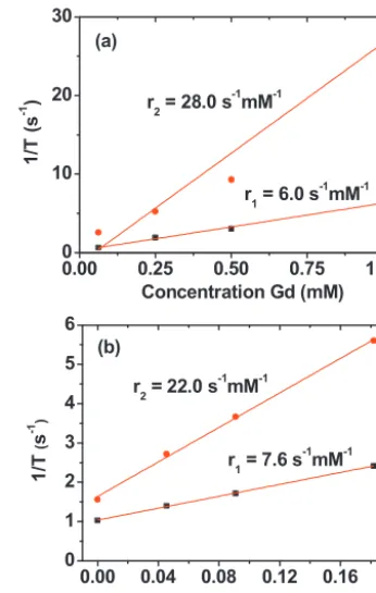
Fig.5.Plotsof1/T1and1/T2oftheaqueoussamplesolutionsof(a)
BSA-PEGD-GNPsand(b)C-BSA-PEGD-GNPsasafunctionoftheGdconcentration.Theslopes correspondtother1andr2values,respectively.
and4.4×1.2×1.2,respectively,assumingacuberootdependence
ofthesizeonthemass.Therefore,C-BSAisnotlargeenoughin massandsizetobindmanyultrasmallGNPs,whichissimilarto thepolymers.
3.3. SuggestedstructuresofBSA-PEGD-GNPand C-BSA-PEGD-GNP
Asmentionedbefore,theconjugationbetweenPEGD-GNPand
BSA(orC-BSA)isanamidebondbetween–COOHofPEGD-GNP
and –NH2 of BSA (or C-BSA). The BSA consists of 607 amino
acids and has amino acids with a free –NH2 [35,36] that can
beusedfortheamidebondingtoPEGD-GNP.In fact,60lysines witha free–NH2 areinBSA.Therefore,thereareplentyoffree
–NH2inBSAwhichcanbeconjugatedtoPEGD-GNPsthroughthe
amidebonding.Asdescribedpreviously,∼9PEGD-GNPswere
esti-matedtobeconjugatedtoeachBSA whereas∼0.2PEGD-GNPs,
to each C-BSA. Based on these results, structures of the
BSA-PEGD-GNP and C-BSA-PEGD-GNP were schematically drawn in
Scheme1c.
3.4. Relaxivitiesandmapimages
Magnetic propertiesof gadolinium oxide nanoparticleshave
been well characterized [37]. They are paramagneticbut have
anappreciable magneticmoment at roomtemperature. Thisis
becauseGd(III)hassevenunpaired4f-electrons(8S
7/2).Therefore, appreciabler1 andr2valuesareexpectedfromsamplesolutions, which were in fact observed in this study. The r1 and r2 val-uesofBSA-PEGD-GNPswere estimatedtobe6.0s−1mM−1 and 28.0s−1mM−1,respectively,fromtheslopesintheplotof1/T
Table1
Waterprotonrelaxivity(r1andr2)aofvariouschemicals.
Chemical dordavgb Nc r1 r2 Hd Te Ref.
Gd-DTPA – – 4.1 – 0.47 38 [5]
BSA-GNP 20–40 – 6.7 38.5 4.7 37 [10]
BSA-PEGD-GNP 2.0 8.8 6.0 28.0 1.5 22 Thiswork C-BSA-PEGD-GNP 2.0 0.21 7.6 22.0 1.5 22 Thiswork
aUnit:s−1mM−1.
bParticlediameteroraverageparticlediameter(nm). c NumberofGNPsconjugatedtoaBSAorC-BSA. d AppliedMRfield(T).
eSamplesolutiontemperature(◦C).
forcomparison.Ther1andr2valuesofBSA-PEGD-GNPsand C-BSA-PEGD-GNPsarelargerthanthose[5,6]ofmolecularGd-DTPA.These increasedrelaxivitieswereattributedtothehighdensityofGd(III) intheNPs.Theselargervaluesgenerallyleadtoahighersensitivity fordetectingdiseasesinthebodythroughcontrastenhancements andcanalsoprovidethesamequalityMRimagesasthoseofthe Gd-chelatesatreduceddoses.Ther2valuesaresignificantlylargerthan thatofmolecularGd-DTPA,whichiswhyonlyNPsareeligibleas
T2MRIcontrastagents,whereasmolecularagentsareonlysuitable as T1 MRI contrastagents. The r1 and r2 values of BSA-PEGD-GNPsandC-BSA-PEGD-GNPsweresimilartothose[10]ofBSA-GNP (d=20–40nm)measuredatahigherappliedMRfield.Ontheother hand,consideringthatthewaterprotonrelaxivitiesincreasewith
increasingappliedMRfield,thoseoftheBSA-PEGD-GNPsand
C-BSA-PEGD-GNPswillbelargerthanthoseofBSA-GNPatthesame appliedMRfield. Thisisdue likelytotheparticlesizeeffectof
theGNP.BothaqueoussolutionsofBSA-PEGD-GNPsand
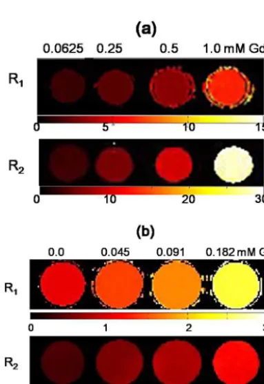
C-BSA-PEGD-GNPsshowedcleardose-dependentcontrastenhancements
intheirR1andR2mapimages(Fig.6aandb),suggestingthatthese NPsarepotentialcandidatesforMRIcontrastagents,whichwere confirmedinamouseexperiment.
Fig.6.R1andR2mapimagesofaqueoussamplesolutionsof(a)BSA-PEGD-GNPs
and(b)C-BSA-PEGD-GNPsasafunctionoftheGdconcentration.
3.5. Invitrocytotoxicity
Theinvitrocytotoxicityoftheaqueoussamplesolutionsof
BSA-PEGD-GNPsandC-BSA-PEGD-GNPsweremeasuredusingDU145
andNCTC1469cellswithGdconcentrationsupto200M(Fig.7a
andb).TheresultsshowedthatC-BSA-PEGD-GNPswereslightly
lesstoxicthanBSA-PEGD-GNPs.Thisisprobablybecausemany
C-BSAlikepolymersencapsulatedthePEG-GNPs,asshownin the
HVEM image (Fig.1c), whereas many PEGD-GNPs were
conju-gatedtoeachBSAonthesurfaceofBSA,asshownintheHVEM
image(Fig.1b).Therefore,PEGD-GNPswerebetterprotectedin C-BSA-PEGD-GNPsthaninBSA-PEGD-GNPs.Thecellviabilityofboth samplesdecreasedgraduallywithincreasingGdconcentration.The cellviabilityofC-BSA-PEGD-GNPsat100MGdreachedmorethan
70%forbothcells,whereasthatofBSA-PEGD-GNPsreached∼60%
forbothcells.Theselevelsofcellulartoxicityaresufficientlylowto carryoutinvivoMRIexperiments.
3.6. Invivo3TT2MRimagesofamouse
BecauseBSAcouldbindmanyultrasmallGd2O3NPs,whereas
C-BSAdidnot,asmeasuredbyTGA,aninvivoMRIexperiment
0 20 40 60 80 100 120
(a) DU145
NCTC1469
Concentration Gd ( M)
0 10 100 200
Ce
ll
Vi
a
bility
(%)
0 20 40 60 80 100 120
Concentration Gd ( M)
Cell
Via
bi
li
ty
(%
)
0 10 100 200 DU145
NCTC1469
(b)
[image:7.612.43.563.74.129.2] [image:7.612.348.528.431.718.2] [image:7.612.72.264.444.722.2]74 Md.W.Ahmadetal./ColloidsandSurfacesA:Physicochem.Eng.Aspects450(2014)67–75
Fig.8.(a)3TT2MRimagesoftheliverofamousebeforeandafterinjectingan
aqueoussamplesolutionofBSA-PEGD-GNPsintoamousetailveinand(b)theplot ofsignalintensityinT2MRimagesasafunctionoftimeafterinjection(0indicates
“beforeinjection”).
wasfurthercarriedoutusinganaqueoussamplesolutionof BSA-PEGD-GNPs.AlthoughGNPsaregenerallyusedasT1MRIcontrast agents,theT2MRimageswereinvestigatedbecausether2value wasa lotlargerthanther1 value,due totheappreciable mag-netizationofultrasmallGNPs atroomtemperature[37].250L
(0.1mmolGd/kg)ofanaqueoussolutionofBSA-PEGD-GNPswas
injectedintoamousetailveinand3TT2 MRimagesoftheliver weretakenbeforeandafterinjectingtheaqueoussample solu-tion.AsshowninFig.8a,appreciablenegative(ordarker)contrast enhancementswereobservedinthemouseliverafterthe injec-tion,whichreturnedtoalmosttheoriginalcontrastafter24hdue likelytotheexcretionofBSA-PEGD-GNPs.Tomoreclearlyseethe timeevolutionofthecontrastchangeinT2MRimages,thesignal intensityinT2MRimageswasplottedasafunctionoftimeupto 24hinFig.8b.Thisplotclearlyshowsthatthenegativecontrast enhancementmaintainedupto91minafterinjectionbutreturned toalmostzeroabove91mindue likelytotheexcretionof BSA-PEGD-GNPs.Theseresultsclearlyindicatethatthesamplesolution functionedasaT2MRIcontrastagent.
4. Conclusions
Insummary,wesynthesizedPEGDcoatedultrasmallGd2O3NPs
(i.e.PEGD-GNPs)whichwerethenconjugatedtoBSAandC-BSA
through amidebonding (i.e. BSA-PEGD-GNPsand
C-BSA-PEGD-GNPs). We characterizedphysical and in vitro MRI properties,
andcytotoxicityofBSA-PEGD-GNPsand C-BSA-PEGD-GNPs,and
obtainedinvivoMRimagesusingBSA-PEGD-GNPs.
(1)BSA (Mn=66.5kD) could bind many ultrasmall PEGD-GNPs
(thecoredavg=2.0nm),showingthatBSAisagoodultrasmall
NP carrier.TheTGA showedthat ∼9 ultrasmall PEGD-GNPs
couldbeconjugatedtoeachBSA.However,C-BSAs(Mn<7kD) couldnotbindmanyultrasmallPEGD-GNPsduetoitsreduced
sizeand mass.Instead manyC-BSAs wereconjugatedtoan
ultrasmallPEGD-GNPlikepolymers.TheTGAshowedthat∼5
C-BSAswereconjugatedtoeachultrasmallPEGD-GNP.
(2)MR relaxivity measurements revealed that both
BSA-PEGD-GNPsandC-BSA-PEGD-GNPshadr1 andr2valueslargerthan thoseofmolecularGd-chelates.
(3)The3TT2MRimagesafterinjectinganaqueoussample solu-tionofBSA-PEGD-GNPsintothemousetailveinshowedclear
negativecontrastenhancements.
(4)Largerelaxivitiesand invivoT2 MR imagesprovethat
BSA-PEGD-GNPsandC-BSA-PEGD-GNPsarepotentialMRIcontrast
agents.
TheseresultssuggestthatbiologicalmoleculessuchasBSAcan beusedtoconjugatemanysurfacemodifiedultrasmallNPswhich canbeappliedtoavarietyofbiomedicalareassuchasMRIcontrast agentsstudiedinthiswork.
Acknowledgments
ThisstudywassupportedbytheBasicScienceResearch
Pro-gram(Grantno.2012R1A1B3004241toKSC,2011-0015353toYC,
and2013R1A1A4A03004511toGHL)andtheBasicResearch
Labo-ratory(BRL)program(Grantno.2013R1A4A1069507)throughthe
NationalResearchFoundationfundedbytheMinistryofEducation, Science,andTechnology,theR&DprogramofMKE/KEIT(Grantno. 10040393,developmentandcommercializationofmolecular diag-nostictechnologiesforlungcancerthroughclinicalvalidation),and theKNUResearchFund(2013).TheauthorswishtothanktheKorea BasicScienceInstitutefortheuseoftheirHVEMandXRD.
AppendixA. Supplementarydata
Supplementarydataassociatedwiththisarticlecanbefound, intheonlineversion,athttp://dx.doi.org/10.1016/j.colsurfa.2014. 03.011.
References
[1]C.J.Ackerson,P.D.Jadzinsky,R.D.Kornberg,Thiolateligandsforsynthesisof water-solublegoldclusters,J.Am.Chem.Soc.127(2005)6550–6551.
[2]R.A.Sperling,W.J.Parak,Surfacemodification,functionalizationand bioconju-gationofcolloidalinorganicnanoparticles,Philos.Trans.R.Soc.A368(2010) 1333–1383.
[3]V.V.Martin,W.H.Ralston,M.R.Hynes,J.F.W.Keana,Gadolinium(III)di-and tetrachelatesdesignedforinvivononcovalentcomplexationwithplasma pro-teins:anovelmoleculardesignforbloodpoolMRIcontrastenhancingagents, Bioconjug.Chem.6(1995)616–623.
[4]T.N.Nagaraja,R.L.Croxen,S.Panda,R.A.Knight,K.A.Keenan, S.L.Brown, J.D.Fenstermacher,J.R.Ewing,ApplicationofarsenzoIIIinthepreparation andcharacterizationofanalbumin-linked,gadolinium-basedmacromolecular magneticresonancecontrastagent,J.Neurosci.Methods157(2006)238–245.
[5]R.B.Lauffer,Paramagneticmetalcomplexesaswaterprotonrelaxationagents forNMRimaging:theoryanddesign,Chem.Rev.87(1987)901–927.
[6]P.Caravan,J.J.Ellison,T.J.McMurry,R.B.Lauffer,Gadolinium(III)chelatesasMRI contrastagents:structure,dynamics,andapplications,Chem.Rev.99(1999) 2293–2352.
[7]W.Xu,K.Kattel,J.Y.Park,Y.Chang,T.J.Kim,G.H.Lee,Paramagnetic nanopar-ticleT1 andT2 MRIcontrastagents, Phys.Chem.Chem.Phys.14(2012) 12687–12700.
[8]T.J.Kim,K.S.Chae,Y.Chang,G.H.Lee,Gadoliniumoxidenanoparticlesas poten-tialmultimodalimagingandtherapeuticagents,Curr.Top.Med.Chem.13 (2013)422–433.
[9]E.J.Lee,W.C.Heo,J.W.Park,Y.Chang,J.-E.Bae,K.S.Chae,T.J.Kim,J.A.Park, G.H.Lee,d-glucuronicacidcoatedGd(IO3)3·2H2Onanomaterialasapotential T1MRI-CTdualcontrastagent,Eur.J.Inorg.Chem.(2013)2858–2866.
[10]M.A.McDonald,K.L.Watkin,Smallparticulategadoliniumoxideand gadolin-ium oxidealbuminmicrospheresasmultimodalcontrastandtherapeutic agents,Invest.Radiol.38(2003)305–310.
[11]J.L.Bloem,J.Wondergem,Gd-DTPAasacontrastagentinCT,Radiology171 (1989)578–579.
[12]T.Kawano,H.Ishijima,T.Nakajima,J.Aoki,K.Endo,Gd-DTPA:apossible alternativecontrastagentforuseinCTduringintraarterialadministration,J. Comput.Assist.Tomogr.23(1999)939–940.
[14]G.DeStasio,D.Rajesh,P.Casalbore,M.J.Daniels,R.J.Erhardt,B.H.Frazer,L.M. Wiese,K.L.Richter,B.R.Sonderegger,B.Gilbert,S.Schaub,R.J.Cannara,J.F. Crawford,M.K.Gilles,T.Tyliszczak,J.F.Fowler,L.M.Larocca,S.P.Howard,D. Mercanti,M.P.Mehta,R.Pallini,Aregadoliniumcontrastagentssuitablefor gadoliniumneutroncapturetherapy?Neurol.Res.27(2005)387–398.
[15]G.DeStasio,P.Casalbore,R.Pallini,B.Gilbert,F.Sanita,M.T.Ciotti,G.Rosi,A. Festinesi,L.M.Larocca,A.Rinelli,D.Perret,D.W.Mogk,P.Perfetti,M.P.Mehta, D.Mercanti,Gadoliniuminhumanglioblastomacellsforgadoliniumneutron capturetherapy,CancerRes.61(2001)4272–4277.
[16]D.P.Gierga,J.C.Yanch,R.E.Shefer,Aninvestigationofthefeasibilityof gadolin-iumforneutroncapturesynovectomy,Med.Phys.27(2000)1685–1692.
[17]R.B.Lauffer,T.J.Brady,Preparationandwaterrelaxationpropertiesof pro-teinlabeledwithparamagneticmetalchelates,Magn.Reson.Imaging3(1985) 11–16.
[18]H.S.Choi,W.Liu,P.Misra,E.Tanaka,J.P.Zimmer,B.I.Ipe,M.G.Bawendi, J.V.Frangioni,Renalclearanceofquantumdots,Nat.Biotechnol.25(2007) 1165–1170.
[19]J.Chen,R.Wan,H.Liu,C.-M.Cheng,Y.-F.Zhao,CleavageofBSAbyadipeptide seryl-histidine,Lett.Pept.Sci.7(2001)325–329.
[20]W.Xu,J.Y.Park,K.Kattel,M.W.Ahmad, B.A.Bony,W.C.Heo,S.Jin,J.W. Park,Y.Chang,T.J.Kim,J.A.Park,J.Y.Do,K.S.Chae,G.H.Lee, Fluorescein-polyethyleneimine coated gadoliniumoxide nanoparticlesasT1 magnetic resonanceimaging(MRI)-celllabeling(CL)dualagents,RSCAdv.2(2012) 10907–10915.
[21]H.Cao,S.-Y.Xu,EDC/NHS-crosslinkedtypeIIcollagen-chondroitinsulfate scaf-fold:characterizationandinvitroevaluation,J.Mater.Sci.:Mater.Med.19 (2008)567–575.
[22]F.Söderlind,H.Pedersen,R.M.PetoralJr.,P.-O.Käll,K.Uvdal,Synthesisand char-acterizationofGd2O3nanocrystalsfunctionalizedbyorganicacids,J.Colloid InterfaceSci.288(2005)140–148.
[23]L.Parca,P.F.Gherardini,M.Helmer-Citterich,G.Ausiello,Phosphate bind-ing sitesidentificationinproteinstructures,NucleicAcidsRes.39(2011) 1231–1242.
[24]A.K.H.Hirsch,F.R.Fischer,F.Diederich,Phosphaterecognitioninstructural biology,Angew.Chem.Int.Ed.46(2007)338–352.
[25]J.R.Jadhav,M.W.Ahmad,H.-S.Kim,SelectiverecognitionofH2PO4−bya cholestane-imidazole-zincensemble,TetrahedronLett.53(2012)2627–2631.
[26]JCPDScardnumber32-0386forGdPO4,PCPDFWIN,Version2.4,2003. [27]C.B.Mendive,T.Bredow,M.A.Blesa,D.W.Bahnemann,ATR-FTIRmeasurements
andquantumchemicalcalculationsconcerningtheadsorptionand photoreac-tionofoxalicacidonTiO2,Phys.Chem.Chem.Phys.8(2006)3232–3247.
[28]O.W.Duckworth,S.T.Martin,Surfacecomplexationanddissolutionofhematite byC1–C6dicarboxylicacidsatpH=5.0,Geochim.Cosmochim.Acta65(2001) 4289–4301.
[29]S.J.Hug,D.Bahnemann,Infraredspectraofoxalate,malonateandsuccinate adsorbedontheaqueoussurfaceofrutile,anataseandlepidocrocitemeasured withinsituATR-FTIR,J.ElectronSpectrosc.Relat.Phenom.150(2006)208–219.
[30]S.J.Hug,B.Sulzberger,InsituFouriertransforminfraredspectroscopicevidence fortheformationofseveraldifferentsurfacecomplexesofoxalateonTiO2in theaqueousphase,Langmuir10(1994)3587–3597.
[31]X. Xu, J.-F.Zhang, Y. Fan, Fabrication of cross-linked polyethyleneimine microfibersbyreactiveelectrospinningwithinsituphoto-cross-linkingbyUV radiation,Biomacromolecules11(2010)2283–2289.
[32]I.Yudovin-Farber,N.Beyth,E.I.Weiss,A.J.Domb,Antibacterialeffectof compos-iteresinscontainingquaternaryammoniumpolyethyleneiminenanoparticles, J.Nanopart.Res.12(2010)591–603.
[33]C.Liu,P.Zhang,X.Zhai,F.Tian,W.Li,J.Yang,Y.Liu,H.Wang,W.Wang,W. Liu,Nano-carrierforgenedeliveryandbioimagingbasedoncarbondotswith PEI-passivationenhancedfluorescence,Biomaterials33(2012)3604–3613.
[34]R.C.Weast,CRCHandbookofChemistryandPhysics,65thed.,CRCPressInc., BocaRaton,FL,1984–1985,pp.B-96.
[35]R.G.Reed,F.W.Putnam,T.PetersJr.,Sequenceofresidues400–403ofbovine serumalbumin,Biochem.J.191(1980)867–868.
[36]R.N.M.Weijers,Aminoacidsequenceinbovineserumalbumin,Clin.Chem.23 (1977)1361–1362.