ISOLATION ANTHOCYANIN FROM ROSELLE PETALS (
Hibiscus sabdariffa
L)
AND THE EFFECT OF LIGHT ON THE STABILITY
Siti Nuryanti
1,*, Sabirin Matsjeh
2, Chairil Anwar
2, and Tri Joko Raharjo
2 1Department of Chemistry, Faculty of Teacher Training and Education, Tadulako University, Jln. Soekarno-Hatta Km 8, Palu, Indonesia
2
Department of Chemistry, Faculty of Mathematics and Natural Sciences, Universitas Gadjah Mada, Yogyakarta, Indonesia
Received November 16, 2011; Accepted January 3, 2012
ABSTRACT
This study was conducted to isolate anthocyanins from roselle petals and testing the stability toward light. Isolation of anthocyanin was accomplished by extracting roselle petals using eluents with different polarity levels. Nonpolar compounds was eliminated using n-hexane, then semipolar compounds extracted with ethyl acetate and isolated anthocyanin by solvent mixtures of methanol-HCl 0.5%. Color test to determine the presence of anthocyanin was performed with NH3vapor, Pb-acetate 1% and Pb-nitrate 5%. The structure of anthocyanin in the roselle flower was determined using UV-Vis spectrophotometer, FT-IR and1H-NMR. Anthocyanin stability test of the influence of light carried out in a room without light conditions (dark room) and light 25 W at 31 °C. The results showed that the roselle petals contain anthocyanin cyanidin-3-glucoside. Light has been found to affect the stability of anthocyanin cyanidin-3-glucoside.
Keywords:anthocyanin; stability; light; Hibiscus sabdariffa L
ABSTRAK
Penelitian ini bertujuan untuk mengisolasi antosianin dari mahkota bunga rosella dan menguji kestabilannya terhadap cahaya. Isolasi antosianin dilakukan dengan mengekstrak mahkota rosella menggunakan pelarut dengan perbedaan tingkat polaritas. Senyawa nonpolar dihilangkan menggunakan n-heksana, kemudian senyawa semipolar diekstrak dengan etil asetat dan antosianin diisolasi dengan pelarut campuran metanol-HCl 0,5%. Tes warna untuk menguji adanya antosianin dilakukan dengan uap NH₃, Pb-asetat 1% dan Pb-nitrat 5%. Struktur antosianin dalam bunga rosella diidentifikasi menggunakan spektrofotometer UV-Vis, FT-IR dan ¹H-NMR. Tes stabilitas antosianin terhadap pengaruh cahaya dilakukan dalam kondisi ruang tanpa cahaya (ruang gelap) dan bercahaya 25 W pada 31 °C. Hasil penelitian menunjukkan bahwa mahkota rosella mengandung antosianin sianidin-3-glukosida. Cahaya memberikan pengaruh terhadap kestabilan antosianin sianidin-3-glukosida.
Kata Kunci:antosianin; stabilitas; cahaya; Hibiscus sabdariffa L
INTRODUCTION
Crown roselle flower (Hibiscus sabdariffa L) has not been widely exploited commercially and only discarded as waste, while the red petals already widely used as a food coloring and also used as a tea is beneficial for maintaining good health. Chen et al. [3] reported on the use of flower petals (calyx) roselle as a drug to lower blood cholesterol levels, lowering blood pressure, lowering blood sugar levels for diabetics and as detoxification (neutralizing poison). Amor and Allaf [2] has reported calyx roselle is used for coloring food, inhibit cancer cell growth, maintain stamina, lowers the level of clumping of fat in the liver, balancing the weight, reduce the heat inside. Another benefit of calyx roselle is
(migraine), treating coughs and diarrhea [4]. In the calyx roselle contains cyanidin-3-rutinoside and delphinidin-3-glucoxyloside as the major anthocyanin [8].
Considering the benefits and the attractive properties of anthocyanin, then isolation and identification of anthocyanins on the crown of roselle flower and stability test of the influence of light are very urgent to do. Furthermore, the result of identification anthocyanin on the roselle flower crown is expected to be harnessed into products that can be commercialized as calix, so it can be enhanced economic value of rosella crown.
EXPERIMENTAL SECTION
Materials
Crown roselle (Hibiscus sabdariffa L) collected from Trisik Kulonprogo and identified in Laboratory of Plant Taxonomy, Faculty of Biology, Universitas Gadjah Mada. Chemicals used in this study are methanol, ethanol, butanol, n-hexane, silica gel F 254, ethyl acetate, hydrochloride acid, aquades, and acetic acid.
Instrumentation
The apparatus used are the extractor (IKA ® KS 130 basic), TLC, rotary evaporator Buchii (R-124), Buchner funnel, pH meter (Hanna HI-8314), Erlenmeyer, brown bottles, magnetic stirrer, vessel developers (chamber), tweezers, analytical balance (Shimadzu Libror EB-330), IR (Shimadzu 8201 PC), UV-Vis (array Miltonroy 3000), 1H-NMR (500 MHz) (JEOL JNM ECA 500 series Serpong LIPI).
Procedure
Extraction of roselle crown (Hibiscus sabdariffa L)
Roselle crown about 500 g cut into small pieces, put into brown or dark bottles. Then 2500 mL n-hexane was added, extracted by maceration for 24 h. The extract was filtered with Whatman paper no.1 through a Buchner funnel. The residue (dregs) was aerated, to remove the solvent. Dry residues extracted again with 2500 mL ethyl acetate for several hours. The extract was filtered followed by aerated. Extracted the dry residue with methanol-HCl 0.5% (v/v) for several hours and then filtered. The filtrate were collected, then concentrated by vacuum rotary evaporator at 60-65 °C until concentrated extract was obtained.
Qualitative test of anthocyanin roselle crown (Hibiscus sabdariffa L)
Identification of anthocyanin with NH3vapor and 1%
Pb-acetate.Concentrated extracts were taken using the capillary tube and placed on the TLC plate, aerated and dried then steamed using NH3 and the color changes
was observed. The same method was applied using reagent Pb-acetate 1%.
Identification of anthocyanin with 5% Pb-nitrate.
Measured 5 mL Pb-nitrate 5% solution, put in Erlenmeyer, and added 1 mL roselle crown extract. The mixture was stirred using a magnetic stirrer for 3 h then the absorbance were measured using UV-Vis
spectrophotometer at λ200-700 nm.
Isolation and identification of the anthocyanin structure on the roselle crown extract (Hibiscus sabdariffa L)
Roselle crown concentrated extract was taken using capillary tube and placed on a TLC plate and then inserted into the vessel developers (chamber) which is filled with solvent. The solvent used is a mixture of n-butanol : 0.5% HCl : ethanol (5:1:4). Elution was performed until the solvent reaches 0.5 cm distance from the upper edge of the TLC plate. The plate was observed under UV light at λ 254 and 366 nm with NH3 vapor. Column chromatography was performed, sample with same Rf was combined, then evaporated, and the structure was identified by UV-Vis spectrophotometer, FT-IR, 1H-NMR as practiced [1].
The stability test of anthocyanin extract isolated from the crown of roselle flower (Hibiscus sabdariffa L) of the influence of light
Weighed as much as 1.0 mg of insulating crystals dissolved in 10 mL of methanol-0.5% HCl. The solution was divided into two parts, 5 mL (part 1) was inserted into transparent bottle (translucent light), then placed in the room lights 25 W. While 5 mL (part 2) pour on a black bottle, put in the opaque box, and placed in the same room with part 1. The next step is measured each piece as much as 20 μL, then diluted to 500 μL with methanol. Furthermore, each solution was analyzed by UV-Vis spectrophotometer at
λ 200-700 nm and the observations were made every single week.
RESULT AND DISCUSSION
Identification of anthocyanins by UV-Vis and color reagents
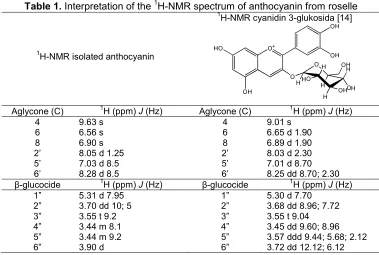
Table 1.Interpretation of the1H-NMR spectrum of anthocyanin from roselle
1
H-NMR isolated anthocyanin
1
H-NMR cyanidin 3-glukosida [14]
Aglycone (C) 1H (ppm)J(Hz) Aglycone (C) 1H (ppm)J(Hz) 3.57 ddd 9.44; 5.68; 2.12 3.72 dd 12.12; 6.12
the blue color due to the reaction between OH group in anthocyanins and NH3.
Identification using reagents of Pb acetate 1% also gave blue color. The use of color reagent Pb-acetate 1% in this study was to differentiate the hydroxyl group attached at position C-3' and C-4' in ring B. Anthocyanin peonidin and malvidin which is not that not bearing hydroxyl group at those positions gives violet color, whereas anthocyanin that has hydroxyl group in those position after reaction with Pb-acetate produces a blue color [6].
Analysis of extract using UV-Vis spectrophotometer after reaction with Pb-nitrate 5% exhibited absorption at
λmax 544 nm. The spectrum indicates red shift of
λmax 536 nm to 544 nm. The interaction between metal Pb with molecule anthocyanin cyanidin-3-monoglucoside give rise to wavelength changes to bathochromic [12]. The color change is due to the formation of metal-anthocyanin complex compounds. Anthocyanins cyanidin form complexes with metallic Pb [13]. Therefore, based on the color change, it can be concluded extract crown roselle contains anthocyanins cyanidin.
Isolation of anthocyanins from the extract crown roselle (Hibiscus sabdariffaL)
Isolation of anthocyanins from the extract of roselle flower crown (Hibiscus sabdariffa L) was done by column chromatography using solvent mixture of butanol : 0.5% HCl : ethanol with a ratio of 5:1:4. The 36 vials that have Rf0.27 was combined, after evaporation, then
dried on exicator which gave a red powder as much as 5.48 mg. The red powder was analyzed by the UV-Vis
and gives absorption at λmax 536 nm. Further identification is using a solution of Pb acetate 1% which produces a blue color. These data indicate that the red powder is the anthocyanins cyanidin.
Identification of anthocyanin structure by FT-IR
Glycosides of anthocyanin are Polyphenolic compounds that absorb infrared radiation to produce absorption bands which quite complex. But according to Ribereau-Gayon [10], the interpretation of infrared absorption bands of anthocyanins aglycone is more easily observed in the region 1800-1380 cm-1. Interpretation of FT-IR spectra from isolated anthocyanins crown roselle (Hibiscus sabdariffaL) has been also conducted using the absorption bands at other areas to support the conclusions.
From FT-IR spectrum, it can be interpreted that there is aromatic CH absorption at 3032 cm-1. Absorption at 1581 cm-1 is characteristic for the stretching of -C=C-. The FT-IR spectrum also presents a broad absorption at 3425 cm-1 which is a characteristic absorption band of hydrogen bonding (-OH). Absorption at 2121 cm-1 showed stretching of the -C=C- bisubstitute of different groups. The emergence of absorption at 2368-2337 cm-1 indicate
characteristic of the group carbonyl (-C=O). With these data it can be ascertained that ether groups contained in
the aromatic ring is emerging in
1635 cm-1[11].
Identification of anthocyanin structure by 1H-NMR
1
H-NMR spectrum indicates, there are 7 types of peaks which describes seven types of protons with different chemical environments. Signal (δ 9.03 ppm, singlet, 1 H) describes the proton at C-4 atom in the ring C. Signal (δ 8.29 to 8.27 ppm, duplet, J = 8.5 Hz, 1 H) and (δ 7.03 to 7.02 ppm, duplet, J = 8.5 Hz 1 H) indicates the existence of two protons on aromatic ring B in ortho position to each other, proton C-5 'and C-6', respectively. While the signal (δ 8.05 to 8.05 ppm, duplet, J = 1.25 Hz, 1 H) derived from the proton C-2’ which have coupling meta with proton C-6'. Furthermore, two singlet peaks of the signal (δ6.90 ppm, singlet, 1 H) and (δ 6.57 ppm, singlet, 1 H) represented the two protons on ring A at position C-8 and C-6. The existence of clusters indicated by the absorption from the sugar group protons with the integration of seven protons in chemical shift from 5.31 to 5.30 ppm and 3.90 to 3.31 ppm. Signal (δ 5.31 to 5.30 ppm, duplet, J = 7.95 Hz, 1 H) derived from the proton H-1'', δ 3.90 ppm signal, duplet 1 H is the proton H-2''. Signal (δ 3.56 to 3.54 ppm, J = 9.2 Hz, triplet, 1 H) describes the proton H-3'', the signal (δ 3.71 to 3.69 ppm, J = 8.1 Hz, multiplet, 1 H) presented a proton from the H-4''. Signal (δ 3.45 to 3.43 ppm, J = 9.2 Hz, multiplet, 1 H) derived from the proton H-5'' andδ 3.31 ppm signal duplet, 2 H indicates protons from H-6''.
The spectrum1H-NMR of the isolation product and anthocyanin cyanidin-3-glucoside [14] were compared, there are similarities (Table 1). Based on data from UV-Vis, FT-IR and 1H-NMR and supported by color test using NH3 vapor, Pb-acetate 1% and 5% Pb-nitrate, it was ensured that in the crown of roselle contain anthocyanin cyanidin-3-glucoside.
The stability of anthocyanins from roselle crown (Hibiscus sabdariffaL) toward light
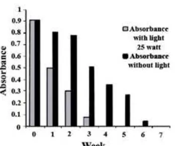
The study uses light 25 W and room temperature 31 °C, a decrease in absorbance at λmax 536 nm for 44.48% in the first week, second week of 66.99% and the third week of the absorbance decrease of 91.50%
Fig 1. UV-Vis spectra anthocyanin cyanidin-3-glucoside of roselle flower (a) before being stored, (b) after being stored for 7 weeks without light, (c) stored for 4 weeks with the influence of light
cle avag
e
Fig 3. Decrease in absorbance caused by the influence of light on anthocyanin cyanidin-3-glucoside
was observed. At the fourth week the color of anthocyanin from roselle crown was already undetectable. Meanwhile, in the absence of light (dark room) at the same temperature conditions (31 °C), only 43.81% decrease in absorbance was observed on the third week. At the sixth week, the decreases in absorbance were 95.25%, and at the seventh weeks anthocyanin was undetectable.
Based on the results of this study, it is proved that the light affects the stability of anthocyanin cyanidin-3-glucoside from roselle crown. To confirm that anthocyanin cyanidin-3-glucoside has been degraded for 7 weeks without light and 4 weeks with light, it was observed using UV-Vis spectrophotometer. The spectra
show that at λmax 536 nm which is characteristic for anthocyanin cyanidin become undetectable (lost) and
new absorption appears at λmax282 and 281 nm (Fig. 1) which is estimated as phloroglucinaldehyde.
The UV-Vis spectra data presented here was resemble to the degradation of anthocyanin cyanidin-3-glucoside isolated from elderberry reported by Patras et al. [9], that absorption of phloroglucinaldehyde occurs at
λmax 280 nm (Fig. 2), which occurs from a ring disconnection process in anthocyanin cyanidin-3-glucoside. Decrease in absorbance can be easily followed through Fig. 3.
CONCLUSION
From the results of this study it can be seen that crown of roselle flower (Hibiscus sabdariffa L) contains
anthocyanin cyanidin-3-glucoside and light has been proved to affect the stability of the anthocyanin. The result of this study is the first step, and further thorough research needs to be done on the application of roselle flower crown as a further thorough product.
REFERENCES
1. Adje, F., Lozano, Y.F., Meudec, E., Lozano, P., Adima, A., N’zi, G.A., and Gaydou, E.M., 2008, Molecules, 13, 1238–1245.
2. Amor, B., and Allaf, K., 2009,Food Chem., 115, 3, 820–825.
3. Chen, C.C., Hsu, J.D., Wang, S.F., Chiang, H.C., Yang, M.Y., Kao, E.S., Ho, Y.C., and Wang, C.J., 2003,J. Agric. Food Chem., 51, 18, 5472–5477. 4. Farombi, E.O., and Fokoya, A., 2005, Mol. Nutr.
Food Res.,49, 12, 1120–1128.
5. Harborne, J.B., 1958,Biochem.J., 70, 1, 22–28. 6. Jackman, R.L., Yada, R.Y., Tung, M.A., and
Speers, R.A., 1987,J. Food Biochem., 11, 4, 279– 308.
7. Janna, O., Khairul, A., Maziah, M., and Mohd, Y., 2006,Afr. J. Biotechnol., 5, 2, 170–174.
8. Maganha, E.G., Halmenschlager, R.C., Rosa, R.M., Henriquez, J.A.P., Ramos, A.L.L.P., and Saffi, J., 2010,Food Chem.,118, 1, 1–10.
9. Patras, A., Brunton, N.P., O’Donnell, C., and Tiwari, B.K., 2010,Trends Food Sci. Technol, 21, 1, 3–11.
10. Ribereau-Gayon P., 1969, Les Composess Phenoliques des Vegetaux Les Anthocyanin, Dunod Paris, 142–147.
11. Silverstein, R.M., Bassler, G.C., and Morrill, T.C., 1991, Spectrometric Identification of Organic Compounds, 5th ed., John Wiley & Sons, Inc., Singapore, 117–118.
12. Smyk, B., Priszka, B., and Drabent, R., 2007,Food Chem.,107, 4, 1616–1622.
13. Ukwueze, N.N., Nwadinigwe, C.A., Okoye, C.O.B. and Okoye, F.B.C., 2009, Int. J. Phys. Sci., 4, 2, 058–062.
![Fig 2. Degradation cyanidin-3-glucoside because the effect of heating [9]](https://thumb-ap.123doks.com/thumbv2/123dok/872474.820932/4.595.329.525.196.516/fig-degradation-cyanidin-glucoside-effect-heating.webp)