* corresponding author: ars_tambunan@yahoo.com
Assessment of maximal urinary flow rate
(Q
max
) of urethral stricture patients three
weeks post internal urethrotomy Sachse in
Dr. Sardjito General Hospital Yogyakarta
Juni Ariston Tambunan1*, Prawito Singodimedjo2, Ishandono Dachlan2
1St. Elisabet Hospital, Batam, Riau Islands Province,2Department of Surgery, Dr. Sardjito
General Hospital/Faculty of Medicine, Universitas Gadjah Mada, Yogyakarta, Indonesia
ABSTRACT
Urethral stricture is a common urologic problem in developing countries including Indonesia due to its high prevalence. Internal urethrotomy is still the gold standard to return patients to a state of normal voiding. To evaluate the outcome of the internal urethrotomy, uroflowmetry assessment can be conducted with its principal variable of maximal urinary flow rate (Qmax). Since 1985, in Dr. Sardjito General Hospital Yogyakarta, the internal urethrotomy has been used as the main treatment modality to manage the urethral stricture. However, its outcome has not been evaluated. The aim of this study was to evaluate Qmax of urethral stricture patients post internal urethrotomy Sachse in Dr. Sardjito General Hospital. This was a cross-sectional study performed starting from November 2009 to April 2010. The Qmax was assessed using the uroflowmeter three weeks after internal urethrotomy. The length and the locations of the patients’ stricture, as well as its correlation with Qmax were also measured and evaluated. Among 24 patients selected, 13 patients who fulfilled the inclusion and exclusion criteria were involved in this study. The mean of the Qmax of patients was 22.3±6.7 mL/s.The mean of Qmax of patients who had the length of urethral stricture of d” 2 cm (14.8±3.8 mL/s) was significantly higher than patients who had length of à 2 cm (6.4±2.6 mL/s) (p=0.03), whereas patients who had the location of urethral stricture on anterior (12.4±5.4 mL/s) were not significantly different compared to patients who had those on posterior (8.5±4.9 mL/s) (p=0.398). In conclusion, the majority of patients returned to a state of normal urinary tract function post internal urethrotomy. The Qmax of urethral stricture patients after internal urethrotomy are influenced by the length of the stricture but not by its location.
ABSTRAK
adalah 22,3±6,7 mL/detik. Rerata Qmax pasien yang mempunyai panjang striktur uretra d” 2 cm (14,8±3,8 mL/detik) lebih tinggi bermakna dari pasien yang mempunyai panjang was à 2 cm (6,4±2,6 mL/detik) (p=0,03), sedangkan rerata Qmax pasien yang mempunyai lokasi striktur uretra pada anterior (12.4±5.4 mL/detik) tidak berbeda bermakna dengan pasien yang lokasinya pada posterior (8,5±4,9 mL/detik) (p=0,398). Dapat disimpulkan, sebagian besar fungsi saluran urin pasien kembali normal setelah uretrotomi internal. Nilai Qmax pasien striktur uretra setelah uretrotomi internal dipengaruhi oleh panjang striktur tetapi tidak oleh lokasi striktur.
Keywords: urethral stricture - urethrography - Sachse - uroflowmetry - Qmax
INTRODUCTION
Internal urethrotomy is still the gold standard procedures to manage the lower grade (I-II) strictures with low morbidity, minimally invasive characteristic and a success rate reaching 69%.1Other treatment modalities of
strictures can be performed with dilatation, end-to-end anastomosis and both urethroplasty graft and flap.2However, these modalities are not as
good as internal urethrotomy. Since 1985 in Dr. Sardjito General Hospital, Yogyakarta the internal urethrotomy has been used as the main treatment modality to manage the urethral stricture. The internal urethrotomy procedure is performed using cold knife to cut the urethral stricture at the twelve o’clock position until all the strictures and healthy tissue, as indicated with bleeding and an appropriately sized Foley catheter, are inserted.3
Wound healing that is expected post the internal urethrotomy is the secondary wound closure, where the wound boundaries left open and eventually expected to close each other after epithelialization and biological process of wound contraction.4,5In the phases of wound
healing, the proliferative phase is one of the most important phases in the wound healing process. The proliferative phase is characteriz-ed by angiogenesis, collagen deposition, granulation tissue formation, epithelialization, and wound contraction. Depending on the wound size, healing conditions and causes of wounds, the proliferative phase generally
begins about three or four days after the wound occurs until day 21.6 Steps in the proliferative
phase do not occur in a series but rather partially overlap in time.At the end of the wound healing process after internal urethrotomy, the maximal urinary flow rate (Qmax) is expected to be better than before internal urethrotomy.
As reported in previous study, the recurrence rate of urethral stricture after surgery is still quite high. Chhetriet al.1reported that
the urethral stricture recurrence rate is 40-50% irrespective to cause, site, severity of stricture, and whatever procedure that is used to manage the stricture. Moreover, Zehri et al.7 also
reported that the urethral stricture recurrence rate is 37% with mean time recurrence of 4.5 months. Stricture length, etiology and site are significant factors of the recurrence. Although, internal urethrotomy to manage urethral stricture has been conducted since 1985 in Dr. Sardjito General Hospital, its recurrence rate has not been evaluated and reported yet.
Uroflowmetry is a simple, non-invasive and safe examination to assess the urinary flow rate electronically during urination. The value of Qmax obtained during the assessment can be used to evaluate the outcome of a urological surgery.8Moreover, the Qmax not only can be
in correlation to age with a decrease of 1-2 mL/ second every 5 years of increase in age.10It is
indicated that the high rate of urethral stricture recurrence after internal urethrotomy is inversely correlated with the urinary flow rate. The study was conducted to evaluate the Qmax value changes on urethral stricture patients three weeks post internal urethrotomy Sachse in Dr. Sardjito General Hospital Yogyakarta. The factors that influenced Qmax changes post the internal urethrotomy Sachse were also investigated.
MATERIALS AND METHODS
Patients
The study was conducted using cross-sectional design performed in Sub-section Poly Urology Surgery, Department of Surgery, Dr. Sardjito General Hospital, Yogyakarta starting in November 2009 to April 2010. Subjects were patients who came to the Poly Surgeon three weeks after underwent surgery for internal urethrotomy Sachse and fulfilled the inclusion and exclusion criteria. The inclusion criteria were 1) patients with urethral stricture after three weeks of internal urethrotomy Sachse, 2) the catheter had been removed from patients at 5th until 21st day (phase III of healing) post
internal urethrotomy Sachse, 3) patient’s bladder was full at the time of the research, 4) patients were willing to be involved in the study by signing a inform consent. The exclusion criteria were patients with urethral stricture residive or underwent repeated surgery.
Protocol of study
From November 2009 to April 2010 among a total of 24 patients, there were only 13 patients who met the inclusion and the exclusion criteria involved in this study. Preoperative examination included anemnesis and physical examination.
The internal urethrotomy Sachse was then performed in all patients. After surgery, a silicon catheter was kept for three weeks. To obtain homogeneity of patients’ conditions, patients were scheduled for uroflowmetry three weeks after the internal urethrotomy Sachse because the different catheter removal of each patients. The uroflowmetry was conducted using the
uroflowmeter (Urodyn-1000®, Dantec,
Skovlunde, Denmark) in Sub-section Poly
Urology Surgery, Department of Surgery, Dr.
Sardjito General Hospital according to
standard operating procedure. All uroflowmetry results assessment including
maximum flow rate (Qmax), average flow rate (Qave), voided volume (Tcomp), voiding time
(T100), flow time (TQ), time to max flow
(TQmax) were recorded and analyzed.
Analysis of data
Data of patient’s characteristics were presented as mean ± standard deviation (SD) or percentage. Uroflowmetry findings were presented as mean ± SD and range. The Qmax value based on the location and length of stricture were calculated with statistical analysis using Mann Whitney or Chi square test. A probability level of p value < 0.05 was considered as significant.
RESULTS
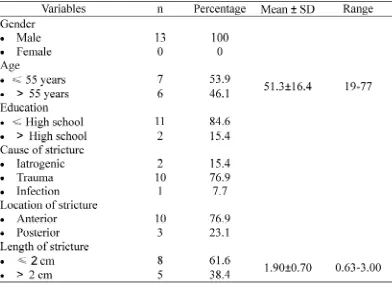
TABLE 1. Characteristics of subjects
All patients involved in this study were male with age 52.3 ± 16.4 years and most of their education (11 patients or 84.6%) were below High School. The most common cause of the urethral stricture was trauma (10 patients or 76.9%). Only 1 patient (7.7%) and 2 patients (15.2%) were caused by an infection and an iatrogenic, respectively. Ten (76.9%) of the stricture were located on the anterior urethral and only 3 (23.1%) were located on posterior. It was indicated that the anterior urethra was the most common site of urethral stricture. Mean
of the length of urethral stricture was 1.90 ± 0.70 cm with range of 0.63-3.00 cm. Eight (61.6%) patients had the length of urethral stricture of2 cm and 5 (38.4%) patients had length of2 cm.
The results of uroflowmetry assessment of patients after three weeks post intenal urethro-tomy Sachse consisting of the mean value of Qmax, Qave, Tcomp, T100, TQ, and Tqmax are presented in TABLE 2. The mean of principle variable of uroflowmetry (Qmax) was 22.3±6.7 mL/s.
The Qmax value of patients based on the location and length of urethral stricture are presented in TABLE 3, while the comparisons
of patients’ Qmax three weeks after internal urethrotomy based on the length and location of urethral stricture are presented in TABLE 4.
TABLE 3. The Qmax value of patients based on the location and length of urethral stricture
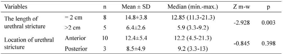
The mean of Qmax of patients who had the length of urethral stricture of2 cm (14.8±3.8 mL/s) was significantly higher than patients who had length of2 cm (6.4±2.6 mL/s) (p=0.03), whereas patients who had the location of
urethral stricture on anterior urethra (12.4±5.4 mL/s) were not significantly different in Qmax compared to patients who had those on posterior urethra (8.5±4.9 mL/s) (p=0.398).
TABLE 4. Comparisons of patients Qmax (in mL/s) after three weeks internal urethrotomy based on the length and location of urethral stricture
DISCUSSION
Urethral stricture is an abnormal narrowing of the tube that carries urine out of the body from the urethra. Urethral stricture may be caused by inflammation or scar tissue from surgery, disease, or injury.11Internal urethro-tomy is still the gold
standard procedures to return patients to a state of normal voiding. To evaluate the outcome of the internal urethrotomy,uroflowmetryassessment can be conducted with its principal variable of Qmax. In this studythe Qmax of urethral stricture patients
three weeks post internal urethrotomy Sachse in Dr. Sardjito General Hospital is reported.
All oftheurethral stricturepatients(13 patients or 100%) involved in this study were male. As reported in previous study, males have higher chances of the urethral stricture than females.1,12,13
The incidence of the urethral stricture on the patients aged55 years (54%) was higher than on those55 years (46%). The mean of patients’ age in this study was 51.3±16.4 years (19-77 years. Some authors reported the different mean age of the patients with urethral stricture in their studies. Zehriet al.7reported
that the mean of patients age involved in their study was 54 years (17-87 years), while the mean patients age involved in the study conducted by Chhetriet al.1was 36.3 years
(10-70 years) and conducted by Prihadi and Sugandi14was 35.7±11.6 years (25-34 years).
Moreover, Santucci et al.15demonstrated an
increase in urethral stricture disease with age, with a marked increase in patients over the age of 55 years.
In this study, trauma (76.9%) was the common cause of the urethral stricture, followed by iotrogenic (15.4%) and infection (7.7%). The trauma was referred to traffic accidents (38.4%), falling from trees (15.4%) and late complication of prostatectomy (23.1%). The common causes of the urethral stricture have been reported by some authors. Chhetriet al.1
reported that the common cause of the urethral stricture is injury-related trauma (67.3%), then iatrogenic (21.3%) and infection (11.4%). Other study also reported trauma (55.1%) to be the common cause of the urethral stricture, followed by iatrogenic (25.9%) and infection (19%).12
The Qmax is principle variable of uroflowmetry besides Qave, Tcomp, T100, TQ, and Tqmax. A Qmax value can be used to evaluate urinary tract function post internal urethrotomy. Several studies have been conducted to classify Qmax value as normal and abnormal. The Qmax value from 20 to 30 mL/s are considered to be normal value of urinary tract function.16-18 The mean of Qmax of the
patients with urethra stricture after three weeks post internal urethrotomy Sachse was 22.3±6.7 mL/s (TABLE 2). Based on this normal Qmax
value, this study showed that the majority of patients returned to a state of normal urinary tract function three weeks post internal urethrotomy.
This study showed that the length of urethral stricture significantly influenced the Qmax of patients. The mean of Qmax of patients who had
the length of urethral stricture of 2 cm
(14.8±3.8 mL/s) was significantly higher than patients who had length of à 2 cm (6.4±2.6 mL/ s) (p=0.03). This findings supported some previous studythat reported the length of urethral stricture was thought to be related with the Qmax of patients.1,7,13,14
The majority of the urethral stricture were found on anterior urethra (10 patients or 76.9%), with only 23.1% (3 patients) were found on posterior urethra. Among the urethral stricture found on anterior urethra, 80.0% (8 patients) were found in bulbous urethra, 20.0% (2 patients) in pendulous urethra, while on posterior urethra it was found all on membranous urethra (3 patients or 23.1%). The similar locations with the different incidence of the urethral stricture have been reported by some authors. Zehriet al.7found in their study
that there were bulbomembranous or bulbous urethra in 61% cases of the urethra stricture, while Prihadi and Sugandi14 found that there
were 77% cases of the urethra stricture on anterior urethra that did not specifically state its locations.
CONCLUSION
correlation of the location or urethral stricture with Qmax.
ACKNOWLEDGEMENTS
Authors would like to thank all patients who have participated in this study.
REFERENCES
1. Chhetri RK, Shrestha GK, Joshi HN, Shrestha RKM. Management of strictures and their outcome. Nepal Med Coll J 2009; 11(1): 5-8. 2. Tonkin JB and Jordan GH. Management of distal
anterior urethral strictures: trauma and reconstruction. Medscape 2009; 2-11.
3. Anonim. Protap uretrotomi interna. Yogyakarta: SMF Urologi RSUP Dr. Sardjito, 2007.
4. Wein AJ. Urethral stricture disease. In: Wein AJ, Kavoussi LR, Novick AC, Partin AW, Peters CA, editors. Campbell-Walsh urology, 9th ed. Philadelphia: WB Saunders, 2007.
5. Schwartz SI, Shires GT, Spencer FC. Anatomi dalam prinsip-prinsip ilmu bedah. Edisi 6. Jakarta: EGC, 2000.
6. Midwood KS, Williams LV, Schwarzbauer JE. Tissue repair and the dynamics of the extracellular matrix. Int J Biochem Cell Biol 2004; 36(6):1031-2.
7. Zehri AA, Ather MH, Afshan Q. Predictors of recurrence of urethral stricture disease following optical urethrotomy. Int J Surg 2009; 7(4): 361-4.
8. Kirby R, Lepor H. 2007. Evaluation and nonsurgical management of benign prostatic hyperplasis. In: Wein AJ, Kavoussi LR, Novick AC, Partin AW, Peters CA, editors. Campbell-Walsh Urology, 9thed. Philadelphia: WB Saunders, 2007. 9. Heyns CF. 2008. Follow-up strategies after urethral stricture treatment. Current clinical
urology: Urethral Reconstructive Surgery. Totowa: Humana Press, 2008: 315.
10. Jorgensen JB, Jensen KM, Bille-Brahe NE, Morgensen P. Uroflowmetry in asymptomatic elderly males. Br J Urol 1986; 58: 390-5. 11. Rosentein DI, Alsikafi NF. Diagnosis and
classification of urethral injuries. Urol Clin N Am 2006; 33:73–85
12. Jalbani MH, Shaik NA. 2002. Experience with cold knife optical internal urethrotomy and temporary dilatation. Pakistan J Med Res 2002; 41(4): 1-5.
13. Ishigooka M, Tomaru M, Hashimoto T, Sasagawa I, Nakada T, Mitobe K. Recurrence of urethral stricture after single intenal urethrotomy. Int Urol Nephrol 1995; 27(1):101-6.
14. Prihadi JC dan Sugandi S. Penilaian perubahan pancaran urin maksimal pada pasien striktur uretra setelah kateter uretra dilepas dan tiga minggu pasca uretrotomi interna. JURI 2004. 11(1): 27-30.
15. Santucci RA, Joyce GF, Wise M. Male urethral stricture disease. In: Litwin MS and Saigal CS. Urologic disease in America. Los Angeles: National Institute of Diabetes and Digestive and Kidney Diseases (U.S.), National Institutes of Health (U.S.), United States. Dept. of Health and Human Services, RAND Health, University of California, 2007:531-51.
16. Haylen BT, Ashby D, Sutherst JR, Frazer MI, West CR. Maximum and average urine flow rates in normal male and female populations-the Liverpool nomograms. Br J Urol 1989; 64(1):30-8. 17. Haylen BT, Parys BT, Anyaegbunam WI, Ashby D,
West CR. Urine flow rates in male and female urodynamic patients compared with the Liverpool nomograms. Br J Urol 1990; 65(5):483-7. 18. Klingele CJ and Webb MJ. Normal function of