VOL. 6, NO. 1, pp. 15-22, January, 2016
Effect of Nigella sativa Extract on Inflammatory Cells, Interleukin-10, Interferon-
γand Histological
of Kidney in Monosodium Glutamate-Induced Rats
Abdalrauf A. Mahmud Yousif1*
, Hidayat Sujuti2
, Edi Widjajanto3
1Master program in Biomedical Sciences concentration in Immunology, Faculty of Medicine, Brawijaya University, Malang, Indonesia 2Department of Biochemistry, Faculty of Medicine, Brawijaya University, Malang, Indonesia
3Department of Clinical Pathology, Faculty of Medicine, Brawijaya University, Malang, Indonesia
ABSTRACT
Overconsumption of food additives monosodium glutamate (MSG), a flavour enhancer was unhealthy. Herbal medicine Nigella sativa (NS) has antioxidant properties able to cure the toxic induced by MSG. This study aimed to evaluate the risks of excessive use of MSG and to study the role of NS to inhibit inflammation and renal dam -age. Treated rats (twenty four male wistar rats) were divided into six group and analyzed by measuring the cells in blood, interleukin-10, interferon-γ serum levels by ELISA method and remove kidneys for histological examina-tion. Histological of kidney for all groups except control, were showed different abnormalities include congestion of some blood vessels, hemorrhage between tubules, widening in the renal tubules, revealed severe dilatation of Bowman's capsule, shrinkage of glomeruli, and huge vacuole area, were observed compared with control. Inter-leukin-10 was reduced in Groups 2,3,4 and 5, whereas increase in group 6 compared with control. Interferon-γ was increased in groups 2,3,4 and reduced in groups 5,6 compared with control. Eosinophil was increased in groups 2,5 and reduced in groups 3,4 and 6. Basophil was reduced in groups 2, 3 and increased in groups 5 and 6 compared with control. This present study showed that administration of MSG to rats induced many changes eff-ffects on inflammatory cells, cytokines and histological of kidneys. NS has benefit in blood parameters, whereas
harmful on kidney at these doses.
Keywords: eosinophil, interleukin-10, interferon-γ, kidney, monosodium glutamate, Nigella sativa
Monosodium glutamate (MSG, C5H8NO4Na, E621) is a type of salt whichis used widely in most countries of the world, MSG one of the additives foods and one of the components of commercially processed foods. MSG added in foods because they can improve the food palatability and flavour enhancer. MSG has a spe-cial flavour that does not provide any victuals or other enhancers [1]. MSG improves the taste of food which is called “umami” taste in Japan [2,3].
Monosodium glutamate is the sodium salt of glu-tamic acid, also known as the fifth taste. The basic ssory function of MSG is attributed to its ability to en-hance the presence of other taste-active compounds.
Recommended for patients with missing desire or ap-petite for food also used in homes and restaurants [4] especially in Asian cuisine [5]. It is added to the food either as a purified monosodium salt or sodium salt as a component of a mixture of amino acids and small peptides resulting from the acid or enzymatic hydroly-sis of proteins [6]. MSG are present in most canned prepared snacks and fast food. Glutamic acid is an amino acid naturally present in vegetables and foods of animal origin, such as cheese, and seafoods [4, 5, 7, 8, 9, 10, 11].
Over consumption of MSG for a long time led to many of the toxic effects, various inflammatory disease, positively related to obesity, and referred to the Chi-nese restaurant syndrome (CRS) [4, 8, 12]. Neuronal cell death is associated with glutamic production of free radicals in various organs. The brain is the most sus-ceptible to oxidation due to a high rate of metabolic ac-tivity of oxidative stress [6, 7, 8, 10]. MSG causes kid-ney dysfunction, renal oxidative stress [13], and
JTLS | J. Trop. Life. Science 15 Volume 6 | Number 1 | January | 2016
INTRODUCTION
*Corresponding author: Abdalrauf A. Mahmud Yousif
Master program in Biomedical Sciences, Faculty of Medicine, Brawijaya University
histopathological alterations in the kidney tissues of rats included revealed severe dilatation of Bowman's capsule, shrinkage of glomeruli, loss of brush border to see the relationship between expression of TLR4 and SRNS produced correlation value of 0.512 and its im-portance p value was 0.013 (p<0.05). There is a differ-ence significant expression of TLR4 on SRNS com-pared to SSNS with its importance value of 0.012 (p<0.05) and there is also a significant correlation be-tween expression of TLR4 and SRNS with its signifi-cance level of 0.013 (p<0.05).
Chemicals
Monosodium glutamate was obtained from local market in Malang, Indonesia and was dissolved in distilled water and given orally by gavage to rats. Black seeds (Nigella sativa) were obtained from local market in Malang, Indonesia. Black seeds will crush by mill mixer braun and its extract was prepare as suspension using distilled water and given orally in different doses by gavage to groups of treated male Wistar rats.
Animals
Healthy male wistar rats (2-3 months old littermate, 210 ± 30 g) were obtained from the Physiological Labo-ratory of Brawijaya University, Malang, Indonesia.Ani-mals were placed on Animal Research Care. Adapta-tion time was done in one week to prepare the rats, that was weighing, examining the health status, and help the rats to adapt with new environment. Animals were housed in polypropylene cages and maintained at 25 ± 2ºC and constant wetness degree according to the alteration of day and night.
Experimental design
Twenty four wistar rats were selected and randomly divided into six groups (four rats each). Group 1 served as control and was orally administered with dis-tilled water throughout the experimental protocol. While Group 2 was orally administered 1 g of MSG at oral dose of 1 g/kg bw in distilled water for 30 days. Groups 3, 4, and 5 were administered 1 g of MSG per rat orally in the morning and NS (100, 200, and 400 mg) in the evening, respectively, for 30 days. Group 6 was orally administered 200 mg/kg bw of NS in dis-tilled water for 30 days. Rats were sacrificed at the end of 30 days to take blood for measured the white blood cells differentiation count % using Sysmex XT- 4000i Automated Hematology Analyzer device. Interleukin-10, interferon-γ levels in serum were measured by
ELISA Sandwich method. The kidneys were carefully dissected and removed. The tissues were fixed in 10% formalin, embedded in paraffin and that tissue sections (5 µm) were stained with hematoxylin-eosin to study their micro architecture by light microscopy.
Ethical issue
The study was approved by Health Research Ethics Committee (Medical Faculty, Brawijaya University) number No.237/EC/KEPK-S2/03/2015.
Statistical analysis
The results were expressed as mean ± SD. Differ-ences in the various parameters between groups were evaluated by one factor, One-way ANOVA test. All mean differences were considered significant if p < 0.05. Analysis were conducted using SPSS for Win-dows® v.17.
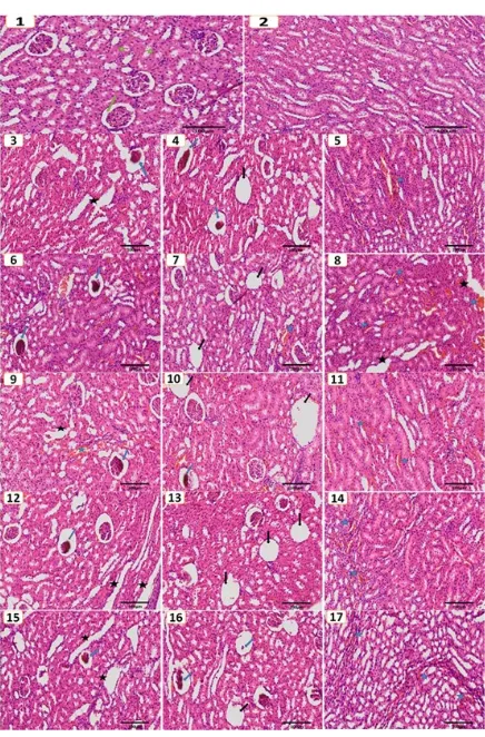
Histological examination of kidney tissue in group 1 as control rats stained with Hematoxylin and Eosin staines (H&E) were normal kidneys with well demar-cated cortex, medulla, normal Bowman’s capsule and glomerulus as well as normal sized renal tubules as shown in (Figure 1; photos 1, 2). While histological ex-amination of the kidney tissue after addition of MSG in group 2 rats shows different abnormalities (Figure1, photos 3, 4, 5). The abnormalities of kidney sections were summarized as congestion of some blood vessels, hemorrhage between tubules (Figure 1, photo 5), widening in the renal tubules (Figure1, photo 3), re-vealed severe dilatation of Bowman's capsule and shrinkage of glomeruli, and/or widening of the Bow-man’s space due to contraction of the renal glomerulus and hyper-cellularity (Figure1; photos 3, 4). In addition to huge vacuole area were also observed (Figure1, photo 4). The histological examination of kidneys tis-sue of groups 3, 4, 5, and 6, showed almost similar in their changed of structure of group 2, but had a lesser degree of these changes in group 3 compared with group 2, group 4 compared with 3, and groups 5,6 compared with 4, as followed figures: group 3 (Figure 1; photos 6, 7, 8), group 4 (Figure1, photos 9,10, 11), group 5 (Figure1; photos 12, 13, 14), and group 6 (Fig-ure 1; photos 15, 16, 17).
Figure 2 shows that the average of interleukin-10 for each group is different each other significantly. The lowest average is in group 3 of 95.70 ± 23.06, and the highest average is in group 6 at 219.85 ± 40.99. Overall average of interleukin-10 is obtained by 134.99 ± 55.77. RESULTS AND DISCUSSION
Figure 1. HE staining examination of histological kidney, Group 1 control group, Group 2 MSG group, Group 3 MSG & NS 100 mg/kg group, Group 4 MSG & NS 200 mg/kg group, Group 5 MSG & NS 400 mg/kg group, and Group 6 NS group (100× magnification). Green ar-row normal Bowman’s capsule and glomerulus. Green stars normal renal tubules. Blue stars congestion of some blood vessels, andhemorrhage between tubules. Black stars widening in the renal tubules. Blue arrow revealed severe dilatation of Bowman's capsule and shrinkage of glomeruli. Black arrow areas of huge vac-uole.
Analysis of variance show a significance level of 0.00 is smaller than α (0.05), and the value of F count (15.12) is greater than F table 5% (2.77). These results indicate that there are differences of average that are significant (real) in the treatment of interleukin-10 factors.
Figure 2 shows also the average interferon-γ each group was look different from each other significantly. The lowest average in group 5 of 264.000 ± 62.844, and the highest average in group 2 at 771.500 ± 329.769. Overall average of interferon-γ 407.500 ± 239.527. Analysis of variance showed a significance level of 0.014 is smaller than α (0.05), and the value of F count (3.932) is greater than F table 5% (2.773). From these
Figure 2. Data of interleukin-10 and interferon-γ levels in serum rats for each group
results indicate that there are differences of average that are significant (real) in the treatment of interferon-γ factors.
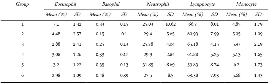
Table 1 shows that the average of each group is not too much different, as the following:
In eosinophil the lowest average is in group 3 of 2.88 ± 2.41, and the highest average is in group 2 at 4.48 ± 2.58. Overall average of eosinophil obtained by 3.28 ± 1.65. Analysis of variance showed a significance level of 0.80 is greater than α (0.05), and the value of F count (0.46) is smaller than F table 5% (2.77). From these results indicate that there are differences of aver-age that are not apparent in the groups of eosinophil factors.
In basophil the lowest average is in group 2 of 0.15 ± 0.10, and the highest average is in group 6 at 0.48 ± 0.39. Overall average of basophil obtained by 0.31 ±0.21. Analysis of variance showed a significance level of 0.37 is greater than α (0.05), and the value of F count (1.15) is smaller than F table 5% (2.77). From these results indicate that there are differences of aver-age that are not apparent in the groups of basophil fac-tors.
In neutrophil the lowest average is in group 1 of 25.03 ± 10.62, and the highest average is in group 5 at 31.85 ± 8.69. Overall average of neutrophil obtained by 28.24 ± 6.88. Analysis of variance showed a significance level of 0.76 is greater than α (0.05), and the value of F count (0.52) is smaller than F table 5% (2.77). From these results indicate that there are differences of aver-age that are not apparent in the groups of neutrophil factors.
In lymphocyte the lowest average is in group 5 of 59.83 ± 8.74, and the highest average is in group 1 at 66.70 ±8.01. Overall average of lymphocyte obtained by 62.98 ± 6.83. Analysis of variance showed a significance level of 0.753 is greater greater than α (0.05), and value
of F count (0.53) is smaller than F table 5% (2.77). From these results indicate that there are differences of average that are not apparent in the groups of lympho-cyte factors.
In monocyte the lowest average is in group 1 of 4.85 ± 1.79, and the highest average in group 5 at 6.20 ± 1.73. Overall average of monocyte obtained by 5.44 ± 1.58. Analysis of variance showed a significance level of 0.833 is g greater than α (0.05), and the value of F count (0.41) is smaller than F table 5% (2.77). From these results indicate that there are differences of aver-age that are not apparent in the groups of monocyte factors.
MSG, a sodium salt of glutamic acid being added to Chinese food, canned vegetables, soups and processed food, acted as a flavour enhancement by stimulate the sensory receptors, thereby improving the palatability of food. Despite of its taste stimulation and appetite en-hancement, various researchers had reported that it was toxic to humans and experimental animals [25]. Our results showed that histological examination of the kidney tissue after oral administration of MSG reveal different abnormalities. The abnormalities of kidney sections are summarized as congestion of some blood vessels, hemorrhage between tubules, widening in the renal tubules, reveal severe dilatation of Bowman's cap-sule and shrinkage of glomeruli. In addition, huge vac-uola were also observed. This result was agreement with the study of Ortiz et al. and Mousa et al. [26, 27].
MSG has hepatoxicity and nephrotoxicity tenden-cies especially when consumed at higher concentra-tions [28]. The effect of MSG addition to renal weretis-sue impairment, swelling of the lining epithelium of glomeruli, hydropic degeneration and vacuolization of the renal convoluted tubules, blood vessels dilatation and focal hemorrhage between the degenerative renal tubules, and many renal tubules of the rat kidneys
showed degenerative lesions under the effect of MSG. These are justifiable since the renal tubules are particu-larly sensitive to toxic influences due to their high oxy-gen consumption and vulnerable enzyme systems, complicated transport mechanisms that may be used for transport of toxin and maybe damaged by such toxin. The tubules also come in contact with toxic chemicals during their excretion and elimination by the kidneys [6]. One possible mechanism for the tubu-lar lesions is the direct toxic effect on the cell function[14]. Alterations in the levels of lipid peroxides and antioxidants such as reduce glutathione, catalase and superoxide dismutase were observed in different organs and systems of adult rat during MSG treatment [6,29]. The effects observed in both liver and kidneys could have occurred because these organs are involved in the metabolism of glutamate [30]. MSG has a toxic effect on many body organs by altering ionic perme-ability of neural membrane and induced persistent de-polarization. The mechanisms of MSG-induced dam-age include the production of free radicals that alter mitochondrial activity and genetic information [14].
Our results showed the toxic effect of N. sativa. Those are supported by Zaghlol et al. who reported that high doses of N. sativa oil had wide range of toxic effects on the kidney and liver. Some of these changes increase with doses additionof N. sativa. The previous study observed that the acute toxic effect of thymo-quinone a main constituent of the N. sativa caused a significant increase in the concentration of serum crea-tinine and blood urea, which indicated serious effects on renal. High doses of N. sativa also caused cellular damage because of the alkylation of proteins and DNA and formation of reactive oxygen species, including su-peroxide and hydrogen su-peroxide, which caused oxida-tive stress function [31]. These results suggest the pos-sible toxicity of lethal doses of N. sativa. Whereas these
Table 1. Data of White Blood Cells Differentiation Count percentage in different groups
Group Eosinophil Basophil Neutrophil Lymphocyte Monocyte
Mean (%) SD Mean (%) SD Mean (%) SD Mean (%) SD Mean (%) SD
1 3.1 1.32 0.33 0.15 25.03 10.62 66.7 8.01 4.85 1.79
2 4.48 2.57 0.15 0.1 29.4 5.65 60.93 7.99 5.05 1.09
3 2.88 2.41 0.25 0.13 25.78 4.04 65.18 4.15 5.93 2.19
4 3.08 1.26 0.33 0.17 29.9 2.84 61.88 5.25 5.13 1.65
5 3.2 1.22 0.35 0.13 31.85 8.69 59.83 8.74 6.2 1.73
were different with previous study which report the protective effect of N. sativa against renal injury in rat kidneys [32].
In the present study, group 2 with only MSG treat-mentis significantly decrease interleukin-10 compared withcontrol group, this result was agreement with Has-san et al. IL-10 is an anti-inflammatory cytokine pro-ducedin large amounts from activated B-lymphocytes and play a regulatory role in suppressing harmful im-mune responses. IL-10 prevents the increase of nuclear factor kappa-light-chain-enhancer in activated B cells (NF-kB) binding activity evoked by glutamate. There is a strong positive correlation among the changes of serum IL-10 levels and the loss of kidney anti-oxidants, this lends credence to the potential role that oxidative stress might be exit for mediating the immunotoxicity of MSG [4]. Our studys showed that group 6 with only NS treatment more increase the interleukin-10 com-paredto control group, this result was agreement with [18, 20, 22, 23, 24]. In contrast, IL-10 has potentialto be anti-inflammatory effects and suppress pro-inflam-matory cytokines. The suggested that mechanism of N. sativa may affect both oxidative stress and inflamma-tory process simultaneously are through the inhibition of NF-kB [20].
Our result showed that oral administration of MSG in group 2 are significantly increase the IFN-γ level compared with other groups. Earlier studies, which have also been confirmed this result on more recent re-ports on the adverse effects of MSG to increase levels of pro-inflammatory cytokines [33, 36]. The anti-in-flammatory cytokines are series of immunoregulatory molecules that control the pro-inflammatory cytokine response [37]. MSG-treated animals develop obesity.In obesity, the intra-abdominal adipose tissue growth pro-motes increase pro-inflammatory cytokines infiltration and activation, which denotes the primary causes for chronic inflammation, morbidity and mortality risk [36].
In the present study, IFN-γ level in groups 3,4 and 5 treated rats, weregradual decline respectively. When-ever the increasing dose of N. sativa would decrease the level of IFN-γ, and all of them were significantly more decrease compared to group 2 with only MSG treatment, it meansthat N. sativa may has strong abil-ity to protect of MSG. In group 6 with only NS treat-ment showed that the IFN-γ level is also significant more decrease compared to group 2 MSG treatment. These results are similar to previous study that N. sativa has been proved experimentally to be an anti-in-flammatory substance and it significantly reduced the
levels of pro-inflammatory mediators [20, 22, 23, 24]. N. sativa extract has been shown to have strong antiox-idant properties to suppress the expression of inducible nitric oxide (NO) synthesis in rat macrophages. It has been shown that N. sativa extract has inhibitory effects on both the cyclooxygenase and the 5-lipoxygenase pathways of arachidonic acid metabolism and on mem-brane lipid peroxidation [38].
In the present study Group 2 was increased the eosinophil percentage compared with other groups. eosinophil possess a battery in their granules and lipid bodies that potent cytotoxic and pro-inflammatory agent [39]. Previous studies found that MSG associated with asthma [4, 8, 27, 29, 40] and autoimmune disease [41, 42] and these diseases may be the main causes to high percentage of eosinophil in blood.Groups 3, 4 and 5 were observed decrease compared with Group 2,it is mean NS has ability to protect of MSG and also we ob-served that at low dose of NS was more effect, and this result agreement with [43, 44]. previous studies re-vealed that an early influx of eosinophils into sites of inflammation precedes that of lymphocytes [45].
In the present study, the percentage of Basophil in Group 2 was decrease compared with other groups. We suggested that may basophils migrate from the cir-culation to the tissues. A decreased of basophil percent-age known as basopenia may be due to severe allergy and Severe injury. Also recent genetic approaches indi-cate that basophils can migrate into lymphoid tissues and, in some circumstances, cooperate with other im-mune cells to promote optimal TH2 cytokine responses in vivo [46]. However it has been very difficult for most laboratories to obtain basophils without major contaminating cell populations, because the percentage of basophils in blood is low (< 1% of total WBC) and they share physicochemical properties with other blood cells. This lack of satisfactory purification protocols has considerably hampered basophil research and nega-tively affected the interests in this cell type. Basophils have also been proposed to play a key role in allergy by directly inducing the switch to the IgE isotope in B cells independently of T cells [47].
In conclusion, short term administration of MSG to rats in present study induced many changes effects on inflammatory cells. Cytokines and histological struc-ture of the kidneys include congestion of some blood vessels. Hemorrhage between tubules. widening in the renal tubules. revealed severe dilatation of Bowman's capsule. shrinkage of glomeruli. and huge vacuole
JTLS | J. Trop. Life. Science 16 Volume 6 | Number 1 | January | 2016
CONCLUSIONS
area. This present study also demonstrated that N. sativa may improve inflammation and reduce oxidative stress in blood rats as well as increased anti-inflamma-tory cytokines as IL-10 and decreased pro-inflamma-tory cytokines as IFN-γ induced by MSG-treated rats. In addition. the result clearly indicate that overdoses of N. sativa may have toxic and harmful effect on the his-tological structure similar with MSG effect.
The authors acknowledge the teaching staffs at Brawijaya University and technical assistance of staffs at Biomedical Laboratory, Pathology Anatomy Labora-tory, and Physiology Laboratory.
1. Husarova V, Ostatnikova D (2013) Monosodium Glutamate Toxic Effects and Their Implications For Human Intake: A Review. IBIMA Publishing JMED Research. DOI: 10.5171/2013.608765.
2. Ilegbedion I, Onyije F, Chibuike O (2013) Infiltration of Inflammatory Cells in the Ovary Following Oral Administration of Monosodium Glutamate. Bangladesh Journal of Medical Science 12: 413-418.
3. Savcheniuk O, Virchenko O, Falalyeyeva T, Beregova T, Babenko L, Lazarenko L, Demchenko O, Bubnov R, Spivak M (2014) The Efficacy of Probiotics for Monosodium Glutamate-Induced Obesity: Dietology Concerns and Opportunities for Prevention. The EPMA Journal 5: 2.
4. Hassan Z, Arafa M, Soliman W, Atteia H, Al-Saeed H (2014) The Effects of Monosodium Glutamate on Thymic and Splenic Immune Functions and Role of Recovery (Biochemical and Histological Study). J Cytol Histol. 5:6. 5. He Ka L, Daviglus M, Dyer A, Horn L, Garside D, Zhu L,
Guo D, Wu Y, Zhou B, Stamler J (2008) Association of Monosodium Glutamate Intake with Overweight in Chinese Adults: The Intermap Study Obesity 16 (8): 1875–1880.
6. Abass M, Abd El-Haleem M (2011) Evaluation of Monosodium Glutamate Induced Neurotoxicity and Nephrotoxicity in Adult Male Albino Rats. Journal of AmericanScience. 7(8): 264-276.
7. Shivasharan, Nagakannan P, Thippeswamy B, Veerapur V (2013) Protective Effect of Calendula officinalis L Flowers Against Monosodium Glutamate Induced Oxidative Stress and Excitotoxic Brain Damage in Rats. Ind J Clin Biochem 28 (3): 292–298.
8. Euteum P, Kim DK, Kim S, Sapkota K, Kim S, Kim CS, Chun HS (2014) Protective Effects of N-Acetylcysteine Against Monosodium Glutamate-Induced Astrocytic Cell
Death. Food And Chemical Toxicology 67: 1–9.
9. Xu L, Sun J, Lu R, Ji Q, Xu J (2005) Effect of Glutamate on Inflammatory Responses of Intestine and Brain after Focal Cerebral Ischemia. World Journal of Gastroenterology 11 (5): 733-736.
10. Yaqub H, Abdel Baky N, Attia H, Faddah L (2008) Hepatoprotective Effect of N-Acetyl Cysteine and/or β
-Carotene on Monosodium Glutamate-Induced Toxicity in Rats. Research Journal of Medicine and Medical Sciences. 3 (2): 206-215.
11. Shimada A, Cairns B, Vad N, Ulriksen K, Pedersen A, Svensson P, Baad-Hansen L (2013) Headache and Mechanical Sensitization of Human Pericranial Muscles After Repeated Intake of Monosodium Glutamate (MSG). The Journal of Headache and Pain 14: 2.
12. Pavlovic V, Sarac M (2010) The Role of Ascorbic Acid and Monosodium Glutamate in Thymocyte Apoptosis. Bratisl Lek Listy 111 (6): 357-360.
13. Sharma A, Wongkham C, Prasongwattanav, Boonnate P, Thanan R, Reungjui S, Cha’on U (2014) Proteomic Analysis of Kidney in Rats Chronically Exposed to Monosodium.
14. Afeefy A, Mahmoud M, Arafa M (2012) Effect of Honey on Monosodium Glutamate Induced Nephrotoxicity (Histological and Electron Microscopic Studies). Journal of American Science 8 (1s): 146-156.
15. Paarakh P, Linn NS (2010) A Comprehensive Review. Indian Journal of Natural Products and Resources 1(4): 409-429.
16. Sandhu KS, Rana AC (2013) A Review of Plant Nigella Sativa: A Brief Consideration of Its Pharmacognostic Characters. Chemical Constituents and Therapeutic Benefits. An International Journal of Pharmaceutical Sciences. 4
17. Salem M (2005) Immunomodulator, Therapeutic Properties of the Nigella sativa L. Seed. International Immunopharmacology 5: 1749–1770.
18. Gholamnezhad Z, Boskabady M, Hosseini M (2014)Effect of Nigella sativa on Immune Response in Treadmill Exercised Rat. BMC Complementary and Alternative Medicine 14: 437.
19. Dollah M, Parhizkar S, Abdul Latiff L Bin Hassan M, (2013) Toxicity Effect of Nigella sativa on The Liver Function of Rats. Advanced Pharmaceutical Bulletin 3 (1): 97-102.
20. Hadi V, Alizadeh M, Hosseini H (2014) Effects of Nigella sativa Oil Extract on Inflammatory Cytokine Response and Oxidative Stress Status in Patients With Rheumatoid Arthritis: ARandomized. Double-Blind. Placebo-Controlled Clinical Trial. Avicenna J Phytomed.
21. Panahi M, Namjoyan F, Shakerin Z (2011) Evaluation of
ACKNOWLEDGMENT
Antioxidant Effects of Nigella sativa Extract on The Ultra Structure of Neural Tube Defects in Diabetic Rats's Offspring. Jundishapur Journal of Natural Pharmaceutical Products 6 (1): 16-23.
22. Fahmy H, Noor N, Faten F, Elsayed A, Radwan N (2014) Nigella sativa as An Anti-Inflammatory and Promising Remyelinating Agent in The Cortex and Hippocampus of Experimental Autoimmune Encephalomyelitis-Induced Rats. The Journal of Basic & Applied Zoology 67: 182– 195.
23. Ibrahim Z, Ishizuka M, Soliman M, Elbohi K, Sobhy W, Muzandu K, Elkattawy A, Sakamoto K, Fujita S (2008) Protection by Nigella sativa Against Carbon Tetrachloride-Induced Downregulation of Hepatic Cytochrome P450 Isozymes in Rats. Japanese Journal of Veterinary Research 56 (3): 119-12.
24. Liua X, Jong-Hyouk P, Abd El-Aty A, Assayed M, Minoru S, Jae-Han S (2013) Isolation of Volatiles From Nigella sativa Seeds Using Microwave-Assisted Extraction: Effect of Whole Extracts on Canine and Murine CYP1A. Biomed. Chromatogr. 27: 938–945.
25. Dixit S, Rani P, Anand A, Khatri K, Chauhan R, Bharihoke V (2014) To Study The Effect of Monosodium Glutamate on Histomorphometry of Cortex of Kidney in Adult Albino Rats. Renfail. 36 (2): 266–270.
26. Ortiz G. Bitzer-Quintero O, Beas Zarate C, Rodriguez-Reynoso S, Larios- Arceo F, Velazquez-Brizuela I, Pacheco-Moises F, Rosales-Corral S (2006) Monosodium Glutamate-Induced Damage in Liver And Kidney: AMorphological and Biochemical Approach.Biomedicine & Pharmacotherapy 60: 86-91.
27. Moussa E, Al Mulhim J (2013)Modulating Effect of Nigella sativa on Renal Structural Changes by Monosodium Glutamate in Female Mice. Egypt. Acad. J. Biolog. Sci. 5 (2): 33-45.
28. Inuwa H. Aina V. Gabi B. Aim Ola I. Ja’afaru L (2011) Determination of Nephrotoxicity and Hepatoxicity of Monosodium Glutamate (MSG) Consumption. British Journal of Pharmacology and Toxicology 2 (3): 148-153. 29. Attia H, Faddah L, Yaqub H (2008) Trans-Retinol
Precursor and/or N-Acetyl Cysteine Protects Against Monosodium Glutamte-Induced Nephrotoxicity in Rats. Journal of Applied Sciences Research 4 (12): 2108-2119. 30. Onaolapo A, Onaolapo O, Mosaku T, Akanji O, Abiodun
OA (2013) Histological Study of the Hepatic and Renal Effects of Subchronic Low Dose Oral Monosodium Glutamate in Swiss Albino Mice. British Journal of Medicine & Medical Research 3 (2): 294-306.
31. Zaghlol D, Kamel E, Mohammed D, Abbas N (2012) The Possible Toxic Effect of Different Doses of Nigella sativa Oil on the Histological Structure of the Liver and Renal
Cortex of Adult Male Albino Rats. The Egyptian Journal of Histology 35: 127-136.
32. Yildiz F, Coban S, Terzi A, Savas M, Bitiren M, Celik H, Aksoy N (2010) Protective effects of Nigella sativa against ischemia-reperfusion injury of Kidneys. Renal Failure 32 (1): 126-31.
33. Alarcon-Aguilar F, Almanza-Perez J, Blancas G, Angeles S, Garcia-Macedo R, Roman R, Cruz M (2008) Glycine Regulates The Production of Pro-Inflammatory Cytokines in Lean And Monosodium Glutamate-Obese Mice. European Journal of Pharmacology 599: 152–158. 34. Chaparro-Huerta V, Flores-Soto M, Gudino-Cabrera G,
Rivera-Cervantes M, Bitzer-Quintero O, Beas-Zarate C (2008) Role of P38 MAPK and Pro-Inflammatory Cytokines Expression in Glutamate-Induced Neuronal Death of Neonatal Rats. Int. J. Devl Neuroscience 26: mRNAExpression of Peroxisome Proliferator-Activated Receptors in Mice. Basic & Clinical Pharmacology & Toxicology 108: 406–413.
36. Hernández-Bautista R, Alarcón-Aguilar F, Escobar-Villanueva M, Almanza-Pérez J, Merino-Aguilar J, Fainstein M, López-Diazguerrero N (2014) Biochemical Alterations During The Obese-Aging Process in Female and Male Monosodium Glutamate (MSG)-Treated Mice. Int. J. Mol. Sci.15: 11473-11494.
37. Zhang J, Jianxiong A (2009) Cytokines. Inflammation and Pain. Int Anesthesiol Clin. 45 (2): 27–37.
38. Keyhanmanesh R, Pejman L, Omrani H, Mirzamohammadi Z, Shahbazfar A (2014) The Effect of Single Dose of Thymoquinone. the Main Constituents of Nigella sativa in Guinea Pig Model of Asthma. Bioimpacts.4(2): 75-81. doi: 10.5681/Bi.2014.006 39. Behm C, Ovington K (2000) The Role of Eosinophils in
Parasitic Helminth Infections: Insights from Genetically Modified Mice. Parasitology Today. 16 (5): 202-209. 40. Stevenson D (2000) Monosodium Glutamate and Asthma.
American Society for Nutitional Sciences. 10678-10738. 41. Nakanishi Y, Tsuneyama K, Fujimoto M, Salunga T,
Nomoto K, Jun-Ling An, Takanoy, Iizuka S, Nagata M, Suzuki W, Shimada T, Aburada M, Nakano M, Selmi C, Gershwin M (2008) Monosodium Glutamate (MSG): A Villain and Promoter of Liver Inflammation and Dysplasia. Journal of Autoimmunity 30: 42-50.
42. Jovic Z, Veselinovic M, Vasic K, Stankovic-Djordjevic D,
Cekic S, Milovanovic M, Sarac M (2009) Monosodium Glutamate Induces Apoptosis in Naïve and Memory Human B Cells. Bratisl Lek Listy. 110 (10): 636-640. 43. Al-Sa'aidi J, Dawood K, Latif A (2012)
Immunomodulatory Effect of Nigella sativa Seed Extract in Male Rabbits Treated With Dexamethasone. Iraqi Journal of Veterinary Sciences. 26.
44. Kamil Z (2014) Effect of Crude Oil of Black Seeds (Nigella sativa) on White Blood Cell and Hematocrit of Male Albino Mice Treated With Low Toxic Dose of Paracetamol. Medical Journal of Babylon. 10(4).
45. You Lu (2013) Eosinophils. Their Progenitors and T Helper Cells in Allergic Airway Inflammation. Institute of Medicine at Sahlgrenska Academy. University of Gothenburg.
46. Siracusa M, Kim B, Spergel J, Artis D (2013) Basophils and Allergic Inflammation. J Allergy Clin Immunol. 132: 789-801.