L
Journal of Experimental Marine Biology and Ecology 248 (2000) 105–119
www.elsevier.nl / locate / jembe
Serological estimation of prey-protein gut-residence time and
quantification of meal size for grass shrimp consuming
meiofaunal copepods
a a ,* a b
M. Hoyt , J.W. Fleeger , R. Siebeling , R.J. Feller a
Department of Biological Sciences, Louisiana State University, Baton Rouge, LA 70803, USA b
Belle W. Baruch Institute for Marine Biology and Coastal Research, Department of Biological Sciences
and Marine Science Program, University of South Carolina, Columbia, SC 29208, USA Received 2 July 1999; received in revised form 13 January 2000; accepted 18 January 2000
Abstract
A series of experiments using serological reagents was conducted to examine predation, ingestion and digestion in a model predator–prey system. The harpacticoid copepod Amphias-coides atopus, obtained from mass culture, was used as prey and the grass shrimp, Palaemonetes pugio, as predator. Bulk-gut passage time in P. pugio was measured by visualization of latex beads and ranged from 0.5 to 4 h in starved and continuously-fed grass shrimp. A polyclonal antibody was prepared from crude extracts of A. atopus; cross reactions with P. pugio and three other crustaceans were either negligible or not detected using slide agar-gel-double-immunodiffusion (AGID) and Western blot preparations. The presence of A. atopus antigens was detected with great sensitivity (e.g., seven copepods, 35 mg dry weight, gave positive results) in grass shrimp gut contents even when proteins of other crustacean prey were present. Prey-proteins could be detected for as long as 4 h with AGID and 8 h with Western blot techniques. Individual grass
21
shrimp that were fed A. atopus and consumed from 0 to 98 copepods h were subjected to Western-blot preparation with chemiluminescence detection and densitometric evaluation. There was a significant curvilinear relationship between protein content and the number of copepod prey ingested. Results suggest that serological techniques can be modified to estimate the mass or abundance of standard-sized prey ingested by field-collected predators. 2000 Elsevier Science B.V. All rights reserved.
Keywords: Immunochemistry; Predator–prey interactions; Feeding; Meiofauna
*Corresponding author.
1. Introduction
Visual analysis of the gut contents of many marine predators is often precluded because prey tissue may be macerated, or predators may ingest only the softer, inner tissue of the prey after rejecting hard parts. The result is unidentifiable, amorphous gut contents. An alternative approach uses immunological reagents to identify the presence of prey-proteins in predator gut contents (e.g., Pickavance, 1970; Feller et al., 1979; Zagursky and Feller, 1988; Feller, 1992). Serological techniques have improved understanding of marine food webs by identifying the presence of targeted prey-proteins in selected predators (Feller, 1984; Feller, 1986; Hunter and Feller, 1987; Goarant et al., 1994; Pape-Lindstrom et al., 1997). Serological tests have also demonstrated differential digestion of different prey types in a single predator (Scholz et al., 1991).
Although serology can be a valuable tool for elucidating food-web relationships, the technique has several limitations. Shared antigenic determinants among putative prey have been identified, indicating serological tests may yield false positives (Feller et al., 1985). Serological techniques are capable of detecting the presence of solubilized prey-proteins in minute quantities but are not easily adapted to quantify the amount (or mass) of prey-proteins in predator gut contents. Furthermore, the residence time of prey-proteins is often unknown, partly because laboratory experiments with the predators and prey of interest are rare. Gut-residence time data are needed to determine the maximum time that a meal of a given prey type can be detected in field-collected predators. Residence data are especially significant for predators that forage across habitats or feed intermittently. Identified limitations must be addressed before serological reagents can be used to estimate ingestion rates or meal size in field-collected predators. Recent advances in biotechnology suggest solutions to limitations in the use of serology in predator–prey studies may be available.
We undertook a series of experiments using serological reagents to examine the interaction between predation, ingestion and digestion in a model predator–prey system. The model system consisted of an important estuarine food-chain link: a harpacticoid copepod (as a representative of meiofauna) as prey and the dagger-blade grass shrimp (Palaemonetes pugio Holthius) as predator. Grass shrimp are known predators of meiofauna, including harpacticoids (Walters et al., 1996; Gregg and Fleeger, 1998). The harpacticoid copepod, Amphiascoides atopus Lotufo and Fleeger, is readily grown in mass culture (Lotufo and Fleeger, 1995; Sun and Fleeger, 1995), and during the course
21
of this research a harvest of 0.5 g dry weight day was available for feeding experiments and immunogen preparations. A. atopus is readily consumed by juvenile and adult grass shrimp, and an exclusive diet of harpacticoids can sustain grass shrimp indefinitely (Nieland et al., 1998; Fleeger, pers. obs.). Previous laboratory experiments revealed that individual adult P. pugio ingest A. atopus at a maximum rate of about 100
21
2. Materials and methods
2.1. Anticrustacean serum preparation
A. atopus adults and late-stage copepodites were collected regularly from filters placed in a recirculating culture system constructed as in Sun and Fleeger (1995). The thousands of individuals (about 80% adults) that accumulated daily were transferred to a beaker with polystyrene beads (to act as an inert food source to stimulate gut clearance) for 2 h. Harpacticoids were then frozen in liquid nitrogen and stored at 2808C until processed to generate crude protein immunogen extracts. Five ml (1.9 g wet weight) of
A. atopus (approximately 500 000 individuals) were concentrated and washed in Tris-buffered saline (50 mM Tris pH 7.5, 150 mM NaCl, 5 mM MgCl ) until supernatant2
fluid was clear. Two ml of chilled Tris-buffer was used to pulverize whole copepods in a Potter Elvejem tissue grinder. An ice bath was utilized to chill the grinder and all components during processing. The homogenized sample was centrifuged at 16 000 g for 10 min to sediment exoskeletons and other debris leaving a soluble crude extract.
21
The extract was standardized to 5 mg protein ml by dilution with Tris-buffer. Crude protein extracts (as described above) were also prepared from additional crustacean species; field-collected Acartia tonsa (a calanoid copepod); Palaemonetes pugio (grass shrimp); laboratory-reared Artemia salina (brine shrimp) and an undescribed harpac-ticoid copepod in the family Laophontidae, also from mass culture.
One ml of crude extract (immunogen) from each taxon was concentrated to a 0.5 ml volume and emulsified in 0.5 ml Freund’s complete adjuvant and used to immunize rabbits. Immunizations were done by intradermal injection in 12 sites on the back of the
21
rabbit (0.05 ml site ) using the protocol described by Stills (1994). Each rabbit was boosted 4 weeks later when 1 ml of crude extract was combined with Freund’s
21
incomplete adjuvant and injected at two subcutaneous sites (0.25 ml site ) and two
21
intramuscular sites (0.25 ml site ). Antiserum was collected 4 weeks later when antibody activity was detected by a ring precipitin test. A sharp precipitin line was visible at the interface of antiserum and antigenic extract. Rabbits were exsanguinated and the blood allowed to clot at room temperature. The serum was removed from the clot, centrifuged at 2000 g to remove residual erythrocytes and stored at 2208C in small aliquots.
Slide agar-gel-double-immunodiffusion (AGID) (Bergdoll and Bennett, 1984) was used to visualize antigen–antibody reactions. The slide immunodiffusion assay when
21
used in this format can detect 0.1mg ml of an antigen (McLandsborough and Tatini, 1991). A 10-ml aliquot of an undiluted antiserum was placed in the center well, and 10
ml of crude A. atopus extract was added to each of the four peripheral (outside) wells. The gel was maintained in a humidified chamber at room temperature for 24 h before examination. When gels were stained with 1% Comassie blue for 15 min after drying, visualization of immune precipitate (bands) was enhanced. All polyclonal antisera were individually tested against each of the crude extracts to detect cross reactions.
extract was added to 10 ml 23loading buffer [distilled water 4 ml, 0.5 M Tris–HCL (pH 6.8) 1.0 ml, glycerol 0.8 ml, 10% (w / v) SDS 1.6 ml, 2 beta-mercaptoethanol 0.4 ml, 0.05% (w / v) bromophenol blue 0.2 ml] and heated to boiling for 4 min. Each sample was electrophoresed in a Mini-Protean II cell (Bio-Rad, Hercules, CA, USA) at 150 V for 45 min. Resultant protein bands were blotted to nitrocellulose for Western blot analysis.
2.2. Grass shrimp gut-clearance processes
Grass shrimp were collected from Spartina alterniflora marshes in the vicinity of Port Fourchon, LA, USA, and transferred to the laboratory for feeding trials. Following collection, shrimp were maintained in aquaria with 25‰ artificial sea water (ASW) and fed pelleted shrimp food. Shrimp used for feeding trials were maintained at room temperature and all food sources were removed 24 h prior to the beginning of a trial. Bulk-gut passage time was examined with the use of nontoxic, orange, fluorescent, 2–4 mm diameter latex beads (Radiant Color) incorporated into food pellets prepared from ground flake fish food and shrimp pellets. Eight grass shrimp were held individually in 500 ml plastic beakers with 25‰ ASW and starved for 24 h. Fluorescent bead-amended food pellets were added to each beaker, and shrimp were allowed to feed for 15 min. Shrimp were then transferred into another container; half were not provided alternative food after transfer (starved shrimp) and half were provided unlabeled, live, day-old Artemia (brine shrimp) nauplii (fed shrimp). One of the shrimp in the continuously-fed group did not ingest labeled food, and it was removed from analysis. Beads were visible within the digestive system by both ultraviolet and ambient light. The times that beads were first detected within the esophagus, proventriculus and hindgut were recorded for each individual shrimp, as were the times of first feces production and complete elimination (all beads eliminated by defecation).
To determine a possible role for the hepatopancreas in digestion, a companion fluorescent bead-food study was conducted. Eighteen P. pugio were held individually in 400-ml beakers in 25‰ ASW for 24 h prior to the introduction of labeled (as above) food. Shrimp were allowed to feed for 15 min. Nonfeeding shrimp were removed from the experiment. After feeding, the shrimp were relocated to beakers (as above) and provided with nonlabeled shrimp pellets. Shrimp were randomly sacrificed at 0 (immediately after feeding for 15 min on labeled food), and at 15, 30, 60, 120 and 240 min postfeeding on labeled food. Shrimp were dissected and the position of beads in the digestive tract was noted by examination with a stereo-dissection microscope. If no beads were observed in the digestive tract, beads were assumed to have been voided by defecation.
2.3. Soluble prey-protein gut-residence time
day-old brine shrimp nauplii (continuously-fed group) as additional prey. A control group, consisting of four nonfeeding shrimp starved for 24 h, was included. Four starved and four continuously-fed grass shrimp were removed, sacrificed at 0, 30, 60, 120, 240, and 480 min postfeeding and were frozen in liquid nitrogen until evaluation. Four replications of the feeding trial (including control) were conducted. Gut content samples were subsequently collected from the proventriculus because the proventriculus was easily sampled, had the longest residence time for ingested substances in the digestive tract and contained a reasonably large volume. Gut fluids were collected with a micropipetter fitted with a sterile tip and transferred to a sterile 1.5 ml microfuge tube. The gut was rinsed with 10ml ice-cold Tris-buffered saline and rinses were added to the same tube. The small size of individual shrimp necessitated the pooling of gut-wash fluids and contents from each group of four grass shrimp.
To determine soluble protein residence times, 10 ml of pooled gut contents were deposited in one outside well of an AGID. Tenml of A. atopus antiserum were placed in the center well. In trials, each AGID slide tested three gut content samples and one control sample (crude protein extract of A. atopus). The resulting gels were incubated for 24 h and examined with a stereo-dissecting microscope before and after staining. Each pooled gut content sample was also tested with antisera prepared against P. pugio,
Artemia and Acartia tonsa.
Western Blot analysis of soluble protein residence times were conducted on 10ml of each pooled gut content sample mixed with 10ml of 23loading dye (Laemmli, 1970) and heated before application to a 12% SDS–PAGE minigel. The gel was electrophor-esed in a Mini-Protean II cell (Bio-Rad) at 150 V. The separated bands of denatured protein were blotted to nitrocellulose and blocked with phosphate buffered saline plus 0.1% Tween (PBST) with 1% bovine serum albumin for 30 min followed by exposure to a 1:10 000 dilution of anti-A. atopus for 1 h at room temperature. Three 10-min washes to remove heterologous serum proteins were followed by a 1:2000 dilution of antirabbit IgG conjugated to alkaline phosphatase. Another three 10-min washes were completed before a development reagent was added for detection of prey-proteins on the nitrocellulose.
2.4. Meal size–protein content relationship
bengal. Subsequently, the number of intact (noningested) A. atopus in each beaker was enumerated with a stereo-dissection microscope. P. pugio rejects the exoskeleton of ingested copepods (Fleeger, pers. obs.), and numerous empty exoskeletons were observed but were not counted. The number of A. atopus ingested by each grass shrimp
21
was calculated by subtraction and ranged from 0 to 98 individuals shrimp .
Immediately before serological testing, each frozen grass shrimp was thawed and the contents of the proventriculus were removed. Shrimp were analyzed individually; gut contents were not pooled. Each sample was prepared for SDS–PAGE electrophoresis as described previously. Gut-washing samples from the eight shrimp in a single trial were loaded onto a 12% separating gel along with a prestained SDS–PAGE low range molecular weight marker (Bio-Rad) and 5 ml of standardized A. atopus extract. The SDS–PAGE gels were electrophoresed at 150 V for approximately 45 min. Samples were analyzed blind as the number of copepods ingested by each shrimp was unknown at the time of electrophoresis. Resulting protein bands were blotted to nitrocellulose from the gel using a Mini-Trans-Blot unit (Bio-Rad). Tris–glycine buffer (25 mM Tris base, 192 mM glycine, 20% v / v methanol, pH 8.3) was used to immerse all components before assembly and for blotting. Filter paper (3M, St. Paul, MN, USA) was placed on each side of the gel and nitrocellulose paper and the fiber pads were placed outside the filter paper. All air pockets were removed from the assembled blot before placing in the holder and transferring to the buffer-filled tank. Blotting was completed at 100 V for 1 h. Western blot analysis of the nitrocellulose containing the protein bands was completed
using a protocol for chemiluminescence that can detect ,1 pg of an antigen (ECL ; Amersham-Pharmacia). The nitrocellulose blot was incubated with a 5% blocking reagent in PBST for 1 h, then washed. The wash format used throughout the procedure consisted of two brief rinses, followed by a 20 min wash with agitation, then three 5-min washes. A 1:10 000 dilution of the primary antibody (20ml) was added to the blot and incubated for 1 h. After a second round of washing, the secondary antibody (goat antirabbit IgG horseradish peroxidase conjugate) diluted 1:2000 was reacted with the blot for 1 h. The blot was washed and then placed on filter paper to remove excess moisture before addition of the chemiluminescent substrate. The blot was exposed to detection reagent containing luminol for 1 min and wrapped in clear plastic wrap before exposure to films (Hyperfilm ECL, Amersham) for 15, 30 and 60 s respectively.
Films exposed by chemiluminescence were developed as directed by the manufacturer and read as light absorbed by a densitometer (Bio-Rad Model 620). Multiple readings were taken on each film to locate a target band (approximately 60 000 Daltons in weight) that was present and consistent in samples of gut contents containing A. atopus protein. The band may represent a protein that is refractory to digestion since it migrated
2
the same distance for A. atopus-positive samples. The area (in mm ) under the peak for each lane was measured by densitometry. A peak generated from a 5-ml sample of
21
prey-protein (from crude extract standardized to 2.5 mg protein ml ) was used as a
2
3. Results
3.1. Antiserum characterizations
To characterize shared antigens and access the potential for cross reactions, crude protein extracted from five crustaceans was examined serologically with its homologous antiserum, and each antiserum was tested with each heterologous extract. Seven precipitin bands were evident when anti-A. atopus serum and soluble A. atopus extract were tested, and a similar number of bands (3–8) was expressed in all other homologous tests. No detectable precipitin bands were observed in AGID format when anti-A. atopus serum was tested against extracts of grass shrimp, Artemia or Acartia tonsa. A minor cross reaction represented by a single faint band was observed when anti-A. atopus serum was tested against an unidentified laophontid harpacticoid extract. No precipitin line was detected when the antilaophontid serum was reacted with A. atopus extract. The gel immunodiffusion format was thus highly specific, and when a line of identity occurred between a well that contained A. atopus extract and a neighboring well that contained gut contents from grass shrimp fed A. atopus, it was considered a positive test. Multiple bands from each of the five crustacean crude extracts were discernable when examined by SDS–PAGE electrophoresis followed by Western blot analysis, suggesting that the antibodies recognized denatured proteins using the Western blot method as well as the native soluble proteins in AGID format. Heterologous reactions were not detected by Western blot analysis at the serum dilution used (1:10 000). For example, anti-A.
atopus serum did not stain the crude protein extract prepared from the other harpacticoid
species tested.
3.2. Grass shrimp gut passage
Bulk-gut passage (assessed by monitoring fluorescent bead movement through the digestive tract of live shrimp) was generally similar in starved and continuously-fed shrimp. In both groups, food particles and beads entered the proventriculus 1–4 min after feeding began, and beads traveled to the hindgut 16–28 min following the initiation of feeding (Table 1). Beads were never observed in the hepatopancreas. Movement of beads through the hindgut was rapid and fluorescent beads were noted in feces as quickly as 3 min after entry into the hindgut. The time of first bead entry into the hindgut was more rapid in continuously-fed shrimp (beads entered the hindgut in a mean time of 16.3 min in continuously-fed shrimp and 25.5 min in starved shrimp). However, time to first feces production was more rapid in starved shrimp (beads were first detected in feces in 39.8 min in starved shrimp and 69.3 min in continuously-fed shrimp). Bulk-gut passage times to complete elimination ranged 1–4 h with both the longest and shortest times occurring among starved shrimp, and times were broadly overlapping in starved and continuously-fed shrimp.
Table 1
Bulk-gut clearance times (as noted by visual inspection) in individual grass shrimp fed fluorescent bead-amended food pellets for 15 min. Values are time from ingestion of beads until beads reached the region indicated or until beads were first detected in feces or until all beads were eliminated. Starved shrimp (n54) were removed from food after the feeding episode and fed shrimp (n53) were provided with a nonlabeled food source
Time (min) elapsed until beads were first detected in:
Treatment Proventriculus Hindgut Feces Eliminated
Starved shrimp
1 3 20 30 180
2 4 27 60 60
3 4 27 38 240
4 2 28 31 180
Fed shrimp
1 4 16 44 180
2 1 16 120 120
3 2 17 44 120
min (15 min after the removal from the labeled food source) had completely eliminated all fluorescent beads while both of the remaining shrimp displayed beads in their proventriculi and hindguts. The same distribution was noted in 45-min observations. The 75-min observations revealed one shrimp with beads remaining in the proventriculus, while two had beads in the hindgut and one had cleared all beads. The 135-min observations found one shrimp with beads remaining in the proventriculus and hindgut, but two shrimp had eliminated all beads. The last observation time (255 min) revealed two shrimp with beads in the proventriculus and hindgut, but one shrimp that had cleared all beads. These results indicate a wide variation in gut retention times among individual grass shrimp; however, beads were never observed in the hepatopancreas nor was there any evidence that other digestive diverticula or ceaca retained beads.
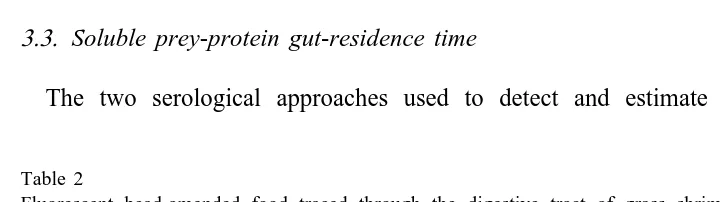
3.3. Soluble prey-protein gut-residence time
The two serological approaches used to detect and estimate A. atopus protein
Table 2
Fluorescent bead-amended food traced through the digestive tract of grass shrimp by microscopy after sacrifice. Elapsed time is in minutes after feeding on labeled food
Elapsed No. shrimp Number of shrimp with beads in: time (min) examined
Proventriculus Hepatopancreas Hindgut No beads found
15 2 2 0 2 0
30 3 2 0 2 1
45 3 2 0 2 1
75 3 1 0 2 1
135 3 1 0 1 2
residence time in shrimp gut contents differed in sensitivity. Both techniques revealed that protein residence times in the proventriculus were similar to or longer than bulk-passage times. AGID, while less sensitive than Western blot analysis, generally detected A. atopus proteins longer in starved grass shrimp compared to continuously-fed grass shrimp (Fig. 1). All groups of grass shrimp had detectable levels of A. atopus proteins in gut contents both immediately after a meal and 30 min following a meal. At
1 and 2 h postfeeding, 100% of the starved grass shrimp groups displayed detectable levels of proteins. This proportion declined to 75% after 1 h and 50% after 2 h in continuously-fed shrimp. No A. atopus protein was detected in continuously-fed shrimp 4 or 8 h postfeeding on A. atopus, but 50% of the groups of starved shrimp had detectable levels at both times. The more sensitive Western blot analysis detected soluble prey-proteins in all groups of starved and continuously-fed shrimp 8 h after feeding. The gut contents of nonfeeding control shrimp showed no evidence of A. atopus protein when tested by either AGID or Western blot assays with A. atopus antisera.
3.4. Meal size–protein content relationship
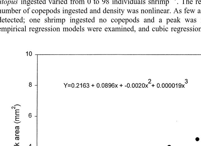
The gut contents of 33 individual grass shrimp from five feeding trials were analyzed by Western blot techniques subjected to chemiluminescence followed by densitometry to determine if the intensity of the antibody–antigen reaction can be related to meal size. Densitometric readings provided an absorbance peak proportional to the staining density of the blot. The area under each peak was measured and plotted against the number of
2
copepods consumed (Fig. 2). Area ranged from 0 to 8.5 mm , and the number of A.
21
atopus ingested varied from 0 to 98 individuals shrimp . The relationship between the number of copepods ingested and density was nonlinear. As few as seven copepods were detected; one shrimp ingested no copepods and a peak was not observed. Several empirical regression models were examined, and cubic regression gave the best fit. The
relationship was significant (P,0.0001); y50.2163(standard error, 1.0061)1 2
0.0896(standard error, 0.0876)x1 20.0020(standard error, 0.0020)x 1 3 2
0.000019(0.00001)x (R 50.68), where y5area, and x5number of copepods ingested during a feeding episode (Fig. 2).
4. Discussion
Our research suggests it is possible to use serological reagents and the powerful tools of biotechnology to estimate prey-protein residence time and meal size in grass shrimp. The sensitive Western blot analysis detected harpacticoid copepod proteins 8 h after ingestion even when shrimp were continuously fed another crustacean. The significant, nonlinear relationship between meal size and antibody–antigen reaction strength (see Fig. 2) strongly suggests meal size in field-collected grass shrimp can be estimated serologically. The number of copepods consumed in our laboratory experiments (0–98
21
individuals h ) was similar to ingestion rates obtained from field studies. Gregg and
21
Fleeger (1998) estimated grass shrimp consume an average of 35 copepods h from the epiphytes of S. alterniflora stems. Our feeding experiments utilized adult and late-stage copepodites of the harpacticoid A. atopus of similar length and weight (copepods used in feeding trials averaged about 0.8 mm in length, 5mg dry wt). Because grass shrimp in the field ingest naupliar, copepodite and adult copepods (Walters et al., 1996; Gregg and Fleeger, 1998), the mass, rather than the number of copepods ingested, of a copepod meal would best be estimated with serological techniques.
Full interpretation of field studies using serological techniques will require knowledge of the lower limit of prey-protein detection and the rate of prey-protein degradation in gut contents. Individual grass shrimp gut contents gave positive results for the presence of A. atopus with Western blot analysis when as few as seven adults, 35mg dry mass, were ingested over a 1-h feeding and digestion interval. Furthermore, our crude extract containing approximately 500 000 undegraded A. atopus (exoskeletons removed) had a
21
protein concentration of 2.5 mg ml (by colorimetric assay); a 1-ml sample of antigen extract was detectable by Western blot analysis with a 1 10,000 dilution of the antiserum, suggesting that it is possible to detect the presence of copepod proteins from a fraction of a whole copepod. Our results, however, also suggest that digestion of soluble proteins may reduce recognition by homologous antisera with increasing gut residence. Soluble protein was extracted from five freshly-killed undigested adult A.
atopus for Western blot analysis, and a strong reaction with chemiluminescent detection
(data not presented) was produced, whereas the comparative reaction with seven copepods from a gut content sample was noticeably weaker. Further experiments are required to determine the rate of change in detection associated with prey-protein degradation, but certainly the mass of a copepod meal consumed for at least 1 h after a meal should be able to be estimated with the techniques developed here.
ingestion. Beads remained in the proventriculus for approximately 20 min before first entering the hindgut; no beads entered the hepatopancreas. Bead residence time in the proventriculus varied greatly among individual shrimp, but beads resided in the proventriculus longer than in any other portion of the gut. The two extremes in this study showed one shrimp completely eliminated (defecated) beads within 30 min of a meal, while two shrimp had beads present in the proventriculus and the hindgut 4 h postingestion (Table 2). These results are generally similar to those of Dall and Moriarty (1983), McLaughlin (1983) and Brunet et al. (1994) who showed that nutrient-rich gut fluid in decapods is circulated in grooves or channels in the foregut (proventriculus) and passes through a ‘‘filter-press’’ to enter the hepatopancreas where most digestion and absorption occurs, and that particles .1 mm do not enter the hepatopancreas and are quickly transported to the hindgut. Overall, bulk-gut clearance times in P. pugio ranged from 0.5 to 4 h, and bulk passage times for starved and continuously-fed shrimp overlapped for the relatively few shrimp we examined; we could not conclusively show that continuous feeding decreased gut passage time, as would be predicted from the ‘‘plug-flow’’ model of many omnivore / carnivore digestive systems (Penry and Jumars, 1986).
Prey-proteins in the proventriculus were detected by serological methods for a longer time and with less variability than might be predicted from the observed bulk passage rates. Detection was also dependent on the technique used. Gel immunodiffusion, the less sensitive technique, detected copepod prey 2 h after feeding in 50% of the shrimp tested. Prey-proteins were detected in starved shrimp after 8 h in 50% of the shrimp examined. Detection in continuously feeding shrimp with immunodiffusion was reduced at all time intervals examined after 1 h (Fig. 1). Western blot analysis, which has greater sensitivity than gel immunodiffusion, detected prey-proteins for the entire 8-h study in pooled samples of continuously-fed and starved shrimp. A. atopus protein bands detected by Western blot became lower in molecular weight and decreased in staining intensity at the longer digestion intervals, but were still present, detectable and quantifiable. Nonfeeding shrimp (controls) showed no discernible A. atopus bands in either AGID or Western blot format. Grass shrimp appear to feed continuously in the field (Morgan, 1980; Fleeger et al., 1999), suggesting that gut contents of field-collected shrimp will contain a long record of information regarding recent meals irrespective of collection time. If the relatively long protein residence time encountered in grass shrimp is typical of other predators, these techniques would be especially useful in animals that feed intermittently by providing a long window of opportunity for prey detection.
antiserum had five precipitin lines visible after staining. The low number of shared antigens is probably related to two factors; (1) the antiserum was made from a single species of harpacticoid from mass culture that provided the large quantities of antigen necessary for antibody preparation, and (2) the immunization schedule and associated techniques may have favored specificity. Furthermore, Western blot assays detected no cross reaction among denatured proteins, and false positives using these techniques appear to be highly unlikely.
The antiserum was derived from a single species of harpacticoid copepod that is unknown from the field; A. atopus was first identified in mass culture. A harpacticoid species-specific antibody is needed to adapt these techniques to detect the presence of harpacticoid prey or quantify the mass of harpacticoids consumed in field-collected predators. Generation of a harpacticoid-specific antibody certainly seems technically feasible. The antibody prepared from purged A. atopus provided sensitive homologous detection with Western blot analysis but failed to detect a second harpacticoid (an unidentified species in the family Laophontidae) in the same denatured protein format. The agar gel immunodiffusion provided faint bands of identity between the antisera and the species from a different family. Possible approaches include (1) finding shared antigens among harpacticoid species, perhaps by using nondenatured proteins in the AGID format, and generating an antiserum to this protein, and (2) using a mass collection of harpacticoids with many species to generate an antibody as has been done by Feller (1991) but with enhanced immunogen procedures. A major concern will be to identify proteins specific to harpacticoids but not shared with other crustacean taxa.
Our results suggest that serological preparations may be enhanced with biotechnology to improve effectiveness in predatory–prey studies. The ability to measure minute quantities of prey-proteins (from as few as seven adult copepods) for as long as 8 h after a meal suggests that serology can provide very detailed information on recent meals of field-collected predators. Furthermore, tests might be designed with different goals in mind. To easily process a large number of predator gut contents, a AGID format spot-plate test could be designed that will detect prey-proteins for up to 4 h after a meal. Greater sensitivity and more information (e.g., protein content) can be achieved with the labor intensive Western blot techniques. The curvilinear relationship determined here between the intensity of the antibody–antigen reaction and meal size may be refined to better predict the mass ingested by grass shrimp. If a harpacticoid-specific antibody can be developed, trials might be conducted that combine traditional enumeration techniques recording decreases in copepod numbers in the presence of a predator (see Gregg and Fleeger, 1998) with the techniques described above to validate estimates of mass consumed. If so, the possibility that serology can be used to estimate ingestion of specific prey by field-collected predators is greatly enhanced. Assuming prey have a similar size and / or protein content, the abundance of ‘‘standard’’ prey could also be estimated in a predator’s gut contents.
Acknowledgements
cultures and for conducting predator–prey experiments. Comments by M. Pace improved an earlier version of this manuscript. This is contribution number 1211 of the Belle W. Baruch Institute for Marine Biology and Coastal Research. [RW]
References
Bergdoll, M.S., Bennett, R.W., 1984. In: Staphylococcal Enterotoxins, Vol. 2, American Public Health Association, Washington DC, pp. 428–457.
Brunet, M., Arnaud, J., Mazza, J., 1994. Gut structure and digestive cellular processes in marine Crustacea. Oceanogr. Mar. Biol. Annu. Rev. 32, 335–367.
Dall, W., Moriarty, D.J.W., 1983. Functional aspects of nutrition and digestion. In: Bliss, D.E., Mantel, L.H. (Eds.), The Biology of Crustacea: Internal Anatomy and Physiological Regulation, Vol. 5, Academic Press, New York, pp. 215–251.
Feller, R.J., 1984. Serological tracers in meiofaunal food webs. Hydrobiologia 118, 119–125.
Feller, R.J., 1986. Immunological detection of Mercenaria mercenaria in a predator and preparation of size-class specific antibodies. Veliger 28, 341–347.
Feller, R.J., 1991. Dietary analysis of penaeid shrimp: The immunoassay approach. Dev. Aquacult. Fish. Sci. 22, 141–156.
Feller, R.J., 1992. Potential applications of immunoassays in studies of flatfish recruitment. Neth. J. Sea Res. 29, 1–5.
Feller, R.J., Gallaher, E.D., 1982. Antigenic similarities among estuarine soft-bottom benthic taxa. Oceologia 53, 305–310.
Feller, R.J., Zagursky, G., Day, E.A., 1985. Deep-sea food web analysis using cross-reacting antisera. Deep Sea Res. 32, 485–497.
Feller, R.J., Taghon, G.L., Gallagher, E.D., Kenny, G.E., Jumars, P.A., 1979. Immunologic methods for food web analysis in a soft-bottom benthic community. Mar. Biol. 54, 61–74.
Fleeger, J.W., Carman, K.R., Webb, S., Hilbun, N., Pace, M.C., 1999. Consumption of microalgae by the grass shrimp, Palaemonetes pugio. J. Crustacean Biol. 19, 324–336.
Goarant, E., Prensire, G., Lair, D.N., 1994. Specific immunological probes for the identification and tracing of prey in crustacean gut contents. The example of cyanobacteria. Arch. Hydrobiol. 131, 243–252. Gregg, C.S., Fleeger, J.W., 1998. Grass shrimp, Palaemonetes pugio Holthius, predation on sediment- and
stem-dwelling meiofauna: Field and laboratory experiments. Mar. Ecol. Prog. Ser. 175, 77–86.
Hunter, J., Feller, R.J., 1987. Immunological dietary analysis of two penaeid shrimp species from a South Carolina tide creek. J. Exp. Mar. Biol. Ecol. 107, 61–70.
Laemmli, U.K., 1970. Cleavage of structural proteins during the assembly of the head of bacteriophage T4. Nature 227, 680–685.
Lotufo, G.R., Fleeger, J.W., 1995. Description of Amphiascoides atopus, a new species (Copepoda: Harpacticoida) from a mass culture system. Proc. Biol. Soc. (Washington) 108, 117–124.
McLandsborough, L., Tatini, S.R., 1991. A 6 h microslide immunodiffusion assay for confirmed detection of
Staphylococcal enterotoxins. Lett. Appl. Microbiol. 12, 81–84.
McLaughlin, P.A., 1983. Internal anatomy. In: Mantel, L.H. (Ed.), The Biology of Crustacea: Internal Anatomy and Physiological Regulation, Vol. 5, Academic Press, New York, pp. 1–41.
Morgan, M.D., 1980. Grazing and predation of the grass shrimp Palaemonetes pugio. Limnol. Oceanogr. 25, 896–902.
Nieland, D.L., Wilson, C.A., Fleeger, J.W., Sun, B., Malone, R.F., Chen, S., 1998. Preliminary evaluation of the use of phosphogypsum for reef substrate. I. A laboratory study of bioaccumulation of radium and six heavy metals in an aquatic food chain. Chem. Ecol. 14, 305–319.
Pape-Lindstrom, P.A., Feller, R.J., Stancyk, S.E., Woodin, S.A., 1997. Sublethal predation: field measurements of arm tissue loss from the ophiuroid Microphiopholis gracillima and immunochemical identification of its predators in North Inlet, South Carolina, USA. Mar. Ecol. Prog. Ser. 156, 131–140.
Pickavance, J.R., 1970. A new approach to the immunological analysis of invertebrate diets. J. Anim. Ecol. 39, 715–729.
Scholz, D.S., Matthews, L.L., Feller, R.J., 1991. Detecting selective digestion of meiobenthic prey in juvenile spot Leiostomus xanthurus (Pisces) using immunoassays. Mar. Ecol. Prog. Ser. 72, 59–67.
Stills, H.F.J., 1994. Polyclonal antibody production. In: The Biology of the Laboratory Rabbit, 2nd Edition, Academic Press, pp. 435–448.
Sun, B., Fleeger, J.W., 1995. Sustained mass culture of Amphiascoides atopus a marine harpacticoid copepod in a recirculating system. Aquaculture 136, 313–321.
Walters, K., Jones, E., Etherinton, L., 1996. Predation on metazoans inhabiting Spartina alterniflora stems. J. Exp. Mar. Biol. Ecol. 195, 251–265.