36 Internaional Journal of Integrated Health Sciences. 2014;2(1):36–9
Original Aricle
Morphometric Analysis of the Corpus, Spinal Canal and Torg Raio Using
Midsagital Cervical Vertebrae Computed Tomography Scan: Indonesian
Populaion
Correspondence:
Farid Yudoyono, Spine Research Laboratory, Department of Neurosurgery, Faculty of Medicine, Universitas Padjadjaran-Dr. Hasan Sadikin General Hospital Jl. Pasteur No. 38, Bandung, Indonesia e-mail: [email protected]
Rully Hanai Dahlan,1 Farid Yudoyono,1 Priandana Adya Eka Saputra,2 Sevline Estheia Ompusunggu,1
Muhammad Zafrullah Ariin,2 Agung Budi Suiono,2 Ahmad Faried2
1Spine Research Laboratory, Department of Neurosurgery, Faculty of Medicine, Universitas
Dr. Hasan Sadikin General Hospital
2Department of Neurosurgery, Faculty of Medicine, Universitas Padjajaran-Dr. Hasan Sadikin General
Hospital
Abstract Objecive:To determine the normal ranges of cervical spinal canal morphometry in Indonesian populaion and to compare the acquired data collected from other populaions
Methods: Computed tomography measurements on the diameter of midsagital spinal canal and corpus of cervical vertebrae and its Torg raio from the lower cervical (C3–C7) canal from 24 normal Indonesian adults were performed at the Radiology Department of Dr. Hasan Sadikin General Hospital. Paients who had cervical spine disorders and those under 20 years old were exluded. We used computed tomography scan midsagital view to measure the aforemenioned parameters.
Results: The average diameter for the cervical spinal canals for the Indonesian populaion is comparable with those of other Asian populaions such as Hongkong and India, albeit with smaller Torg raio.
Conclusions: This study reports the normal radiological anatomy of the midsagital spinal canal and corpus of cervical vertebrae as well as Torg raio from the lower cervical vertebrae among Indonesian populaion. The measurements result of this study shows that, although slightly smaller, the measurement results for those parameters are idenical with other Asian populaions.
Keywords: Corpus cervical vertebrae, midsagital cervical spinal canal, Torg raio IJIHS. 2014;2(1):36–9
Introducion
One of the predisposing factors for neck problems is cervical spinal canal stenosis, a condiion in which the diameter of the cervical spinal canal is less than the normal measurement for the relevant age or sex of the individuals.1–5 So far, there are several radiological and morphological anatomic studies on the size of spinal canal in
diferent populaions in the world.1–6
Plain lateral x-ray is usually used to determine the canal diameter. However, there are many
limitaions found in terms of value intepretaion
when using this method.1,3,7,8 We used computed
tomography (CT) scan imaging to measure the parameters being studied, i.e. midsagital spinal canal and corpus of cervical vertebrae diameters and the Torg raio from lower cervical (C3–C7) canal. The method of analyis used was Torg and Pavlov canal-to-corpus raio, in which
the magniicaion factor could be omited.2,3,9
Cervical CT scan was used because it gives beter image of the bone; thus, allows us to gain more accurate measurements compared to manual
measurement.9,10
To our knowledge, unil recently, there have been no report or study menioning the cervical spinal canal morphometry for Indonesian populaion. Therefore, this study aimed to
Internaional Journal of Integrated Health Sciences. 2014;2(1):36–9 37 determine the normal ranges of cervical spinal
canal morphometry in Indonesian populaion and to compare the acquired data from other populaions.
Methods
Computed tomography measurements of the midsagital spinal canal and corpus of cervical vertebrae diameters and the Torg raio from lower cervical (C3–C7) canal were performed on 24 normal Indonesian adults at the Department of Radiology, Dr. Hasan Sadikin General Hospital. Paients with cervical spine disorders and those under 20 years old were excluded. We used CT scan of midsagital cervical to measure aforemenioned parameters.
Results
The means of the cervical spinal canal diameter are presented below (Table 1). In terms of Torg
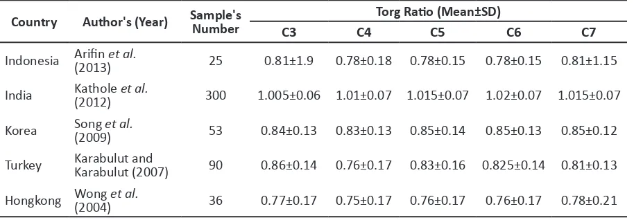
raio criteria, the Hongkong populaion has the smallest value in all spinal level and India has the biggest value in all spinal level. Although slightly smaller, the measurement results for those parameters in this study are idenical with those from other Asian populaion (Table 2).
Discussion
The cervical spines consist of eight vertebras. The third unil eight cervical vertebras are “common” cervical vertebra, while C1 (atlas) and C2 (axis) are the atypical ones. The cervical spinal canal is an opening within cervical vertebras in which the cervical spinal cord runs. Its boundaries are vertebral bodies, bilateral pedicles and laminae, and spinous processes. The canal follows the normal contour of spine. The shape of the cervical spinal canal is triangular, which is the same as the lumbar region. The diameter of the cervical spinal canal is fairly large in the upper cervical region but narrows down from C3 and inferiorly, where the diameter is approximately 12–13 mm in the
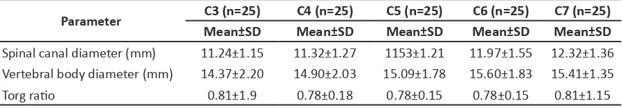
Table 1 Cervical Vertebrae Spinal Canal Diameters, Vertebral Body Diameters and Torg Raio in Indonesian populaion
Parameter C3 (n=25) C4 (n=25) C5 (n=25) C6 (n=25) C7 (n=25)
Mean±SD Mean±SD Mean±SD Mean±SD Mean±SD
Spinal canal diameter (mm) 11.24±1.15 11.32±1.27 1153±1.21 11.97±1.55 12.32±1.36 Vertebral body diameter (mm) 14.37±2.20 14.90±2.03 15.09±1.78 15.60±1.83 15.41±1.35
Torg raio 0.81±1.9 0.78±0.18 0.78±0.15 0.78±0.15 0.81±1.15
Spinal Canal Computed Tomography Scan (Fig. 1a) Measurement of Midsagital Cervical Corpus (Fig. 1b)
Fig. 1
a b
38 Internaional Journal of Integrated Health Sciences. 2014;2(1):36–9 anteroposterior plane. The space in the spinal
canal allows for the free movement of the canal contents without tension or pressure during these movements. Therefore, the normal size of the canal is important. An abnormal reducion in the size of the spinal canal could predispose the individual to neck pain. Due to the narrowing, the lower cervical cord is paricularly vulnerable to a variety of pathological eniies, one of which is canal stenosis, that could compromise the cord within the vertebral canal. The stenosis of the cervical spinal canal is an anatomical anomaly which is more common than realized. Individuals with this anomaly tend to remain neurologically asymptomaic unil a complicaing feature, such as osteophytes or herniated intervertebral discs, develops.
Several studies have been performed to prove that measurement of the sagital diameter of the cervical canal in plain lateral radiography is a useful method for detecing cervical spinal stenosis.2,4,7,11 However, comparison of published data reveals that the range of variaion in the
reported sagital diameter is considerable.1,3–6,10
Those diversity is partly due to the variaion in the radiographic technique (focus-to-ilm distance) and variaion in the body build of the subjects (afecing the object-to-ilm distance). In an atempt to ind a soluion to these discrepancies, Torg et al.12devised a measurement raio that compares the sagital diameter of the spinal canal with the anteroposterior width of the vertebral body, in which both of those are equally
afected by radiological magniicaion factors.2,6,9
Table 2 Torg Raio on Lateral Radiographs of Cervical Spine
Country Author's (Year) Sample's Number Torg Raio (Mean±SD)
C3 C4 C5 C6 C7
Indonesia Ariin (2013)et al. 25 0.81±1.9 0.78±0.18 0.78±0.15 0.78±0.15 0.81±1.15
India Kathole (2012) et al. 300 1.005±0.06 1.01±0.07 1.015±0.07 1.02±0.07 1.015±0.07
Korea Song (2009)et al. 53 0.84±0.13 0.83±0.13 0.85±0.14 0.85±0.13 0.85±0.12
Turkey Karabulut and Karabulut (2007) 90 0.86±0.14 0.76±0.17 0.83±0.16 0.825±0.14 0.81±0.13
Hongkong Wong (2004)et al. 36 0.77±0.17 0.75±0.17 0.76±0.17 0.76±0.17 0.78±0.21
a
b
Ilustraion of Torg Raio = b/a, (a) Anterior Posterior Corpus Length, (b) Spinal Canal Length
Fig. 2
Internaional Journal of Integrated Health Sciences. 2014;2(1) 39
References
1. Maqbool A, Athar Z, Hussain L. Midsagital diameter of cervical spinal canal and Torg’s raio of the cervical spine in Pakistanis. Pakistan J Med Sci. 2003;19(3):203–10.
2. Wong TM, Leung HB, Wong WC. Correlaion between magneic resonance imaging and radiographic measurement of cervical spine in cervical myelopathy paients. J Orthop Surg. 2004;12 (2):239–42.
3. Song KJ, Choi BW, Kim SJ, Kim GH, Kim YS, Song JH. The relaionship between spinal stenosis and neurological outcome in traumaic cervical spine injury: an analysis using Pavlov raio, spinal cord area and spinal canal area. Clin Orthop Surg. 2009;1(1):11–8.
4. Tossel G. Dimension of the cervical spinal canal in the South African Negroid populaion. [dissertaion]. South Africa: School of Medicine, Faculty of Health Science, University of Pretoria; 2005.
5. Tjahjadi D, Onibala MZ. Torg raios based on cervical lateral plain ilms innormal subjects. Universa Medicina. 2010;29(1):8–13.
6. Kathule MA, Joshi RA, Herekar NG, Jadhav SS. Dimensions of cervical spinal canal and their relevance in clinical pracice. Int J Recent Trends
Sci Tech. 2012;3(2):54–8.
7. Karabulut Ő, Karabulut Z. The variaions of Torg Raio with gender in paients with neck pain. Dicle Med J. 2007;34(4):272–4.
8. Choudhary S, Kaimuthu P, Kataria SK. A radiographic study of sagital diameter of cervical spinal canal in adult male populaion of Rajasthan. Asian J Med Res. 2013;2(1):7–9. 9. Gour KK, Shrivastava SK, Thakare AE. Size of
cervical vertebral canal-measurements in lateral cervical radiographs & dried bones. Int J Biol Med Res. 2011;2(3):778–80.
10. Suk KS, Kim KT, Lee JH, Lee SH, Kim JS, Kim JY. Reevaluaion of the Pavlov raio in paients with cervical myelopathy. Clin Orthop Surg. 2009;1(1):6–10.
11. Kang SK, Park JY, Chin DK, Kim KH, Kuh SU, Kim
KS, et al. A PET/CT-based study of spinal canal in
Korean young adults: anteroposterior diameter from cervical vertebra to sacrum. Korean J Spine. 2012;9(3):165–9.
12. Torg JS, Corcoran TA, Thibault LE, Pavlov H, Sennet BJ, Naranja RJ Jr, et al. Cervical cord neurapraxia: classiicaion, pathomechanics, morbidity, and management guidelines. J Neurosurg. 1997;87(6):843–50.
:36–9 This raio is obtained by dividing the midsagital diameter of the cervical spinal canal at any paricular cervical segment by the midsagital diameter of the corresponding vertebral body. It has been reported that, using the raio method as standard, a measurement of less than 0.80 indicates signiicant spinal stenosis.
Usefulness of Torg’s raio in the diagnosis of cervical spinal canal stenosis has also been conirmed by several other studies.1,7,10 Studies have also been performed in order to determine the mean anteroposterior diameter of the cervical spinal canal at diferent vertebral levels in normal subjects making it possible, clinicians
to diagnose cervical spinal stenosis by consuling
these reference values.1–4,7 Comparing those
studies with ours, it is revealed that our measurement data is similar with those of other studies.
In conclusion, this study reports the normal radiological anatomy of the midsagital spinal canal and corpus of cervical vertebrae and the Torg raio from the lower cervical vertebrae among adult Indonesian. The measurements results of this study show that, although the Torg raio is slightly smaller, they are similar and comparable with those from other Asian populaions.