Available online at http://medpet.journal.ipb.ac.id/
*Corresponding author: E-mail: irusmana@ipb.ac.id
Characteristics of Lactic Acid Bacteria Isolated from Gastrointestinal Tract of
Cemani Chicken and Their Potential Use as Probiotics
S. N. Jannaha, A. Dinotob, K. G. Wiryawanc, & I. Rusmanaa,*
aDepartment of Biology, Faculty of Mathematics and Natural Science, Bogor Agricultural University bResearch Center for Biology, Indonesian Institute of Sciences (LIPI)
Jln. Raya Jakarta- Bogor Km. 46, Cibinong, 16911, West Java, Indonesia
cDepartment of Nutrition and Feed Tecnology, Faculty of Animal Science, Bogor Agricultural University. #Jln. Agatis, Kampus IPB Darmaga, Bogor, 16680, West Java, Indonesia
(Received 09-05-2014; Reviewed 03-06-2014; Accepted 23-10-2014)
ABSTRACT
The aims of this study were to screen and characterize lactic acid bacteria (LAB) isolated from gastrointestinal (GI) tract of Cemani chicken, one of Indonesian local chicken and to investigate their potential use as probiotics. LAB were isolated from GI tract using MRSA and GYPA media and incubated anaerobically. Selected LAB were determined their probiotic properties with several assays. Identification of selected LAB was based on 16S rDNA sequences, morphological and bio -chemical characteristics. Ninety five bacteria were isolated and characterized as lactic acid bacteria (Gram positive, catalase negative, non sporeforming and acid producing). Twenty four isolates of LAB demonstrated antimicrobial activity against Escherichia coli JCM 1649 and Salmonella enteritidis B2586, and three selected isolates, i.e. CCM011, CSP004, and CVM002 showed the highest inhibition activity. The isolates had characters of high cell surface hydrophobicity and inter-isolate coaggrega-tion ability of LAB, high survival at low pH, high phytase and protease activity (but no amylase and lipase activity), weak coaggregation with pathogen and no resistance to the examined antibiotics. The isolates were identified based on sequence analysis of 16S rRNA gene as Lactobacillus salivarius, however, each isolate had different profiles of sugar fermentation. Therefore the three LAB isolates had potential application as probiotics for chicken.
Key words: Cemani chicken, gastrointestinal tract, lactic acid bacteria, probiotic
ABSTRAK
Tujuan penelitian ini adalah untuk menyeleksi dan mengarakterisasi bakteri asam laktat (BAL) yang diisolasi dari saluran pencernaan ayam Cemani, salah satu ayam asli Indonesia dan untuk me-ngetahui potensi penggunaannya sebagai probiotik. BAL diisolasi dari saluran pencernaan dengan menggunakan medium MRSA dan GYPA dan diinkubasi secara anaerobik. BAL terpilih kemudian ditentukan karakter probiotiknya melalui serangkaian percobaan. Identifikasi isolat BAL terpilih berdasarkan pada sekuen 16S rDNA, karakter morfologi dan biokimianya. Sembilan puluh lima isolat diisolasi dan dikarakterisasi sebagai bakteri asam laktat (Gram positif, katalase negatif, tidak membentuk endospora dan menghasilkan asam). Dua puluh empat isolat BAL memperlihatkan ak-tivitas antimikrob terhadap E. coli JCM 1649 dan S. enteritidis B2586, dan tiga isolat terpilih, yaitu CCM011, CSP004, dan CVM002 menunjukkan aktivitas penghambatan yang tertinggi. Isolat-isolat tersebut mempunyai karakter pelekatan terhadap permukaan sel yang tinggi, mempunyai kemam-puan koagregasi antar BAL, daya tahan yang tinggi pada pH rendah, menunjukkan aktivitas enzim fitase dan protease yang tinggi (tetapi tidak mempunyai aktivitas amilase dan lipase), koagregasi yang lemah terhadap bakteri patogen dan tidak resisten terhadap antibiotik uji. Isolat-isolat tersebut diidentifikasi berdasarkan analisis sekuen gen 16S rRNA sebagai Lactobacillus salivarius, tetapi tiap isolat mempunyai profil fermentasi terhadap gula yang berbeda. Ketiga isolat BAL tersebut berpo -tensi sebagai probiotik pada ayam.
December 2014 183 INTRODUCTION
Increasing consumption of chicken and their products contributed to an increasing use of antibiotics in a poultry farm. Antibiotics that are used to improve
chicken growth performance and to protect chicken from pathogenic microorganisms are known as
anti-biotic growth promoters (AGPs) (Gaggia et al., 2010).
However, application of AGPs in poultry can cause
development of bacterial resistance to antibiotics and it
can affect human health, due to the residues in chicken products. In European countries, application of AGPs in poultry feed is prohibited.
There are several potential alternative ways
in-stead of using AGPs, one of them is using probiotics. Probiotics are live microorganisms which, when admin
-istered in adequate amount, confer a health benefit to the host (FAO/WHO, 2002). The aims of using probiotics
in chickens are to prevent and combat gastrointestinal
disorders based on competitive exclusion of potentially pathogenic bacteria, such as Salmonella enteritidis and Escherichia coli, to stimulate host immune response, and
to secrete antimicrobial substances (Corcionivoschi et al.,
2010).
Many studies reported that LAB could be used as probiotics for chicken (Torshizi et al., 2008; Sofyan et al.,
2012). LAB probiotics showed beneficial effect by inhib
-iting growth of pathogen bacteria, such as Escherichia coli (Istiqomah et al., 2013) and Salmonella sp (Nouri et al.,
2010). LAB have been used for fermentation of certain
foods, so they are non-pathogenic bacteria and
recog-nize as GRAS (Generally Regarded as Safe) status. These bacteria are also found normally in the gastrointestinal
tract of a chicken.
Probiotics using indigenous LAB isolated from
gastrointestinal tract of Cemani chicken are important as potential probiotics for Indonesian local chicken.
Indonesia has thirty one local chickens with various genotypic and phenotypic characteristics (Nataamijaya, 2010). Cemani chicken is one of the Indonesian local chicken that has unique characteristics with black color on the whole body including nails, tongue, comb, beak, feet, eye-balls, legs, feathers, skin, muscles, bones, and internal organs. This chicken is recognized to have high
disease resistance, and high adaptability to
environ-mental conditions (Sulandari et al., 2009; Nataamijaya,
2010). The chicken was sometimes utilized as traditional medicine (Sartika et al., 2011). Therefore, this study was
conducted to screen and characterize LAB isolates from gastrointestinal (GI) tract of Cemani chicken for poten -tial application as probiotics in chicken.
MATERIALS AND METHODS
Bacterial Cultures and Growth Condition
Two strains of intestinal pathogenic bacteria as
indicators of bacterial strains (Escherichia coli JCM 1649 and Salmonella enteritidis B2586) were used in this ex
-periment. These indicator bacterial cultures were grown
in brain heart infusion broth (Becton Dickinson) at 37 °C for 24 h. All strains were subcultured twice prior to
experiments.
Isolation of LAB
Three healthy Cemani chickens (9 to 12 months old;
1.0 to 1.5 kg body weight) were obtained from Mranggen
district, Central Java, Indonesia. Those chickens received no antibiotic-feed containing rice bran and leftover rice
before experiments. The chickens lumen contents includ
-ing crop, ventriculus, ileum, and cecum were collected aseptically. Lumen samples were serially diluted in 0.85% (w/v) sterile NaCl solution and plated onto MRS (HiMedia Laboratories, India) agar and glucose yeast ex
-tract peptone (GYP) agar supplemented with 0.5% (w/v)
CaCO3 to distinguish acid producing bacteria. The plates
were incubated in anaerobic jars (Merck, Germany) for 48 h at 37 °C with Anaerocult A (Merck, Germany). After incubation, the bacterial colonies were enumerated and purified on MRS agar up to three times to obtain pure LAB isolates. All isolates were examined for colony
morphology and cell shape and catalase assay. For long-term storage, the bacterial isolates were kept at -80 °C in
20% (v/v) glycerol until further use (Guerin-Danan et al.,
1999).
Antimicrobial Activity Assay
For detection of antimicrobial activity, the well
diffusion assay described by Taheri et al. (2009) was
performed. The cultures were grown anaerobically overnight in MRS broth at 37 °C to achieve cell concen -tration of 108 CFU/mL. Bacterial culture, cell-free super
-natant, and neutralized cell-free supernatant (pH 7.0, added with 2M NaOH) of different LAB isolates were
determined for antimicrobial activity against indicator
bacterial strains. Inhibitory zones around the wells were screened for each strain after overnight incubation at 37 °C. The experiment was carried out three times and data were displayed as the mean of radius of inhibitory zone.
pH and Bile Salts Tolerance Assay
Overnight selected LAB cultures were centrifuged
at 7,500 × g for 5 min and washed twice with sterile
phosphate buffer (PBS, pH 7.0). The washed cell density were adjusted to OD600= 0.5-0.7 using spectrophotometer. For pH tolerance assay, 1 mL of cell suspensions were resuspended with 5 mL PBS at pH 2.0, 4.0, and 6.5, and incubated at 37 °C for 90 min. Meanwhile, for bile salts tolerance assay, 1 mL of the washed cell suspension was resuspended in the sterile PBS containing 0.05%, 0.08%, 0.1%, and 0.3% bile salt (Sigma) and incubated at 37 °C for 5 h. The bacterial survival under different
pH conditions and concentrations of bile salts were
determined by plated 0.1 mL suspension onto MRS agar and incubated in anaerobic jars for 48 h at 37 °C (Taheri
Cell Surface Hydrophobicity Test
Assay for microbial surface hydrophobicity was
performed based on the adherence to the non polar
sol-vent (Taheri et al., 2009). Cell suspensions were prepared
as above and then 3 mL of washed cells suspensions were added with 1 mL of toluene and mixed by stirring on a vortex for 2 min. The optical density of mixture was measured at 600 nm using spectrophotometer. Hydrophobicity was calculated as follows:
Hydrophobicity (%)= [(OD600 before mixing − OD600 after mixing)/ OD600 before mixing] × 100
Co-Aggregation Test
Equal volume (2 mL) of suspension of each indica
-tor bacteria and the LAB isolates were placed together in a test tube and mixed by vortexing. The OD600 of the
bacterial mixture was measured after incubation at 37 °C for 5 h (Taheri et al., 2009). The percentage of co-aggrega
-tion was calculated by using the equa-tion as follows: Co-aggregation (%) = {[(Ax + Ay)/2− A(x+y)]/[(Ax+ Ay)/2]}
× 100
which representing absorbance value (A) at each of the two strains examined in the control tube (x and y), and at their mixture (x + y).
Enzymatic Activities Assay
The selected LAB isolates were assayed for the presence of dietary enzymes, i.e. amylase, protease, lipase, and phytase (Taheri et al., 2009). To detect the amylase, lipase, and phytase activities, the isolates were
subcultured in MRS broth and then spot-inoculated onto relevant agar-based media (starch agar, skim milk agar, phytic acid enriched agar and lipid hydrolysis agar). After anaerobic incubation for 48 h at 37 °C, a clear zone surrounding each colony was measured. Lugol’s solution was added over the plate surface for clear zone
detection of amylase activity.
Antibiotic Sensitivity Test
Antibiotic susceptibility of selected LAB isolates was determined by using Kirby-Bauer disc method (Taheri et al., 2009). As much as 0.1 mL of LAB cell
suspensions were spread over the entire surface of the plates containing MRS agar. Subsequently, paper discs
containing antibiotics of amoxicillin 10 µg, ampicillin 10 µg, chloramphenicol 30 µg, cefadroxil 30 µg, doxy-cycline 30 µg, erythromycin 15 µg, lincomycin 15 µg, rifampin 5 µg, spiramycin 30 µg, and tetracycline 30 µg
were placed on the plates and incubated anaerobically at 37 °C for 24 h. Diameter of a clear zone was measured to
determine antibiotic sensitivity of the isolates.
Identification of LAB Isolate
Genomic DNAs from the selected LAB isolates were extracted by using Xprep Stool DNA Mini Kit (PhileKorea Technology, INC, Korea) according to the
manufacturer’s instructions. DNA pellet was then resus
-pended in 50 µL TE buffer and stored at -20 °C. A PCR mixture was prepared from each sample using a TaKaRa PCR Thermal Cycler with forward primer 27F (5’-AGAGTTTGATCCTGGCTCAG-3’) and reverse primer 1492R (5’-GTTACGACTTCACCCTCCT-3’) (Zhang, et al.,
2007). PCR reaction mixture consisted of 25 µL GoTaq GreenMaster Mix (Promega, USA), 2 µL of each primer (10 pmol), and destilated water in a final volume of 50 µL and 100 ng DNA template in the final concentration. PCR condition was set up with an initial denaturation at
94 oC for 1 min 30 s, followed by 25 cycles of denatur
-ation temperature at 95 oC for 30 s, annealing
tempera-ture of 50 oC for 30 s, and extension temperature at 72 oC for 1 min 30 s and then final elongation at a tempera
-ture of 72 oC for 5 min. PCR products were confirmed
by electrophoresis by using 1% agarose gel in 1x TAE buffer and visualized with ethidium bromide staining. Purification of PCR products and sequencing were con
-ducted by a company providing sequences services. The DNA sequences were compared with available sequenc
-es in GenBank using the BLASTN tools through the National Center for Biotechnology Information (NCBI). Sequence homology of more than 97% was regarded as belonging to the same species (Tannock, 1999). The sequence was aligned with the clustal X program, then the alignment was manually edited to the construction
of phylogenetic tree. The phylogenetic tree was
con-structed by neigbour-joining method in MEGA program version 4. The values of branches of phylogenetic tree were determined using booststrap analysis based on 1000 resamplings (Felsenstein, 1985).
Biochemical Characterization of LAB
Biochemical characterization of the selected LAB was determined by using API® 50 CHL for performance
of carbohydrate metabolism tests (bioMérieux, Inc, Durham) according to the manufacturer’s instructions. Fermentation profiles of the isolates were determined by using the API web software (Pelinescu et al., 2009).
Statistical Analysis
All quantitative data were subjected to ANOVA analysis by using IBM SPSS statistics 21.0. A test of least significant differences was used to separate means; dif -ferences between means were considered statistically
significant P<0.05.
RESULTS AND DISCUSSION
Population of Culturable LAB in Chicken Gastrointestinal Tracts
By culturing method, the content of gastrointes
-tinal tract of chicken had LAB number 7.12±0.63 up to 9.07±0.17 log CFU g-1 and the highest number was found
December 2014 185 as diet, age, presence of barrier in the intestine and
dif-ferent physiological functions within the organ system (Dumonceaux et al., 2006).
Ninety-five LAB colonies were randomly picked based on clear zone and colony morphology on MRSA or GYPA medium supplemented with CaCO3. All
iso-lates had specific characteristics of lactic acid bacteria, such as positive Gram stain, catalase negative reaction
and no endospore forming, and mostly bacterial cell
had rod-shaped (88 isolates, 92.6%) and 7 isolates (7.4%) had round shaped.
Assessment of Probiotic Characteristics
For antimicrobial activity assays, Escherichia coli and Salmonella enteritidis were used, because they were common potentially intestinal pathogenic bacteria
causing problem of gastro intestinal tract in chicken. Antimicrobial activity of 24 LAB isolates against E. coli and S. enteritidis was shown in Table 2. All bacterial
cultures had antimicrobial activity against E. coli and S. enteritidis, however there were only 10 LAB isolates
(41.7%) and 7 LAB isolates (29.2%) that their cell-free neutralized supernatant exhibited antibacterial activity
against E. coli and S. enteritidis, respectively.
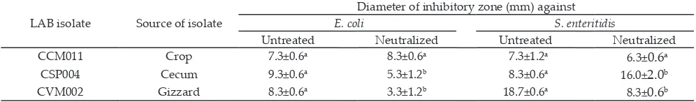
Three LAB isolates, i.e. CSP004, CCM011 and CVM002 that had the highest inhibition zone of their
cell-free neutralized supernatant were selected for fur
-ther assays. The neutralized supernatant of these isolates
were able to inhibit the growth of the Gram negative
pathogen tested in this study. The antimicrobial activity of neutralized cell-free supernatants of CSP004 isolate
to S. enteritidis was higher than that of non neutralized
supernatant. Antimicrobial activity of neutralized super
-natant of CCM011 isolate was not significantly different from that of non neutralized supernatant (Table 3). According to Nouri et al. (2010) and Heravi et al. (2011)
study, L. salivarius had the highest inhibitory activity against S. enteritidis and E.coli.
LAB isolates showed ability to inhibit pathogen growth possibly through cell competitiveness, decreas
-ing pH environment and produc-ing organic acids and bacteriocin. Organic acids produced by LAB such as ace -tic and lac-tic acid inhibited the growth of many bacteria, especially pathogenic gram-negative types, like E. coli and S. enterica, due to its ability to undissociate to pen
-etrate the cytoplasmic membrane, resulting in reduction of intracellular pH and disruption of the transmembrane proton motive force (Alakomi et al., 2000).
Survival assay under acid condition showed that all isolates survived at pH 4 and pH 2, however, there were
decreasing viability at pH 2 comparing with control
(pH 6.5). The CVM002 isolate had the highest survival ability at low pH (Table 4). Most LAB grow more slowly at low pH, probably caused by acid that can damage and loss of cell viability. However, LAB had ability to regulate their cytoplasmic or intracellular pH at near neutral during growth or storage at low extracellular pH (Konings et al., 1997).
Survival assay under bile salts condition showed that all isolates did not survive under 0.3% bile salts. However the isolates could survive under 0.1% bile salts. The survival of the isolates under 0.1% bile salts was decreased after 5 hours incubation at 0.1% bile salts suspension (Table 4). Iniguez-Palomares et al. (2007)
reported that LAB isolates had no resistance to CPBS (conjugated porcine bile salts) at concentration more
than 0.1% bile salt. Bile released in the small intestine,
could damage bacteria because its destroying effect to
cell membrane. Some bacteria, like lactic acid bacteria
had bile salt hydrolase enzyme (BSH), so that had abil
-ity hydrolyzing bile salt and reducing their solubil-ity. To enhance survival of bacterial passage through upper gastrointestinal tract could be conducted by encapsulat -ing bacteria with alginate and skim.
One benefit of probiotics is production of extracellu
-lar enzymes supporting the host to digest their nutrients.
Table 2. Number of lactic acid bacteria having antimicrobial activity against E. coli and S. enteritidis using diffusion agar test
Bacterial indicator
Number of lactic acid bacteria having antimicrobial activity (n (%)) from: Bacterial cell Supernatant Neutralized supernatant
E. coli 24(100) 20(83.3) 10(41.7)
S. enteritidis 24(100) 21(87.5) 7(29.2)
Note: Means in the same row with different superscripts differ significantly (P<0.05) LAB isolate Source of isolate
Diameter of inhibitory zone (mm) against
E. coli S. enteritidis
Untreated Neutralized Untreated Neutralized
CCM011 Crop 7.3±0.6a 8.3±0.6a 7.3±1.2a 6.3±0.6a
CSP004 Cecum 9.3±0.6a 5.3±1.2b 8.3±0.6a 16.0±2.0b
CVM002 Gizzard 8.3±0.6a 3.3±1.2b 18.7±0.6a 8.3±0.6b
Table 3. Inhibitory activity of cell-free supernatants of selected lactic acid bacteria (LAB) isolates against E. coli and S. enteritidis Table 1. Total culturable lactic acid bacteria incubated in MRS
agar for 48 h and pH from gastrointestinal tract of In-donesian Cemani chicken
Sample log cfu/g +SD (n=3) pH +SD (n=3)
Crop 9.07+0.17 5.05+1.48
Ventriculus 7.12+0.63 4.50+0.71
Ileum 7.25+0.22 6.20+0.14
186 December 2014
Chicken feed especially grains contains many substrates like starch, protein and fats, and other components such as mannans, cellulose, lignin and phytic acid, which are difficult to be digested by monogastric animals like chicken. API 50 CHL assay confirmed that all isolates did not grow on starch agar (Table 5). The isolates also did not have lipase enzyme. However, the isolates could
produce protease and phytase (Table 6). Musikasang et al. (2012) found that LAB isolates showed the proteinase
activity, but neither starch nor lipid digestions were
detected.
Selected LAB isolates can produce phytase that hydrolyzes phytic acid to myo-inositol and phosphoric acid. This enzyme is needed for chicken, because chick
-en do not capable to metabolize phytate phosphorus due to the lack of digestive enzymes hydrolyzing the substrate. In addition, phytic acid that is the storage form of phosphorus in cereal, oil and legume, act as an antinutritional agents forming complexes with proteins and various metal ions, thereby decreasing the dietary bioavailability of these nutrients (Raghavedra & Halami, 2009). Bifidobacterium dentium, L. reuteri L-M15 and L. salivarius L-ID15 isolated from gastrointestinal tract of chickens showed the highest phytate degrading activity
(Palacios et al., 2007).
Microbial adhesion to hydrocarbon of the LAB iso
-lates indicated the hydrophobicity of cell surface prop
-erties. The LAB isolates exhibited high hydrophobicity that was determined by microbial adhesion to toluene in
the range of 77%-82%. The strong hydrophobicity ability
was shown by CVM002 isolate. As much as 82% cells of the isolate could adhere to hydrocarbon toluen. It was suggested that CVM002 isolate showed high adhesion ability to mucus and epithelial cells in the intestine. L.
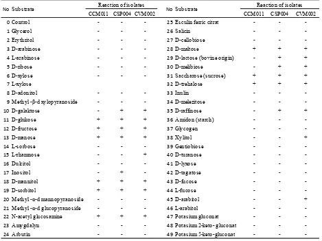
Table 5. Sugar fermentation pattern of selected lactic acid bacteria (LAB) isolates using API 50 CHL Kit
1
+++ = clear zone ≥ 3 mm
2
++ = clear zone ≤ 3 mm
3
4
5
6
API 50 CHL Kit
7
8
9
10
CCM011 CSP004 CVM002 CCM011 CSP004 CVM002
0Control - - - 25 Esculin ferric citrat - -
-1Glycerol - - - 26Salicin - -
-2Erythritol - - - 27D-cellobiose - -
-3D-arabinose - - - 28D-maltose + + +
4L-arabinose - - - 29D-lactose (bovine origin) - + +
5D-ribose - - - 30D-melibiose - + +
6D-xylose - - - 31Saccharose (sucrose) + + +
7L-xylose 32D-trehalose + + +
8D-adonitol - - - 33Inulin - -
-9Methyl -β-d xylopyranoside - - - 34D-melezitose - -
-10D-galaktose - + + 35D-raffinose - + +
11D-glukose + + + 36Amidon (starch) - -
-12D-fructose + + + 37Glycogen - -
-13D-manose + + + 38Xylitol - - +
14L-sorbose - - - 39Gentiobiose - -
-15L-rhamnose - - + 40D-turanose - -
-16Dulcitol - - - 41D-lyxose - -
-17Inositol - + - 42D-tagatose - -
-18D-mannitol + + + 43D-fucose - -
-19D-sorbitol + + + 44L-fucose - -
-20Methyl -α-d mannopyranoside - - - 45D-arabitol - - +
21Methyl -α-d glucopyranoside - - - 46L-arabitol - -
-22N-acetyl glucosamine + + + 47Potasium gluconat - -
-23Amygdalyn - - - 48Potasium 2-keto- gluconat - -
-24Arbutin - - - 49Potasium 5-keto-gluconat - -
-Note : + : positive reaction, - : negative reaction
Reaction of isolates Reaction of isolates
Substrate Substrate
No No
Note: Means in the same column with different superscripts differ sig-nificantly (P<0.05); ND = Not detected.
Inhibitory condition of
Viable LAB isolate (Log10 CFU/mL)
CCM011 CSP004 CVM002
pH
pH 2.0 5.71±0.01a 4.68±0.03a 5.85±0.01a
pH 4.0 6.11±0.10b 5.79±0.01b 6.16±0.02b
pH 6.5 6.73±0.02c 6.25±0.07c 6.2±0.040b
Bile salts
0% 6.73±0.11a 6.60±0.31a 6.87±0.03a
0.05% 3.68±0.06b 4.04±0.62b 4.04±0.37b
0.08% 1.45±0.04c 2.39±0.30c 1.57±0.04c
0.10% 0.74±0.06d 0.59±0.16d 0.45±0.21d
0.30% ND ND ND
December 2014 187 salivarius has capability to produce bacteriosin and
exopolysaccaharide (EPS) that helps to adhere to intes
-tinal mucus (Raftis et al., 2011). The ability of bacteria to
produce biofilm and adhesion are able to increase the gut residence time of commensal bacteria and promote pathogen exclusion.
Inter-isolate coaggregation ability of LAB was needed to evaluate the effectiveness of probiotics adhe
-sion in the intestine. Meanwhile, coaggregation between LAB isolates and bacterial pathogens to avoid adhesion
of pathogen on host intestinal cells. Inter-isolate
coaggre-gation of LAB was from 26.64% to 81.56%, and the high
-est coaggregation was found on CSP004 and CVM002 isolates (Figure 1). Coaggregation ability between patho -genic bacteria, E. coli and CCM011 isolates showed weak coaggregation, however there was no coaggregation of
E. coli with CSP004 and CVM002 isolates (Figure 2). In
addition, coaggregation of LAB isolates with S. enteritidis
was 4%-20% (Figure 2).
Antibiotic resistance assay of the LAB isolates
showed that there was no resistant to examined
antibio-tics, but each isolate at least showed intermediate status to one of antibiotics. Isolates of CCM011, CSP004, and CVM002 were detected to be intermediate resistant to
lincomycin, tetracyclin and erythromycin, respectively.
Identification of Selected LAB Isolates
Genotypically, the three selected LAB isolates i.e. CCM011, CSP004 and CVM002 were identified based on sequence analysis of 16S rRNA gene and showed that DNA fragments of amplification products were about 1500 bp (Figure 3). The analysis of 16S rRNA gene of isolates has been successfully sequenced, aligned and compared. The isolates were identified as Lactobacillus salivarius with sequences similarity 99% (GenBank acces
-sion number, KC020621.1 and AB612967.1). Phylogenetic tree based on 16S rRNA gene sequences analysis showed that the isolates were clustered in one group (Figure 4).
L. salivarius was known as the indigeneous strain of gas
-trointestinal tract and has probiotic properties (Nouri et al., 2010, Raftis et al., 2011).
Phenotypic identification based on carbohydrate fermentation assay by the API 50 CH system showed
that the three selected isolates were closely related to L. salivarius with a 99.9% similarity. This result confirmed
genotypic identification. However, there were traits dif
-ferences in consuming various carbon sources among the LAB isolates. CSP004 and CVM002 isolates showed relatively similar carbon fermentation profiles, however they had different carbon fermentation profiles from CCM011 isolate. CCM011 isolate was lack ability to use d-galactose, d-lactose, d-mellibiose, and d-raffinose as carbon sources. L. salivarius CCM011 lacked ability to
use galactose and lactose. It was possible that CCM011 did not have an intracellular transport system to take up the lactose and cannot produce the enzyme β-galactosi -dase/ lactase.
Meanwhile, there were traits differences between CSP004 and CVM002 isolates. CVM002 could use rham
-nose, xilitol and arabitol for carbon sources, however CSP004 could use inositol for carbon source (Table 6).
1
Figure 1. Inter-isolate coaggregation ability of the LAB isolates
Figure 2. Coaggregation ability of LAB isolates (n=3) and indi -cator bacteria (E. coli (■) and S. enteritidis (□)).
1
Bacterial isolates Figure 3. Agarose gel (1%) analysis of PCR amplification prod -ucts of 16S rRNA gene of lactic acid bacteria isolates. M= Molecular marker (1 kb ladder), lane 1= CSP004; lane 2= CCM011 and lane 3= CVM002 isolate.
3000 bp Table 6. Enzymatic activities of selected lactic acid bacteria
CONCLUSION
The three selected LAB isolates (CCM011, CSP004, and CVM002 demonstrated antimicrobial activity
against E. coli and S. enteritidis, resistance to low pH, strong hydrophobicity to hydrocarbon and
coaggrega-tion of inter-LAB isolates, but weak coaggregacoaggrega-tion with the bacterial pathogens and susceptible to examined antibiotics. The isolates were identified as Lactobacillus salivarius with 99% similarity. However, there were
different characteristics of sugar fermentation profiles
among the isolates. Considering the characteristics
above, the isolates may be used as probiotics in poultry
farm. In vivo assay will be required for evaluation of the
isolates as a probiotic supplement in chicken.
ACKNOWLEDGEMENT
This work was supported by the scholarship grants from Directorate General of Higher Education, Ministry of Education and Culture, Indonesia, and Research Center for Biology-Indonesian Institute of Sciences, and the SATREPS JST-JICA projects.
REFERENCES
Alakomi, H. L., E. Skytta, M. Saarela, T. Mattila-Sandholm, K. Latva-Kala, & I. M. Helander. 2000. Lactic acid permeabi -lizes gram-negative bacteria by disrupting the outer mem
-brane. Appl. Environ. Microb, 66:2001-2005. http://dx.doi. org/10.1128/AEM.66.5.2001-2005.2000
Corcionivoschi, N., D. Drinceanu, I. M. Pop, D. Stack, L. Stef, C. Julean, & B. Bourke. 2010. The Effect of Probiotics on Animal Health. Anim. Sci. and Biotechnol. 43:35-41.
Dumonceaux, T. J., J. E. Hill, S. M. Hemmingsen, & A. G. Van Kessel. 2006. Characterization of intestinal microbiota and response to dietary virginiamycin supplementation in the broiler chicken. Appl. Environ. Microbiol., 72:2815-2823. http://dx.doi.org/10.1128/AEM.72.4.2815-2823.2006 FAO/WHO – Food and Agriculture organization of the United
Nations/World Health Organization (2002): Guidelines for evaluation of probiotics in food. Available at: http:// www.who.int/foodsafety/fs_management/en/probiotic_ guidelines.pdf
Felsenstein, J. 1985. Confidence limits on phylogenies : an ap -proach using the bootstrap. Evolution 39: 783-791.
Gaggia, F., P. Mattarelli, & B. Biavati. 2010. Review: Probiot
-ics and prebiot-ics in animal feeding for safe food produc -tion. Int. J. Food Microbiol. 141: S15–S28. http://dx.doi. org/10.1016/j.ijfoodmicro.2010.02.031
Guerin-Danan, C., A. Andrieux, & O. Szylit. 1999. Storage of Intestinal bacteria in samples frozen with glycerol. Micro -biol. Ecol. in Health & Disease 11:180-182. http://dx.doi. org/10.1080/089106099435772
Heravi, R. M., H. Kermanshahi, M. Sankian, M. R. Nassiri,
A. H. Moussavi, L. R. Nasiraii, & A. R. Varasteh. 2011.
Screening of lactobacilli bacteria isolated from gastroin-testinal tract of broiler chickens for their use as probiotic. African J. Microbiol. Res. 5:1858-1868.
Iniguez-Palomares, C., R. Perez-Morales, & E. Acedo-Felix. 2007. Evaluation of probiotic properties in Lactobacillus iso-lated from small intestine of piglets. Microbiol. 49:46-54. 1
2 3 4 5 6 7
8 9 10
Figure 4. Phylogenetic tree derived from fulllength of 16S rDNA sequences analysis of LAB isolates showing the position of Lactoba-cillus salivarius CCM011, CSP004 and CVM002 among selected lactobacilli. The tree was generated by the neigbour-joining method and Clostridium perfringens was used as the out group. Bootstrap value based on 1000 replications are given at nodes. Bar 0.01 substitutions per nucleotide position. Accession numbers of sequences obtained from NCBI database.
December 2014 189 Istiqomah, L., S. N. Hayati, E. Damayanti, H. Julendra, A. A.
Sakti, & T. Untari. 2013. Performance and Meat Quality of Broilers Infected with Escherichia coli and Administered with Bio Additive, Probiotic, and Antibiotic. Med.Pet. 36:14-20. http://dx.doi.org/10.5398/medpet.2013.36.1.14
Konings W. N., J. S. Lolkema, H. Bolhuis, H. W. Van Veen, B. Poolman, & A. J. M. Driessen. 1997. The role of trans-port processes in survival of lactic acid bacteria. Antonie van leeuwenhoek 71:117-128. http://dx.doi.org/10.1023/ A:1000143525601
Musikasang H., N. Sohsomboon, A. Tani, & S. Maneerat. 2012. Bacteriocin-producing lactic acid bacteria as a probiotic potential from Thai indigenous chickens. Czech J Anim. Sci., 57: 137–149.
Nataamijaya, A. G. 2010. Pengembangan potensi ayam lokal untuk menunjang peningkatan kesejahteraan petani. J. Litbang. Pertan. 29: 131-138.
Nouri, M., F. Rahbarizadeh, D. Akhmadvand, F. Moosakhani,
E. Sadeqzadeh, S. Lavasani, & V. K. Vishteh. 2010.
In-hibitory effects of Lactobacilus salivarius and Lactobacillus crispatus isolated from chicken gastrointestinal tract on
Salmonella enteritidis and Escherichia coli growth. Iranian J. Biotechnol. 8: 32-37.
Palacios, M. C., M. Haros, Y. Sanz, & C. M. Rosell. 2008. Se-lection of lactic acid bacteria with high phytate degrad-ing activity for application in whole wheat breadmakdegrad-ing. LWT- Food Sci. Technol. 41:82-92. http://dx.doi.org/10.1016/ j.lwt.2007.02.005
Pelinescu, D. R., E. Sasarman, M. C. Chifiriuc, I. Stoica, A. M.
Nohit, I. Avram, F. Serbancea, & T. V. Dimov. 2009.
Isola-tion and identificaIsola-tion of some Lactobacillus and Enterococ-cus strains by a polyphasic taxonomical approach. Roma-nian Biotech. Lett. 14:4225-4233.
Raftis, E. J., E. Salvetti, S. Torriani, G. E. Felis & P. W. O’Toole.
2011. Genomic diversity of Lactobacillus salivarius. Appl. Environ. Microbiol. 77:954-965. http://dx.doi.org/10.1128/ AEM.01687-10
Raghavendra, P. & P. M. Halami. 2009. Screening, selection and characterization of phytic acid degrading lactic acid bacteria from chicken intestine. Int. J. of Food Microbiol. 133:129–134. http://dx.doi.org/10.1016/j.ijfoodmicro.2009.0 5.006
Sartika, T., S. Sulandari, & M. S. A. Zein. 2011. Selection of Mx gene genotype as genetic marker for Avian Influenza resistance in Indonesia native chicken. BMC Proceedings 5:S37.
Sofyan, A., M. Angwar, H. Herdian, E. Damayanti, L. Is-tiqomah, A. Febrisiantosa, H. Julendra, M. H. Wibowo, & T. Untari. 2012. Performance Enhancement and Immunity Profile of Broiler Treated Feed Additive Containing Lactic Acid Bacteria and Ganoderma lucidum. Med. Pet. 35:
201-206. http://dx.doi.org/10.5398/medpet.2012.35.3.201 Sulandari S., M. S. A. Zein, D. Astuti, & T. Sartika. 2009.
Ge-netic polymorphisms of the chicken antiviral Mx gene in a variety of Indonesian indigenous chicken breeds. J Vet.
10: 50-56.
Taheri, H. R., H. Moravej, F. Tabandeh, M. Zaghari, & M. Shivazad. 2009. Screening of lactic acid bacteria toward their selection as a source of chicken probiotic. Poult. Sci. 88:1586–1593. http://dx.doi.org/10.3382/ps.2009-00041 Tannock, G. W. 1999. Identification of Lactobacilli and Bifido
-bacteria. Current Issues Molec. Biol. 1: 53-64
Torshizi, M. A. K., S. H. Rahimi, N. Mogjani, S. Esmaeilkha-nian, & J. L. Grimes. 2008. Screening of indigenous strains of lactic acid bacteria for development of a probiotic for poultry. Asian-Australian J Anim. Sci. 21:1495-1500. http:// dx.doi.org/10.5713/ajas.2008.80081