This content has been downloaded from IOPscience. Please scroll down to see the full text.
Download details:
IP Address: 114.125.41.120
This content was downloaded on 10/04/2017 at 16:47
Please note that terms and conditions apply.
Preparation and Characterization of Cellulose Microcrystalline (MCC) from Fiber of Empty
Fruit Bunch Palm Oil
View the table of contents for this issue, or go to the journal homepage for more 2017 IOP Conf. Ser.: Mater. Sci. Eng. 180 012007
(http://iopscience.iop.org/1757-899X/180/1/012007)
You may also be interested in:
Comparison of microcrystalline characterization results from oil palm midrib alpha cellulose using different delignization method
S Yuliasmi, T R Pardede, Nerdy et al.
Production of cellulose phosphate from oil palm empty fruit bunch: Effect of chemical ratio R Rohaizu and W D Wanrosli
Hydrothermal pre-treatment of oil palm empty fruit bunch into fermentable sugars M D Muhd Ali, P Tamunaidu, A K H Nor Aslan et al.
Effect of Turning Frequency on Composting of Empty Fruit Bunches Mixed with Activated Liquid Organic Fertilizer
B Trisakti, J Lubis, T Husaini et al.
Devolatilization studies of oil palm biomass for torrefaction process optimization D Daud, A Abd Rahman and A H Shamsuddin
Electrical Conductivity of Carbon Pellets from Mixtures of Pyropolymer from Oil Palm Bunch and Cotton Cellulose
Mohamad Deraman, Sarani Zakaria, Ramli Omar et al. Resistivity of carbon from oil palm bunches: percolation theory M Deraman
Effect of Stitching on the Tensile Mechanical Property of Empty Fruit Bunch Oil Palm Fiber Reinforced Epoxy Composites
Preparation
and
Characterization
of
Cellulose
Microcrystalline (MCC) from Fiber of Empty Fruit Bunch
Palm Oil
H Nasution
1*, Yurnaliza
2, Veronicha
1, Irmadani
1, S Sitompul
11Department of Chemical Engineering, Faculty of Engineering, University of Sumatera
Utara, North Sumatra, Medan, 20155, Indonesia
2Department of Biology, Faculty of Mathematics and Natural Sciences, University of
Sumatera Utara, North Sumatra, Medan, 20155, Indonesia
Abstract. Alpha cellulose which was isolated from cellulose of fiber empty fruit bunch palm oil was hidrolized with hydrochloric acid (2,5N) at 80oC to produce microcrystalline cellulose
(MCC). Microcrystalline cellulose is an important additional ingredient in the pharmaceutical, food, cosmetics, and structural composites. In this study, MCC, alpha cellulose, and cellulose were characterized and thereafter were compared. Characterizations were made using some equipment such as x-ray diffraction (XRD), Fourier transform infrared (FTIR), scanning electron microscopy (SEM) and thermogravimetry analyzer (TGA). X-ray diffraction and infrared spectroscopy were studied to determine crystallinity and molecular structure of MCC, where scanning electron microscopy images were conducted for information about morfology of MCC. Meanwhile, thermal resistance of MCC was determined using thermogravimetry analyzer (TGA). From XRD and FTIR, the obtained results showed that the crystalline part was traced on MCC, where the –OH and C-O groups tended to reduced as alpha cellulose has changed to MCC. From SEM the image showed the reduction of particle size of MCC, while the thermal resistance of MCC was found lower as compared with cellulose and alpha cellulose as well, which was attributed to the lower molecular weight of MCC.
1. Introduction
starting material for the production of microcrystalline cellulose. Microcrystalline cellulose (MCC) is a polymer has been widely used in a variety of commercial applications in the food, structural composite, and pharmaceutical industries. This polymer has a high amount of amorphous regions that occurs as a white, odourless, tasteless, crystalline powder composed of porous particles [3]. The MCC was obtained by partially hydrolyzing cellulose with mineral acid to remove amorphouse regions in forming microcrystal.
A conventional method for producing MCC from cellulose is acid hydrolysis process. The hydrolysis of cellulose can be accomplished using mineral acid [4-10], enzymes [11], and microorganism [12]. However, different properties of MCC such as crystallinity, moisture content, molecular weight, surface area and porous structure, thermal resistance were also obtained from different sources of raw material. Reports have shown that MCC can be produced from kenaf fibers [12], rice hull and bin hull [3], cotton linters [13], sawdust [4], rice straw [14], Oil Palm Empty Fruit Bunches [15], groundnut shells [16], corn cobs [17], sisal fibers [5], cotton rags [6].
This article reports the preparation and characterization of MCC by acid hydrolysis method using hydrochloric acid. Characterization was studied by FTIR, XRD, SEM and TGA.
2. Methode
2.1. Raw material
Cellulose derived from the EFBPO which was isolated earlier was kindly supplied by Oil Palm Research Centre in Medan, Indonesia. Sodium hydroxide (NaOH), sodium hypochloride (NaOCl), and hydrochloric acid (HCl) (37%) were supplied by Sigma-Aldrich and used as received.
2.2. Production of alpha cellulose
Alpha cellulose was obtained from the cellulose using 17,5% sodium hydroxide (NaOH) at 80oC for 0,5
h. Bleaching was then carried out with 3,2% sodium hypochloride (NaOCl) at 100oC for 1,5 h.
2.3. Production of MCC
The alpha cellulose powder derived from cellulose was hydrolized using 2,5N hydrochloric acid (HCl) at 80oC for 15 min. The hydrolized cellulose was thoroughly washed with cold distilled water until
neutral to litmus paper and then dried.
2.4. Characterization
2.4.1. Fourier Transform Infrared (FTIR).Infrared spectroscopy of cellulose, alpha cellulose and MCC were carried out using Shimadzu IR-Prestige 21. Bands were recorded in the region from 4000 to 500 cm-1.
2.4.2. X-ray diffraction (XRD). X-ray diffractometry patterns of cellulose, alpha cellulose and MCC were pressed to form pellets and recorded in 6100 Shimadzu.
The crystalline index (Ic) was determined by Eq (1) using the intensity ofI002peak at about 2= 21,9o andIamis the intensity corresponding to the peak at about 2= 12o-18o
= 100 (1)
2.4.3. Morphologycal characteristic. Scanning electron microscopy (SEM) was used to examine the microscopic structure and the surface morphology of the prepared cellulose, alpha cellulose and MCC. For SEM measurement the instrument used for morphological observation was SEM EVO MA 10 ZEISS.
2.4.4. Thermal analysis.Thermogravimetry analysis was utilized using Shimadzu Simultanous TGA/DTA Analyzer DTG-60 to investigate the thermal properties of cellulose, alpha cellulose and MCC.
3. Results and Discussion
3.1. Infrared spectroscopy
The molecular structure of the cellulose isoated directly from EFBPO, alpha cellulose and MCC. Are shown in Figure 1.
Figur 1.FTIR spectra of a. Cellulose; b. Alpha cellulose; c. MCC
FT-IR spectra of the different samples of cellulose were recorded in the range of 4000–500 cm-1. A
slight difference is observed in the region of the intermolecular hydrogen bonding (3200–3400 cm-1).
The shift of the maximum absorbance band of stretching vibration of OH group of MCC to lower wave number (3340 cm-1) as compared with other two samples of cellulose (3363 cm-1and 3400 cm-1for alpha
cellulose and cellulose, respectively). This shift proves that MCC is more crystalline than both samples. Moreover, the characteristic intermolecular and intramolecular OH stretching vibration band in the spectrum of MCC appeared broader than those of alpha cellulose and cellulose. This is due to the degradation of the hydrogen bond between the cellulosic chains during the hydrolysis process. Here, the first part hydrolyzed and degraded by acid were an amorphous region.
The C–H stretching vibration absorbance intensity in MCC (2892 cm−1) is slightly decreased upon
acid hydrolysis of alpha cellulose; this is due to the presence of –CH2moieties in the samples [18]. The
absorption bands at 1635 cm−1 corresponds with 1639 cm−1 and 1631 cm−1 were attributed to O-H
bending, the vibration of adsorbed water molecules [19]. This peak could be due to the presence of small amounts from hemicellulose. The presence of this peak may be arising from the oxidation of the C–OH groups.
The peaks related to –CH or C-O bending vibrations (1369, 1374 cm−1) in the polysacharide aromatic
at 1064 cm-1corresponds with 1061 and 1053 cm-1appears is due to -CH
2-O-CH2pyranos ring stretching
vibration.In addition, the band at about 898 cm−1in the spectrum of MCC is attributed to the asymmetric
out of plane ring stretching in cellulose due to theglicosidic linkage between glucose unit in cellulose. It is noted the linkage stands for the increase in crystallinity of the material [21].
3.2. X-ray diffraction
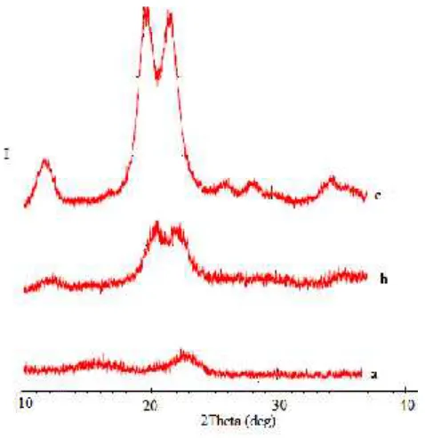
Figure 2 shows the x-ray diffraction spectra of cellulose, alpha cellulose and MCC. .
Figur 2.X ray diffraction of a. Cellulose; b. Alpha cellulose; c. MCC
The powder x-ray diffraction spectra of the three cellulose samples exhibit different diffraction patterns. Cellulose derived from oil palm empty fruit bunch fibre was highly amorphous, as indicated by the less peaks in the diffractogram (Fig 1a). An amorphous region implies a more disordered structure, resulting in a low crystal region.
The crystallinity is traced on the alpha cellulose diffractogram (Fig. 1b). This is due to removal of hemicellulose and lignin, which existed in amorphous region leading to realignment of cellulose molecules. However, These two peaks are smeared and appear as one broad peak. A smeared out diffractogram was observed for samples, especially the peaks that appeared at diffraction angles ranging from 20o-24ofor cellulose and from 18o-24ofor alpha cellulose indicating a low degree of order. This
results are similar with the study have reported by Mat Soom et.al [15]. However, the higher peak of alpha cellulose in comparison to cellulose, indicating that the crystalline behaviour of alpha cellulose was higher than for cellulose. The presence of amorphous aromatic compounds such as lignin, polysacharide polymers and many others was attributed to this behaviour.
Figure 1c shows the x-ray diffraction spectra of MCC. The highest crystal structure was obtained for MCC due to the removal of the amorphous regions of alpha cellulose by acid hydrolysis, realeasing individual crystallites. It is interesting to note that the MCC in the present case shows doublet in the intensity of the main peaks corroborating the coexistence of cellulose I and cellulose II allomorphs. The large different spectra between MCC with cellulose and alpha cellulose results in the appearance of two significant peaks indicating the highly crystalline structure of MCC. High crystallinity indicates an orderedcompact molecuar structure, when the crystalline cellulose content is high, these two peaks are
more pronounced.
The peak intensity of MCC appeared to be higher than that of alpha cellulose indicating that the MCC is more crystalline than alpha cellulose.The peaks at MCC spectra are more defined suggesting that the hydrolysis acid process removed some of the amorphous material from the alpa cellulose. The process was initiated in the fast removal of amorphous cellulose near the surface of macrofibrils, which leads to the exposure of microfibril bundles. An amorphous cellulose near surface is hydrolyzed first and followed by crystalline cellulose near surface. The amorphous cellulose deeply buried in the bulk leach out from macrofibrils during hydrolysis at slower rates due to barriers caused by the microfibril bundles. This process repeats during the hydrolysis process until cellulose degradation occurs [22]. During processing to MCC, the alpha cellulose was hydrolized and depolymerized to remove a large portion of the amorphous region, leaving the crystallinecellulose.The diffractograms for MCC exhibits diffraction patterns typical of cellulose, with diffraction peaks of the 2angles at 12,2; 20,2 and 21,9o.
The index crystallinity (CI) which is calculated according to the equation (1) gives a quantitative measure of the crystallinity in powders. Here the crystallinity index of MCC from hydrolization process is 73%. Since cellulose from different source differs in propeties, different properties of MCC obtained from source are expected and the condition of hydrolysis process also affect the properties. Some studies about the effect of sources on the percent crystallinity of MCC have been reported such as Kenaf is 70% [12] cotton linter is 76% [14], groundnut shells is74% [16], baggas is 76% [10], rice straw is 78% [10], cotton stalks is 77% [10] and sisal is 60% [5].
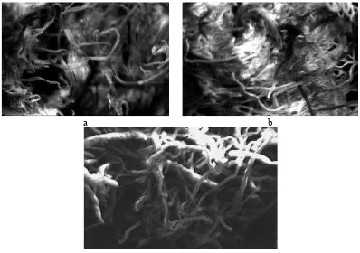
3.3. Scanning Electron Microscopy (SEM)
Figure 3 shows the surface morphology of cellulose, alpha cellulose and MCC using SEM.
a b
c
Figure 3.SEM images (mag. of 5000 ) of a. Cellulose; b. alpha cellulose; c. MCC
the macrofibril interconnections were occured as the hemicellulose could not well dissolved during treatment.
The SEM micrographs for the alpha cellulose (Figure 3b), showed a smaller diameter as compared to the cellulose (Figure 34a). On subsequent treatment with alkali, the hemicelluloses, which was still remain in cellulose, was hydrolyzed and becomes water soluble. These help in defibrillation of the fibrils and result in micrograph, whereby the diameter of the fibrils is reduced to a great extent. However, the interconnections amongst fibrils were still occured.
On the other hand, the MCC image shows a fibrous structure and individualized (Figure 3c). The MCC obtained showing fibers strands which appear like rod-shaped. The MCC appeared to be irregular fiber fragments and also show a network-structure (13). In this case, after macrofibrils were hydrolyzed and rinsed, the volume of amorphous cellulose was occupied by water molecules that were then removed; the remaining macrofibrils contain large amounts of naked microfibril bundles.
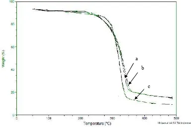
3.4. Thermal Resistance
Figure 4 below shows the thermal resistance of cellulose, alpha cellulose and MCC.
Figure 4.SEM images (mag. of 5000 ) of a. Cellulose; b. alpha cellulose; c. MCC
The thermograms curves of cellulose (a), alpha cellulose (b), and MCC (c) are shown ini Fig...The initial weight losses starting at 60oC for all samples may be attributed to the evaporation of loosely
bound moisture on the surfaces of these samples.
The thermogravimetry curve for cellulose and alpha cellulose follow similar degradation pattern. The first stage degradation for cellulose and alpha cellulose occurs at a temperature range of 225-350oC and
220-3550C, respectively with a total weight loss of 80% for cellulose and 75% for alpha cellulose. The
second stage started at about 350oC for cellulose and 355oC for alpha cellulose and reached a maximum
at 480oC for both samples with a total weight loss of 85%.
In the alpha cellulose the removal of all non cellulosic materials helps to make the cellulose structure more dense and compact and hence a slightly rise in the temperature of degradation have occured.
a b
c
The degradation of MCC appears to follow different mechanism where the temperature at a range of 285-340oC with a total weight loss of 85%, which is due to the degradation of cellulose. The rest of
degradation occured during heating at 340-480oC with a total weight loss of 90%.
A notable difference, in particular between MCC sample and two other samples (cellulose and alpha cellulose), is observed at the temperature at which 20% weight loss of the samples is degraded. This temperature is lower for cellulose and alpha cellulose (275oC) as compared to the MCC (285oC). This
result might be attributed to the higher crystallinity and lower moisture contents of the MCC. The rearangement and reorientation of the crystals in MCC offers to raise the onset temperature of degradation. Additionally higher onset temperature are associated with high thermal stability. However, start at a temperature of 325oC the degradation of MCC shows more drastic as compared with two other
samples. This temperature correspons with 40% weight loss of MCC, while the other two samples (cellulose and alpha cellulose) show 35% of weight losses. It may attributed to the reduction in molecular weight of MCC during hydrolysis process.. Hydrolysis process have made the MCC is more susceptible to degrade when temperature increases. It is also believed that hydrolysis of cellulose not only dissolves the amorphous regions, but also some crystalline regions (21). Similar results have been reported by El-Sakhawy and Hassan (10) when producing MCC from baggase fibers.
4. Conclusion
The microcrystalline cellulose (MCC) was obtained from alpha cellulose which was isolated from
cellulose derived from EFBPO. The three samples were then compared in terms of molecule structure, crystallinity, morphology and thermal stability. MCC was prepared by hydrolysis acid using hydrochloric acid (2,5N). Some structures of the molecules have been changed due to the hydrolysis process. In addition, the x-ray diffractograms proved that the MCC samples are more crystalline compared to two other samples (cellulose and alpha cellulose). On the other hand, it was noticed by the SEM that hydrolysis treatment affected the morphological structure of the resulting microfibrillated cellulose. However, the thermal stability of the MCC samples starting at temperature at 325oC were
lower as compared to the corresponding other two samples which was attributed to the reduction of molecular weight of MCC.
5. Acknowledgements
The authors gratefully acknowledge that the present research is supported by Ministry of Research and Technology and Higher Education Republic of Indonesia. The support is under the research grant BP-PTN USU of year 2016 Contract Number 6049/UN5.1.R/PPM/2016.
References
[1] Li X, Tabil L G and Panigrahi S C 2007 Chemical treatments of natural fibre for use in natural fibre-reinforced composites: a reviewJ. Polym. Environ.15pp 25–33.
[2] Klemm D, Philipp B, Heinze T, Heinze U and Wagenknecht W 1998Comprehensive cellulose chemistryWeinheim Wiley–VCH pp 9–25.
[3] Adel A M, El-Wahab Z H A, Ibrahim A A and Al-Shemy M T 2011 Characterization of microcrystalline cellulose prepared from lignocellulosic materials. Part II: Physicochemical propertiesCarbohydrate Polymers83pp 676–687.
[4] Oyeniyi Y J and Itiola O A 2012 The Physicochemical characteristic Of microcrystalline cellulose, derived from sawdust, agricultural waste productsInt J Pharm Pharm Scivol 4 pp 197-200.
[5] Bhimte N A and Tayade P T 2007 Evaluation of Microcrystalline cellulose prepared from sisal fibers as a tablet excipient: A Technical Note, AAPS PharmSciTech 8 Article 8 (http://www.aapspharmscitech.org).
[7] Hafiz M K M, Eichhorn S J, Hassan A and Jawaid M 2013 Isolation and characterization of microcrystalline cellulose from oil palm biomass residueCarbohydrate Polymers93pp 628– 634.
[8] Ejikeme P M 2008 Investigation of the physicochemical properties of microcrystalline cellulose from agricultural wastes I: orange mesocarpCellulose15pp 141–147.
[9] Laka M and Svetlana C 2007 Obtainning microcrystalline from softwood and hardwood pulpBio Resourrces2(3) pp 583-589.
[10] El-Sakhawy M and Hassan M L 2007 Physical and mechanical properties of microcrystalline cellulose prepared from agricultural residuesCarbohydrate Polymers67pp 1–10
[11] Maha M I, El-Zawawy W K, Juttke Y, Koschella A and • Heinze T 2013 Cellulose and microcrystalline cellulose from rice straw and banana plant waste: preparation and characterization,CelluloseDOI 10.1007/s10570-013-9992-5.
[12] Keshk S M A S and Haija M A 2011 A new method for producing microcrystalline cellulose from Gluconacetobacter xylinus and kenafCarbohydrate Polymers84pp 1301–1305.
[13] Nada A M A, El Kady M Y, El-Sayed E S and Amine F M 2009 Preparation and characterization of microcrystalline CelulloseBio Resources4(4) pp 1359-1371.
[14] Nawar G A M, Hassan F A M, Ali K E, Kassem J M and Mohamed S H S 2010 Utilization of microcrystalline cellulose prepared from rice straw in manufacture of yoghurt Journal of American Science6(10) pp 226-231.
[15] Mat Soom R, Aziz A A, Wan Hasan W H and Mat Top A B G. 2009 Solid state characteristics of microcrystall cellulose from oil palm empty fruit bunches Fibre Journal of Oil Palm Researchvol 21 pp 613-620.
[16] Azubuike C P, Odulaja J O and Okhamafe A O 2012 Physicotechnical, spectroscopic and thermogravimetric properties of powdered cellulose and microcrystalline cellulose derived from groundnut shellsJ. Excipients and Food Chem.3(3).
[17] Azubuike C P and Okhamafe A O 2012 Physicochemical, spectroscopic and thermal properties of microcrystalline cellulose derived from corn cobs International Journal Of Recycling Of Organic Waste in Agriculturehttp://www.ijrowa.com/content/1/1/9.
[18] Khalil H P A, Ismail H, Rozman H D and Ahmad M N 2001 The effect of acetylation on interfacial shear strength between plant fiber and various matricesEuropean Polymer Journal
37(5) pp 1037–1045.
[19] Nacos M, Katapodis P, Pappas C, Daferera D, Tarantilis P A ad Christakopoulos P 2006. Kenaf xylan—A source of biologically active acidic oligosaccharidesCarbohydrate Polymers,66(1) pp 126–134.
[20] Troede, M, Sedan D, Peyratou, C, Bonnet J, Smith A and Guinebretiere R 2008 Influence
of various chemical treatments on the composition and structure of hemp fibers
Composites Part A-Applied Science and Manufacturing
39
(3) pp 514–522.
[21] Mandal .A and Chakrabarty D 2011 Isolation of nanocellulose from waste sugarcane bagasse
(SCB) and its CharacterizationCarbohydrate Polymers86pp 1291– 1299.