Directory UMM :Data Elmu:jurnal:B:Brain Research:Vol880.Issue1-2.2000:
Teks penuh
Gambar
![Fig. 1. Representative autoradiograms of [ H]muscimol binding in horizontal rat brain sections](https://thumb-ap.123doks.com/thumbv2/123dok/3140037.1382879/3.612.94.502.427.706/fig-representative-autoradiograms-muscimol-binding-horizontal-brain-sections.webp)
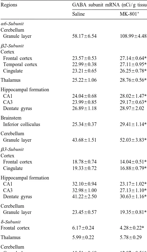
![Table 1Changes in [ H]muscimol binding to brain regions in rats chronically](https://thumb-ap.123doks.com/thumbv2/123dok/3140037.1382879/4.612.100.494.455.707/table-changes-muscimol-binding-brain-regions-rats-chronically.webp)
![Table 2Changes in [ H]flunitrazepam binding to brain regions in rats chronically](https://thumb-ap.123doks.com/thumbv2/123dok/3140037.1382879/5.612.307.550.91.378/table-changes-unitrazepam-binding-brain-regions-rats-chronically.webp)
![Fig. 3. Representative autoradiograms of [ S]TBPS binding in horizontal rat brain sections](https://thumb-ap.123doks.com/thumbv2/123dok/3140037.1382879/6.612.45.284.409.691/fig-representative-autoradiograms-tbps-binding-horizontal-brain-sections.webp)
Dokumen terkait
On the Notably, cells in the contralateral sensory cortex of ani- other hand, a shift in the potential of half-maximal mals with a severe infarction showed also only
This Our results demonstrate increased plasminogen activa- exceeding plasminogen activation was mainly located tion in the basal ganglia and the cortex following perma- around
In the present paper we investigated the role of the noradrenergic projection from the locus coeruleus on the expression of the immediate early gene zif268 in the visual cortex of
Areas of the brain was not the likely explanation for failing to find regional thought to contribute to visual function include frontal, CF avidity differences of the magnitude of
Wrathall, Changes in NMDA with the hypothesis that GluR2 expression is not regulated receptor subunit expression in response to contusive spinal cord by activity-dependent means
AHA, anterior hypothalamic area; Arc, arcuate nucleus; AT, area triangularis; BST, bed nucleus of the stria terminalis; DB, nucleus of the diagonal band; DCx, dorsal cortex;
Task Number of % with Activation All the tasks produced functional activation in the basal studies basal ganglia Size ganglia, motor cortex, and supplementary motor area (right / left
Brain changes associated with early-onset major depression have been reported in the hippocampus, amygdala, caudate nucleus, putamen, and frontal cortex, structures that are
