What is the best internal fixation in pelvic fracture models with
open-book injury and anterior sacroiliac joint disruption?
Keywords: anterior sacroiliac joint disruption, biomechanical study, internal fixation, open-book injury pISSN: 0853-1773 • eISSN: 2252-8083 • http://dx.doi.org/10.13181/mji.v24i4.1233 • Med J Indones. 2015;24:239–44
• Received 30 Mar 2015 • Accepted 13 Dec 2015
Correspondence author: Ismail H. Dilogo, [email protected]
Copyright @ 2015 Authors. This is an open access article distributed under the terms of the Creative Commons Attribution-NonCommercial 4.0 International License (http://creativecommons.org/licenses/by-nc/4.0/), which permits unrestricted non-commercial use, distribution, and
reproduction in any medium, provided the original author and source are properly cited.
Ismail H. Dilogo, Immanuel P. Sitorus
Department of Orthopaedic and Traumatology, Faculty of Medicine, Universitas Indonesia, Cipto Mangunkusumo Hospital, Jakarta, Indonesia
Clinical Research
ABSTRAK
Latar belakang: Teknik operasi yang terbaik untuk cedera panggul tipe open-book yang disertai gangguan ligamen
sakroiliaka anterior (klasifikasi OTA/AO B1.1) masih diperdebatan. Studi biomekanis ini bertujuan untuk mencari teknik fiksasi internal yang terbaik untuk cedera tersebut.
Metode: Fraktur tipe open-book dengan gangguan ligamen sakroilika anterior disimulasikan pada 25 tulang panggul buatan yang kemudian dikelompokkan ke dalam
lima kelompok berbeda. Panggul buatan dalam masing-masing kelompok difiksasi dengan 5 teknik fiksasi berbeda: 1). 1SP+1IS; 2). 2SP; 3). 2SP+2SIP; 4). 1SP+2IS S1, dan 5). 1SP+1IS S1+S2. Pengukuran biomekanis kekuatan aksial dan
anteroposterior diukur menggunakan Tensilon® AMD RTF-1310. Makna translasi tekanan dan beban titik kegagalan
yang terukur dianalisa menggunakan one-way ANOVA diikuti
dengan uji post-hoc Bonferroni.
Hasil: Nilai rerata beban kegagalan tertinggi untuk tekanan
aksial dihasilkan oleh kelompok yang mendapat fiksasi dengan
satu piring symphyseal dan satu sekrup iliosacral di S1 dan S2
(1490,36 N). Terdapat perbedaan bermakna dari nilai rerata
beban kegagalan antara kelompok yang mendapat satu piring symphyseal dan satu sekrup iliosacral dibandingkan dengan kelompok yang mendapat satu plat symphyseal dan dua sekrup iliosacral di S1 dan S2 (p<0,05).
Kesimpulan: Penambahan fiksasi di sendi sakroiliaka posterior
dengan piring atau sekrup akan menambah kekuatan mekanis terhadap beban aksial. Berdasarkan kekuatan mekanis yang diciptakan untuk menahan beban aksial, fiksasi dengan satu
plat symphyseal disertai sekrup ilioscaral di S1 dan S2 adalah
formasi fiksasi yang terbaik untuk fraktur tipe open-book yang
disertai gangguan sendi sakroiliaka anterior.
ABSTRACT
Background: The best operative management for open-book pelvic injury with anterior sacroiliac disruption (OTA/ AO B1.1 classification) is still debated. This biomechanical study aimed to find the best internal fixation technique for
such injury.
Methods: Open-book injury with anterior sacroiliac joint disruption was simulated on 25 artificial pelvic bones. Twenty five artificial pelvic bones were divided into 5 groups (n=5 /group) and fixated with five different fixation techniques: 1). 1SP+1IS; 2). 2SP; 3). 2SP+2SIP; 4). 1SP+2IS S1, and 5). 1SP+1IS S1+S2. Biomechanical properties of each fixation technique were assessed using Tensilon® RTF-1310
to measure the resistance to translation and load to failure.
Data were statistically analyzed using one-way ANOVA followed by post-hoc Bonferroni test.
Results: The highest mean load to failure of axial forces (1490.36 N) was achieved by the fixation technique using one symphyseal plate and two iliosacral screws located at S1 dan S2. The addition of one iliosacral screw significantly increased the mean load to failure for axial compression (p<0.05).
Conclusion: The addition of sacroiliac joint posterior
Pelvic fracture is usually caused by high energy trauma and is associated with high mortality rate.1,2 Managements of pelvic fracture are based on the pelvic stability, direction of traumatic force and pathoanatomy.3-6 The best management of the open-book pelvic injury with anterior sacroiliac ligament disruption [Orthopaedic Trauma Association (OTA) classification/AO B1.1] is still debated.7 Different
techniques for treating partial stable pelvic fracture [Young and Burgess classification of anteroposterior compression (APC) II, lateral compression (LC) II and III] includes anterior fixation alone, anterior and posterior fixation, or double plate (anterior) fixation on partial stable pelvic fracture. Biomechanical study on tile B pelvic fracture showed that optimal result can be achieved by combining the anterior and posterior fixation [one plate narrow dynamic compression plate(DCP) 4.5-mm]. However, there was no clinically significant difference between fixation using one narrow plate DCP4.5-mm (two holes) or one screw 7.0-mm on sacroiliac joint.8,9 In addition, van den Bosch et al10 did not find the
significance on the stability of pelvic with addition of posterior fixation and one screw iliosacral on tile B pelvic fracture.10
Failure of open reduction and internal fixation of diastasis symphysis as a result of trauma was described by Putnis et al.11 Fifteen patients (31%)
experienced anterior shifting of plate or screw, 10 patients experienced screw loosening or damage. Anterior fixation alone was the most frequent cause of treatment failure (n=7; 47%), followed by anterior fixation with unilateral sacroiliac fixation (n=6; 40%) and bilateral sacroiliac fixation (n=2; 13%). Fixation failure was not observed on patients with double-plate anterior fixation with or without sacroiliac fixation. It was postulated that undetected ligamentous injury with microinstabilty was one of the main cause of fixation failure.11
Combination of anterior plate fixation and percutaneous sacroiliac screw in partial stable pelvic injury showed excellent fracture reduction and improvements of functional outcomes.12 The
use of posterior fixation alone with sacroiliac screw showed good result with non-union rate of 6% in posterior injury and 8% in unstable rotational pelvic injury.13 Screw malposition
(4%) is one of factor causing non-union.13
Additional anterior stabilization is needed if there is a secondary dislocation.14 The outcomes of each internal fixation technique were reported based on
the presence of complications in a limited number of patients. Quantitative data that measure the biomechanical properties of each fixation technique are not available. Such information is crucial to objectively determine the best internal fixation method of pelvic fracture. The objective of this study is to quantify the biomechanical strength of five different internal fixation techniques that were commonly used to treat open-book pelvic injury with anterior sacroiliac joint disruption. Mechanical measurements were based on translational rigidity and load to failure. Results from this study will provide a fundamental reasoning of choosing a particular type of internal fixation technique.
METHODS
Model preparation
This biomechanical study used 25 artificial pelvic bone (Synbone®); 0.43 kg in weight, 305 mm in
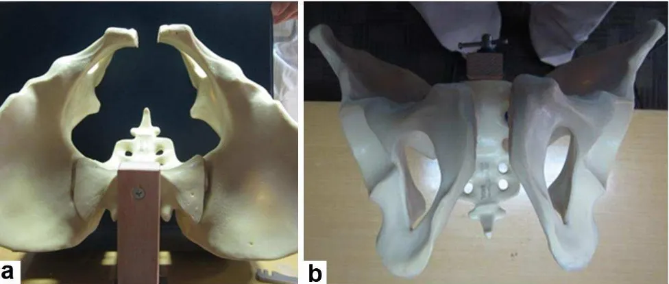
width, and 160 mm height. Open-book fracture with
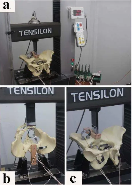
Figure 1. Overview of the biomechanical test procedure. a)
anterior sacroiliac disruption (OTA classification/ AO B1.1) was consistently simulated in all artifical pelvic bones (Figure 1).
Fracture models were divided into five groups with different fixation techniques: 1). Group I (n=5): one symphyseal plate (SP) and one posterior iliosacral screw (IS); 2). Group II (n=5): double symphyseal plate; 3). Group III (n=5): double symphyseal plate on anterior pubic and double sacroiliac plate (SIP); 4). Group IV (n=5): one symphyseal plate and two iliosacral screws on the first sacrum (S1), and 5). Group V (n=5): one symphyseal plate and two iliosacral screws, each on the first (S1) and second sacrum (S2) (Figure 2 and 3).
Biomechanical testing
Resistance of each fixation technique to axial and anteroposterior compression force was measured using Tensilon® RTF-1310 (A&D Company Ltd.,
Figure 2. Biomechanical simulation of open-book pelvic fracture and anterior sacroiliac joint disruption on a artifical pelvic bone Synbone®. a) axial view; b) anteroposterior view
Japan) connected to registrar LabView® Signal
Express (National Instruments, Texas, USA) through a series of TML® strain gauge (Tokyo
Sokki Kenkyuo Co. Ltd., Japan). Five sensors (strain gauge circuit TML®) were attached to each pelvic
bone to measure the translation (displacement) of the anterior symphyseal joint and posterior sacroiliac pelvic cavity. Axial loading was given on the first sacral vertebral plate in four models per group. Anteroposterior compression load was given on the pubic symphysis in one model per group (Figure 4).
An initial 100 newton (N) compression force was given to all fixated pelvic models with increments. The compression force gradually increased until the sacroiliac pubic symphysis joint translation reached ≥2.0 mm on craniocaudal, mediolateral or anteroposterior axis. Load to failure was the amount of force causing translation of the sacroiliac or symphysis pubic joint by ≥2.0 mm on any of
the axis. Biomechanical tests were performed in the Mechanical Engineering laboratory, School of Mechanical Engineering and Aerospace, Bandung Institute of Technology, Bandung, Indonesia.
Statistical analysis
Comparison between each group were assessed with analysis of variance (ANOVA) followed by Bonferroni post hoc test. P<0.05 was considered to be statistically significant. Data were presented as mean. Statistical analysis was performed using SPSS v.20 (SPSS Inc., Chicago, Illinois).
RESULTS
Mechanical strength of fixation with the axial force
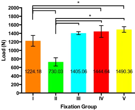
The highest load to failure for axial compresison was obtained by group V (1490.36 N), followed by group IV (1444.64), group III (1405.06 N), group I (1224.18 N) and group II (730.03 N). There were statistically significant differences between group I vs group II (p<0.001), IV (p=0.047) and V (p=0.014). The other statistically significant differences were found between group II vs group III (p<0.001), IV (p<0.001) and V (p<0.001). The load to failure in anterior fixation with double symphyseal plates had a lower score compared to the group with anterior and posterior fixation. The results of axial compression analysis were summarized in figure 5.
Mechanical strength of fixation with the anteroposterior force analysis
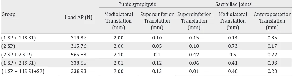
The highest load to failure to anteroposterior compression was obtained by group III (565.83 N), followed by group V (338.93 N), group IV (338.65 N), group I (319.37 N) and group II (315.76 N). There were greater superoinferior,
Figure 4. Outlet projection of pelvic X-ray demonstrating fixation types in each group. a) Group I, fixation with one symphyseal plate and posterior ilisacral screw; b) Group II, double plate pubic symphysis fixation; c) Group III, fixation with double plate of the pubic symphysis and double plate in the sacroiliac joints; d) Group IV, stabilized with one symphyseal plate and two screws at S1 level of sacroiliac joint, e) Group V, stabilized with one symphyseal plate and one screw each at S1 and S2 level sacroiliac joint fixation
I II III IV V
0 200 400 600 800 1000 1200 1400 1600 1800 2000
Fixation Group
Lo
ad
(N
)
1224.18 730.03 1405.06 1444.64 1490.36
*
*
Figure 5. Comparison of load to failure upon axial forces among fixation groups. Data are presented as mean±SD. n=4 /group, *p<0.05
mediolateral, and anteroposterior translations in double symphyseal plate and double plates in the sacroiliac joints (0.42 mm, 0.5 mm, 0.22 mm) than group IV (0.06 mm, 0.41 mm, 0.03 mm) or group V (0.01 mm, 0.4 mm, 0.2 mm). Results of the anteroposterior compression test are summarised in table 1.
DISCUSSION
Additional screw placement in the sacroiliac joint also increases mechanical stability, as shown by the significant differences between group I vs groups IV dan V.
Sacroiliac joint fixation between one or two pieces iliosacral screws showed significant differences in fixation stability for fracture classification tile-C. The highest mechanical stability to axial compression was obtained by group V (1 SP+1 IS S1+S2). This finding is similar to the cadaveric study conducted by van Zwienen et al.15 Percutaneous iliosacral screw fixation
is a definitive and rapid fixation procedure for posterior pelvic ring injuries with minimal risk of bleeding. Sacroiliac screw fixation in S2 is considered safe by the fixation technique using fluoroscopy C-arm.13 This study shows that one
iliosacral screws each at S1 and S2 level was the best configuration of fixation. This fixation has clinical advantages when applied using with C-arm fluoroscopy compared by group with plate fixation in anterior sacroiliac joints. This formation is not only the most stable fixation, but also easier, quicker and less bleeding procedure of surgery. The use of double symphyseal plates was not significantly different compared by the single plate of symphysis.16 The highest stability can be obtained by using an anterior fixation (pubic symphysis) and posterior (sacroiliac joints).17 Similar results were also shown in this
study. In spite of using two symphyseal plate, the load to failure was higher in the group stabilized with anterior and posterior fixation.
In addition, the group stabilized with symphysis pubic plate and sacroiliac joint fixation had higher load to failure than group with symphisis pubic fixation only.8 Mechanical strength with anteroposterior force treatment in double
Group Load AP (N)
Table 1. Anteroposterior (AP) load for each fixation group
symphyseal plates and double plates in the anterior sacroiliac joints group almost as twice as of the mechanical strength of the double symphyseal plate group. Anteroposterior force is clinically insignificant compared to axial forces. The axial forces can be interpreted as weight bearing force in daily settings. Therefore, in cases such as pelvic fractures complicated with sacroiliac joint disruption, axial force test is considered adequate to evaluate the mechanical strength of different types of fixations.
One limitation of this study was the limited number of model tested for the anteroposterior compression analysis. Data from this biomechanical study can not be directly translated into clinical practice. Cadaveric study will provide more accurate information by involving the ligamentous and other pelvic connective tissues strength.
In conclusions, we found significant differences of biomechanial properties between the anterior fixation and anteroposterior fixation. The results from this biomechanical study suggested that one symphyseal plate with two iliosacral screws in S1 and S2 is the best fixation technique for treating open-book pelvic injury with anterior sacroiliac disruption. Further study is required to assesss the functional outcome and rate of complication before routine clinical application of this technique.
REFERENCES
1. Balogh Z, King KL, Mackay P, McDougall D, Mackenzie S,
Evans JA, et al. The epidemiology of pelvic ring fractures: a
population-based study. J Trauma. 2007;63(5):1066–73. 2. van Vugt AB, van Kampen A. An unstable pelvic ring. The
killing fracture. J Bone Joint Surg Br. 2006;88(4):427–33. 3. Tile M. Pelvic ring fractures: should they be fixed? J Bone
Joint Surg Br. 1988;70(1):1–12.
4. Isler B, Ganz R. Classification of pelvic ring injuries. Injury. 1996;27(suppl 1):S-A3-12.
5. Tile M. Acute pelvic fractures: II. Principles of management. J Am Acad Orthop Surg. 1996;4(3):152–61. 6. Agarwal A. Pelvic Ring Fractures. In: Brown CM,
Heckman JD , editors. Rockwood and Green’s Fractures
in Adults. 8th ed. Philadelphia, PA: Lippincott Williams
and Wilkins; 2015.1828–78.
7. Furey AJ, O’Toole R, Nascone JW, Copeland CE, Turen C, Sciadini MF. Surgeon variability in the treatment of pelvic ring Injuries. Orthopedics. 2010;33(10):714. 8. Simonian PT, Routt ML Jr, Harrington RM, Tencer AF.
Internal fixation of the unstable anterior pelvic ring:
a biomechanical comparison of standard plating
techniques and the retrograde medullary superior pubic ramus screw. J Orthop Trauma. 1994;8(6):476–82.
9. Dujardin FH, Roussignol X, Hossenbaccus M, Thomine
JM. Experimental study of the sacroiliac joint micromotion in pelvic disruption. J Orthop Trauma. 2002;16(2):99–103.
10. van den Bosch EW, van Zwienen CM, Hoek van Dijke GA, Snijders CJ, van Vugt AB. Sacroiliac screw fixation for tile B fractures. J Trauma. 2003;55(5):962–5.
11. Putnis SE, Pearce R, Wali UJ, Bircher MD, Rickman MS. Open reduction and internal fixation of a traumatic diastasis of the pubic symphysis: one-year radiological and functional outcomes. J Bone Joint Surg Br. 2011;93(1):78–84.
12. Adams JE, Davis GG, Alexander CB, Alonso JE. Pelvic trauma in rapidly fatal motor vehicle accidents. J Orthop Trauma. 2003;17(6):406–10.
13. Osterhoff G, Ossendorf C, Wanner GA, Simmen HP, Werner CM. Percutaneous iliosacral screw fixation in S1 and S2 for posterior pelvic ring injuries: technique and perioperative complications. Arch Orthop Trauma Surg. 2011;131(6):809–13.
14. Osterhoff G, Ossendorf C, Wanner GA, Simmen HP, Werner CM. Posterior screw fixation in rotationally unstable pelvic ring injuries. Injury. 2011;42(10):992–6.
15. van Zwienen CM, van den Bosch EW, Hoek van Dijke GA, Snijders CJ, van Vugt AB. Cyclic loading of sacroiliac screws in tile C pelvic fractures. J Trauma. 2005;58(5):1029–34.
16. MacAvoy MC, McClellan RT, Goodman SB, Chien CR, Allen WA, van der Meulen MC. Stability of open-book pelvic fractures using a new biomechanical model of single-limb stance. J Orthop Trauma. 1997;11(8):590–3. 17. Simonian PT, Routt ML Jr, Harrington RM, Tencer AF.
The unstable iliac fracture: a biomechanical evaluation