www.elsevier.com/locate/ibmb
Nadph-diaphorase activity in corpus allatum cells of the cockroach,
Diploptera punctata
Ann-Shyn Chiang
*, Chih-Jen Wen, Chih-Yung Lin, Chien-Hung Yeh
Department of Life Science, National Tsing-Hua University, Hsinchu, Taiwan 300, Republic of China
Received 31 October 1999; received in revised form 31 December 1999; accepted 25 January 2000
Abstract
Using the fixation insensitive NADPH-diaphorase reaction as a histochemical marker for the enzyme nitric oxide synthase (NOS), we investigated the possible sites of putatively NOS-related NADPH-diaphorase in the brain and retrocerebral complex of the cockroach, Diploptera punctata. In the cerebral ganglion, NADPH-diaphorase expression was localized in antennal lobes, optic lobes, mushroom bodies and neurosecretory cells. The highest NADPH activity was detected in the corpora allata (CA). Spectropho-tometric quantitation indicated that NADPH-diaphorase activity first increased and then decreased (cycled) in the CA of mated females. In addition, during the first ovarian cycle, NADPH-diaphorase activity fluctuated concurrently with cyclic changes in the size of corpus allatum cells. In virgin females, NADPH-diaphorase activity remained at a low level, but it increased if the neural connectives between CA and brain were severed, indicating that the brain inhibited NADPH-diaphorase expression in the CA. Although nerve terminals were abundant in the CA, NADPH-diaphorase was clearly endogenous and synthesized by glandular cells, as was shown by histochemical staining of the cytosol in all dissociated cells of the CA. We have also demonstrated NADPH-diaphorase activity in the CA of the American cockroach Periplaneta americana, the house cricket Acheta domesticus, the lepidop-teran Leucania loreyi, and the fruit fly Drosophila melanogaster, suggesting that NOS occurs in the CA of most, if not all insects. It is therefore possible that corpus allatum cells release NO, along with juvenile hormone, which presumably can function as a messenger molecule. 2000 Elsevier Science Ltd. All rights reserved.
Keywords: Corpora allata; Juvenile hormone; Nitric oxide
1. Introduction
It has been well established that nitric oxide (NO) generated by NOS acts as a signaling molecule in the
nervous system of both mammals and insects
(Garthwaite et al., 1988; Mu¨ller, 1997). The gaseous NO can diffuse through the plasma membrane of cells and act on the soluble form of the enzyme guanylyl cyclase (Garthwaite, 1991). In insects, NOS has been charac-terized in the brains of the honeybee Apis mellifera, fruit fly Drosophila melanogaster, and locust Schistocerca
gregaria and has been suggested to play a role in both
chemosensory processing and memory formation
(Elphick et al. 1993, 1995; Mu¨ller and Hildebrandt, 1995; Mu¨ller, 1996). It is also believed that NO has a
* Corresponding author. Tel.: +886-3-5715621; fax: + 886-3-5715934.
E-mail address: [email protected] (A.-S. Chiang).
0965-1748/00/$ - see front matter2000 Elsevier Science Ltd. All rights reserved. PII: S 0 9 6 5 - 1 7 4 8 ( 0 0 ) 0 0 0 4 6 - 1
variety of functions in non-nervous tissues. In particular, NO is believed to promote haematophagy in the blood-sucking bug Rhodnius prolixus (Ribeiro et al., 1993), and control epithelial fluid secretion in the Malpighian tubules and the size of body structures in the imaginal discs of D. melanogaster (Dow et al., 1994; Kuzin et al., 1996).
A histochemical method involving NADPH-diaphor-ase has been used to specifically stain NOS-containing cells in tissues fixed with paraformaldehyde (Hope et al., 1991). In this reaction, the oxidation of b-NADPH by diaphorase is coupled to the reduction of nitro blue tetra-zolium chloride (NBT), which as a result of the reaction precipitates as dark blue formazan. The utility of this detection technique is presumably attributable to the selective persistence of NOS after paraformaldehyde fixation. And critically, biochemical studies have con-firmed that in A. mellifera, D. melanogaster and R.
pro-lixus NOS activity and NADPH-diaphorase activity are,
in mammals, cells expressing NOS in insects can be reliably identified with NADPH-diaphorase histochem-ical techniques (Mu¨ller, 1994; Ribeiro and Nus-senzveig, 1993).
The corpora allata (CA) synthesize and release juven-ile hormone (JH), which controls insect growth and reproduction. In adult females of the viviparous cock-roach, Diploptera punctata, the corpora allata exhibit a distinct cycle of JH synthesis during each ovarian cycle (Stay and Tobe, 1977). An increase and subsequent decrease in the synthesis of JH coincide with synchron-ous growth and atrophy of corpus allatum cells: the cells grow and shrink as a result of massive proliferation and autophagy of organelles (Cheng and Chiang, 1995; Yang and Chiang, 1997). Cell growth does not occur in the CA of virgin females, which produce little JH, but is stimulated by mating or by the severance of nerves pro-jecting from the brain to the CA (Chiang et al., 1998). Insect CA are considered analogous to the anterior pituitary of vertebrates (Scharrer, 1987). While several signaling products, including NO, are known to be syn-thesized and released from the anterior pituitary (Ceccatelli et al., 1993; Rettori et al., 1993), it is gener-ally believed that JH is the only product of corpus alla-tum cells. Herein, we demonstrate high NADPH-diapho-rase activity in the corpus allatum cells of D. punctata mated females. Neural regulation of putatively NOS-related NADPH-diaphorase activity in the CA during ovarian cycle is also addressed.
2. Material and methods
2.1. Insects
All cockroach colonies were reared at 27±0.5°C under a 12 h light/12 h dark photoperiodic regime and provided with pelleted Purina rat chow(number 5012) and water ad libitum. Newly emerged adult females (day 0 females) were collected daily from the colony and main-tained in groups of 10–20. Under these conditions, females mated on day 0 ovulated mostly on day 8.
2.2. Histochemical localization of NADPH-diaphorase
Cockroaches were chilled on ice and brain-corpora cardiaca-corpora allata complexes were dissected in ice-cold cockroach saline solution containing 9.27 g NaCl, 1.314 g KCl, 0.324 g NaHCO3, 0.189 g NaH2PO4.H2O, 1.206 g Na2HPO4, and 2.7 g glucose per liter, pH 7.4. This solution is iso-osmotic with adult D. punctata hem-olymph (360 mOsM per Kg H2O) measured with a micro osmometer (Advanced Instruments, Massachusetts). The complexes were fixed in 4% paraformaldehyde in phos-phate buffered saline (PBS, 360 mOsM per kg H2O, pH 7.4) on ice for 2 h. They were then washed 3 times in
ice-cold PBS, 10 min each wash. The tissues were per-meabilized by incubating them for 16 h at 4°C in PBS containing 1% Triton X-100. After being washed with PBS, fixation-insensitive NADPH-diaphorase activity was visualized by incubating the tissues at 27°C for 2 h in 100µl Tris-HCl (50 mM, pH 7.4) containing 1% Tri-ton X-100, 1 mM b-NADPH and 0.5 mM NBT. Blue formazan precipitates out of the solution and, in so doing, indicates sites where NADPH-diaphorase has reacted. Non-specific background precipitation was removed by extensive washing of tissues for 72 h in Tris-HCl buffer containing 1% Triton X-100. After cle-aning the surrounding connective tissues, we mounted the brain-corpora cardiaca-corpora allata complexes in Aqua-Poly/Mount mountant (Polysciences, Warrington, PA) in a concave slide, and viewed and photographed the tissue with a Zeiss Axiophot (Carl Zeiss, Jena, Germany) compound microscope using Nomarski optics. A similar procedure, but excluding the permeabilization step, was applied to dissociated cells of the CA. Details of dissociation of the CA into single cells are provided elsewhere (Chiang et al., 1989). Photographic negatives or slides were digitized on a Nikon LS-1000 film scanner (Nikon Corporation, Tokyo). Negatives were inverted and all images were scaled and histogram-corrected in Adobe Photoshop 4.0 (Adobe System, Mountain View, CA).
2.3. Spectrophotmetric measurement of NADPH-diaphorase activity
NADPH-diaphorase activity was quantitated using
2.4. Chemicals
2,3-Bis-(2-methoxy-4-nitro-5-sulfophenyl)-2H-tetrazolium-5-carboxanilide (XTT) was obtained from Molecular Probe (Eugene, OR), rat neuronal NOS with specific activity of 0.5 µmol mg21 min21 at 37°C was obtained from Oxford Biomedical Research (Oxford, MI), the Aqua-Poly/mount was obtained from Polysci-ences (Warrington, PA). Other reagents were obtained from Sigma (St. Louis, MO).
3. Results
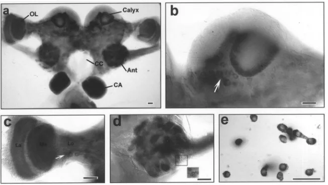
Analysis for NADPH-diaphorase activity in the brain-corpora cardiaca-brain-corpora allata complex of 6-day-old mated adult females revealed that, in the cerebral gang-lion the highest NADPH-diaphorase activity was in the chemosensory neuropiles of the antennal lobes (Fig. 1A). Intermediate activity was detected in the neuropiles of the optic lobe and low activity in the central brain as indicated by NBT-formazan precipitation (Fig. 1A). In the protocerebral ganglion, the calyx of the mushroom bodies exhibited some NADPH-diaphorase activity (Fig. 1B). Presumably, what was labeled in the neuropile had originated from extrinsic neurons since Kenyon cell somata of the mushroom bodies showed no detectable staining. Several neurosecretory cells at the pars inter-cerebralis were stained with variable intensities (Fig. 1B). In the optic lobe, NADPH-diaphorase staining was clearly demonstrated in lamina, medulla and lobula (Fig. 1C). Several somata bellow the lobula region were also stained. The highest NADPH-diaphorase activity in the
Fig. 1. Histochemical localization of NADPH-diaphorase, as made visible by NBT-formazan precipitation, in the brain-corpora cardiaca-corpora allata complex of a 6-day-old, mated adult female. (a) Posterior view of the complex. (b) NADPH-diaphorase staining in the calyx of the mushroom body. Arrow indicates somata of a group of neurosecretory cells at pars intercerebralis. (c) NADPH-diaphorase staining in the optic lobe consisting of lamina (La), medulla (Me) and lobula (Lo). Arrow indicates several stained somata bellow the lobula region. (d) Frontal view of diaphorase staining in the antennal glomeruli. Inset, a cluster of cell bodies in the ventral part of the antennal at different focal plane. (e) NADPH-diaphorase staining in dissociated corpus allatum (CA) cells. Scale bar, 50µm.
brain was found in the glomeruli of the antennal lobe (Fig. 1D). The majority of clustered stained cell bodies found in the dorsal and ventral region of the antennal lobe send their neurites into the glomeruli. In the retro-cerebral complex, while the corpora cardiaca showed no staining, the CA exhibited the highest NADPH-diaphor-ase activity of all regions in the complex (Fig. 1A). Although nerve terminals are abundant in the CA, NADPH-diaphorase expression in the CA was clearly endogenous occurring in every cell, as was indicated by histochemical staining in the cytosol of dissociated cor-pus allatum cells (Fig. 1E). Control experiments, involv-ing histochemical staininvolv-ing without the addition of b -NADPH, showed that NADPH-diaphorase staining of dissociated corpus allatum cells was, in fact, dependent on b-NADPH (data not shown). Since cells exhibited NADPH-diaphorase activities in the brain-retrocerebral complex were located at difference focal planes, the NBT-formazan intensity can only be easily compared under microscope. Visual differentiation of various regions was especially clear when NADPH-diaphorase has reacted for only 1.5 h (data not shown). In summary, NADPH-diaphorase activities in the brain-retrocerebral complex of day-6 mated females exhibited following orders: CA.antennal lobes.optic lobes.mushroom bodies . neurosecretory cells.
Expression of NADPH-diaphorase in the CA of D.
punctata appears to be constitutive since it was found in
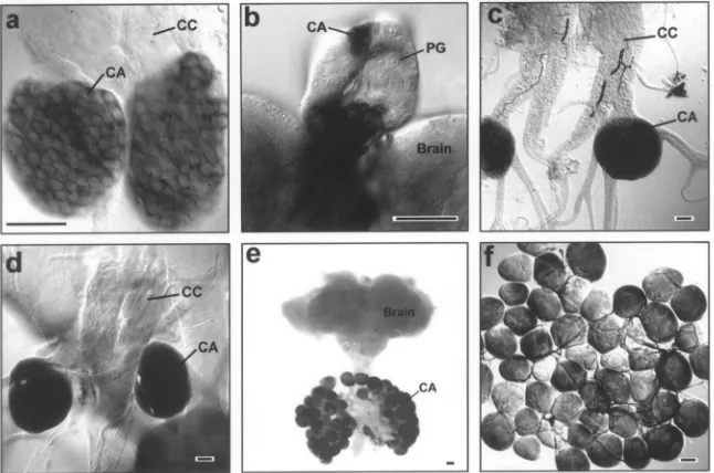
Fig. 2. Whole-mount NADPH-diaphorase histochemistry in the CA of various insects, as made visible by NBT-formazan precipitation. (a) Corpora cardiaca-corpora allata complex from a D. punctata embryo. (b) The ring gland of a final instar larva of the fruit fly, D. melanogaster. (c) Corpora cardiaca-corpora allata complex of the oviparous cockroach, P. americana (d) Corpora cardiaca-corpora allata complex of the house cricket, A.
domesticus. (e) Brain-retrocerebral complex of an adult male of the lepidopteran, L. loreyi. (f) Monolayer of giant corpus allatum cells of L. loreyi.
CA, corpus allatum; CC, corpus cardiacum; PG, prothoracic gland. Scale bar, 50µm.
Fig. 3. Changes in NADPH-diaphorase activity in CA of D. punctata adults. (a) NADPH-diaphorase histochemistry showing intensity of NBT-formazan precipitation in various CA of mated adult females. (b) Quantitative changes in NADPH-diaphorase activity, detected using XTT as a substrate, in the CA of mated adult females, unmated adult females, and adult males. (c) Correlation between NADPH-diaphorase activity and terminal oocyte length. NADPH-diaphorase activity and terminal oocyte length were determined from the same insects. Each data point represents the mean±SEM with the number of measurements indicated. Scale bar, 50µm.
in the larval ring gland of D. melanogaster also con-tained putatively NOS-related NADPH-diaphorase. NADPH-diaphorase histochemistry clearly indicated a tissue specific NADPH-diaphorase staining in corpus allatum cells, which was not found in prothoracic gland cells of the larval ring gland (Fig. 2B). NADPH-diaphor-ase activity was also demonstrated in the American cockroach (Periplaneta americana), the house cricket (Acheta domesticus) and a lepidopteran (Leucania
loreyi), which indicates a common occurrence of
puta-tively NOS-related NADPH-diaphorase in the CA of most, if not all insects (Fig. 2C–F).
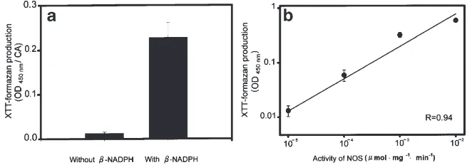
decreased thereafter. In contrast, in virgin females and adult males, the CA in the first 12 days of adulthood stained very poorly (data not shown), with a staining intensity similar to that of the CA of day 0 adult females. The changes in NADPH-diaphorase activity found with histochemical staining were verified by measuring XTT-formazan production, which is water soluble and more suitable for spectrophotometric quantitation than insoluble NBT-formazan. To validate the use of XTT as a substrate for the detection of fixation-insensitive NADPH-diaphorase activity, we first demonstrated that XTT-formazan production by fixed CA is b-NADPH dependent (Fig. 4A). Without presence ofb-NADPH in the reaction solution, only minimal background forma-zan was detected. Moreover, neuronal NOS, purified from rat brain, induced a dose-dependent XTT-formazan production (Fig. 4B). Therefore, XTT was a suitable substrate for the detection of fixation-insensitive NADPH-diaphorase activity. Using XTT as a substrate, we found that NADPH-diaphorase activity in the CA of mated, adult females increased gradually from day 0 to day 6 and then decreased slowly back to the initial level by day 12 (Fig. 3B). The highest NADPH-diaphorase activity occurred in the CA of mated females with ter-minal oocytes 1.5 mm in length (Fig. 3C). By contrast, NADPH-diaphorase activity in the CA of both unmated adult females and adult males remained low throughout the first 12 days of adulthood (Fig. 3B).
In adult females of D. punctata, the synthesis of JH by the CA is neurally inhibited, a neural inhibition that can be removed by mating or severing the nerve connec-tions between the CA and brain (Tobe and Stay, 1985). To examine whether neural signals affected NADPH-diaphorase activity in the CA, we transected the neural connection between the right corpus cardiacum and its adjacent corpus allatum, while the neural connection from the left corpus cardiacum to its adjacent corpus allatum was left intact. Females that were operated on just after adult emergence were kept virgin and exam-ined on each of the following 12 days. In unilaterally NCA-I-transected females, the basal oocytes grew in
Fig. 4. Validation of the use of XTT as a substrate for quantitation of NADPH-diaphorase activity. (a) Dependence of water-soluble XTT-formazan production onb-NADPH. A so-called split-gland design was used: one corpus allatum of a pair, per day-6 mated female, was incubated with XTT andb-NADPH, while the other of the same pair served as control and was incubated with XTT but withoutb-NADPH. Student t-test, p,0.001.
N=10 CA pairs. (b) Dose-dependent XTT-formazan production using NOS purified from rat brain. N=7 for each data point.
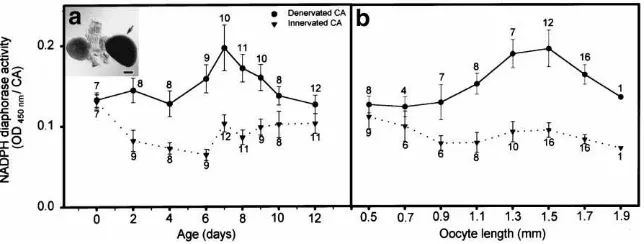
length from 0.66 ± 0.02 mm (n=5) on day 0 to 1.17 ± 0.03 mm (n=9) on day 4, indicating that the denervated right corpus allatum was synthesizing a considerable amount of JH. The denervated gland exhibited a cycle of NADPH-diaphorase activity as indicated by XTT-for-mazan production that peaked on days 6–8 when oocyte length reached to 1.3–1.5 mm and declined gradually thereafter (Figs. 5A and B). In contrast, NADPH-diapho-rase activity in the contralateral intact CA remained at a low level during the 12 days following the surgery. The significantly higher XTT-formazan production in the denervated corpus allatum than in the intact corpus allatum of day-7 virgin females was also verified by NBT-formazan staining (Fig. 5A, inset).
4. Discussion
NADPH-diaphorase has been used as a reliable marker enzyme in the identification of NOS-producing cells in vertebrates and insects (Hope et al., 1991; Mu¨ller, 1994). As has already been described for mam-malian tissue (Matsumoto et al., 1993), the selectivity of the histochemical staining is presumably caused by the resistance of NOS to paraformaldehyde fixation. Thus, it is likely that the cytoplasmic NADPH-diaphorase staining of neuronal and corpus allatum cells in the cock-roach D. punctata reflects NOS expression. In most stud-ies on insects it has been assumed that the NADPH-diaphorase histochemical technique, which we used in this study, is detecting NOS. But, unless NO release is measured, it may be that NADPH-diaphorase staining in the CA is detecting enzyme activities other than NOS. It must be realized, however, that fixation-insensitive NADPH-diaphorase activity in insects has not been dem-onstrated for enzymes other than NOS.
Using NADPH-diaphorase staining, we localized putative NOS expressing neurons in the brain of D.
punctata adult females (Figs. 1A–D) and found it to be
Fig. 5. Neural inhibition of NADPH-diaphorase activity in virgin females. (a) Quantitative changes in NADPH-diaphorase activity in CA of adult virgin females after unilateral CA denervation since day 0 using XTT as a substrate. Inset, NADPH-diaphorase staining of a corpus allatum pair from a 7-day-old virgin adult female using NBT as a substrate. The right corpus allatum of the female was denervated on day 0, while the left corpus allatum’s neural connectives were left intact. The denervated CA was indicated with an arrow. Scale bar, 50µm. (b) Correlation between NADPH-diaphorase activity and terminal oocyte length from the same insects. Each data point represents the mean±SEM with the number of measurements indicated.
Mu¨ller, 1997; Bicker, 1998). In this report, we focused on NADPH-diaphorase activity in the CA, something that has not been reported in any other insect species. We examined the CA of five different insect species, P.
americana, D. punctata, A. domesticus, L. loreyi and D. melanogaster, and in all the CA showed distinct
NADPH-diaphorase activity. Even more interesting, is the fact that NADPH-diaphorase activity was present in all cells of the CA. In the anterior pituitary of the rat, an analogue of the insect CA, NOS activity is limited to gonadotroph and in folliculo-stellate cells, which regu-late the hormone secretion, presumably through the action of NO (Ceccatelli et al., 1993). It seems unlikely, though, that NO is involved in the release of JH from the CA, since this terpenoid hormone readily diffuses through the plasma membrane shortly after it is synthe-sized.
What might the function of NADPH-diaphorase be in corpus allatum cells? Cyclic changes in NADPH-diapho-rase activity in the CA of mated females, but not in those of virgin females or males (Fig. 3), suggests that this enzyme may be involved, in some way, in regulating the activity of the CA. Following cyclic events occur in the CA of females mated on day 0 and raised under our colony conditions: DNA synthesis peaks in the gland on day 2, mitosis peaks on day 3, JH synthesis peaks on day 4, cell size peaks on day 5, and autophagic activity peaks on day 6 (Cheng and Chiang, 1995; Chiang et al. 1996, 1998). Since NADPH-diaphorase activity in the CA slowly increases through day 6 and thereafter gradu-ally diminishes, it is tempting to speculate that this enzyme may be involved in autophagy and, by exten-sion, cell atrophy, which results in a long-term arrest of JH synthesis. Our findings with the denervated corpus allatum of virgin females support such a hypothesis. NADPH-diaphorase activity in the denervated corpus allatum of a virgin female increased and decreased, as in mated females, but the enzyme’s activity remained at a low level in the contralateral innervated corpus allatum
of the same female (Fig. 5A). This indicates that the brain inhibits NADPH-diaphorase activity. It is notable, however, that the highest level of NADPH-diaphorase activity in the denervated CA of a virgin female never approached the peak level in mated females (Figs. 3B and 5A). Thus, if NADPH-diaphorase is responsible for cellular autophagy and atrophy, cells in the denervated CA of virgin females should decrease in size at a slower rate than those in intact CA of mated females. And indeed, this is exactly what happens (Chiang et al., 1998).
Acknowledgements
The authors thank Prof. G.L. Holbrook (Pennsylvania State University) for critical reading of the manuscript, and the National Science Council of the Republic of China for financially supporting this research under Con-tract 033 and NSC89-2311-B007-014-B12.
References
Bicker, G., 1998. NO news from insect brains. Trends Neurosci. 21, 349–355.
Ceccatelli, S., Hulting, A.-L., Zhang, X., Gustafsson, L., Villar, M., Hokfelt, T., 1993. Nitric oxide synthase in the rat anterior pituitary gland and the role of nitric oxide in regulation of luteinizing hor-mone secretion. Pro. Natl. Acad. Sci. USA 90, 11292–11296. Cheng, H.-W., Chiang, A.-S., 1995. Autophagy and acid phosphatase
activity in the corpora allata of adult mated females of Diploptera
punctata. Cell Tissue Res. 281, 109–117.
Chiang, A.-S., Gadot, M., Schal, C., 1989. Morphometric analysis of corpus allatum cells in adult females of three cockroach species. Mol. Cell. Endocrinol. 67, 179–184.
Chiang, A.-S., Tsai, W.-H., Holbrook, G.L., Schal, C., 1996. Control of cell proliferation in the corpora allata during the reproductive cycle of the cockroach Diploptera punctata. Arch. Insect Biochem. Physiol. 32, 299–313.
Dow, J.A.T., Maddrellet, S.H.P., Davies, S.-A., Skaer, N.J.V., Kaiser, K., 1994. A novel role for the nitric oxide-cGMP signaling path-way: the control of epithelial function in Drosophila. Am. J. Phy-siol. 266, R1716–R1719.
Elphick, M.R., Green, I.C., O’Shea, M., 1993. Nitric oxide synthesis and action in an invertebrate brain. Brain Res. 619, 344–346. Elphick, M.R., Rayne, R., Riveros-Moreno, V., Moncada, S., O’Shea,
M., 1995. Nitric oxide synthesis in locust olfactory interneurones. J. Exp. Biol. 198, 821–829.
Garthwaite, J., 1991. Glutamate, nitric oxide and cell-cell signaling in the nervous system. Trends Neurosci. 14, 60–67.
Garthwaite, J., Charles, S.L., Chess-Williams, R., 1988. Endothelium-derived relaxing factor release on activation of NMDA receptors suggests roles as intercellular messenger in the brain. Nature 336, 385–388.
Hope, B.T., Michael, G.J., Knigge, K.M., Vincent, S.R., 1991. Neu-ronal NADPH-diaphorase is a nitric oxide synthase. Proc. Natl. Acad. Sci. USA 88, 2811–2814.
Kuzin, B., Roberts, I., Peunova, N., Enikolopov, G., 1996. Nitric oxide regulates cell proliferation during Drosophila development. Cell 87, 639–649.
Matsumoto, T., Nakane, M., Pollock, J.S., Kuk, J.E., Fo¨rstermann, U., 1993. A correlation between soluble brain nitric oxide synthase and NADPH-diaphorase activity is only seen after exposure of the tissue to fixative. Neurosci. Lett. 155, 61–64.
Mu¨ller, U., 1994. Ca2+/calmodulin-depend nitric oxide synthase in
Apis mellifera and Drosophila melanogaster. Eur. J. Neurosci. 6,
13362–13370.
Mu¨ller, U., 1996. Inhibition of nitric oxide synthase impairs a distinct form of long-term memory in the honeybee Apis mellifera. Neuron 16, 541–549.
Mu¨ller, U., 1997. The nitric oxide system in insects. Prog. Neurobiol. 51, 363–381.
Mu¨ller, U., Hildebrandt, H., 1995. The nitric oxide/cGMP system in the antennal lobe of Apis mellifera is implicated in integrative pro-cessing of chemosensory stimuli. Eur. J. Neurosci. 7, 2240–2248. Rettori, V., Belova, N., Dees, W.L., Nyderg, C.L., Gimeno, M., McCann, S.M., 1993. Role of nitric oxide in the control of luteiniz-ing hormone-releasluteiniz-ing hormone release in vivo and in vitro. Proc. Natl. Acad. Sci. USA 90, 10130–10134.
Ribeiro, J.M.C., Hazzard, J.M.H., Nussenzveig, R.H., Champagne, D.E., Walker, F.A., 1993. Reversible binding of nitric oxide by a salivary heme protein from a bloodsucking insect. Science 260, 539–541.
Ribeiro, J.M.C., Nussenzveig, R.H., 1993. Nitric oxide synthase activity from a hematophagous insect salivary gland. FEBS Lett. 330, 165–168.
Roehm, N.W., Rodgers, G.H., Hatfield, S.M., Glasebrook, A.L., 1991. An improved colorimetric assay for cell proliferation and viability utilizing the tetrazolium salt XTT. J. Immunol. Meth. 142, 257– 265.
Scharrer, B., 1987. Insects as models in neuroendocrine research. Annu. Rev. Entomol. 32, 1–16.
Stay, B., Tobe, S.S., 1977. Control of juvenile hormone biosynthesis during the reproductive cycle of a viviparous cockroach. I. Acti-vation and inhibition of corpora allata. Gen. Comp. Endocrinol. 33, 531–540.
Tobe, S.S., Stay, B., 1985. Structure and regulation of the corpus alla-tum. Adv. Insect Physiol. 18, 305–432.