www.elsevier.com/locate/ibmb
Molecular characterization of a cDNA from the true armyworm
Pseudaletia unipuncta
encoding
Manduca sexta
allatotropin
peptide
1Peter F. Truesdell
a, Peter M. Koladich
b, Hiroshi Kataoka
c, Kuniaki Kojima
c, Akinori
Suzuki
c, Jeremy N. McNeil
d, Akira Mizoguchi
e, Stephen S. Tobe
b, William G.
Bendena
a,*aDepartment of Biology, Queen’s University, Kingston, ON, Canada bDepartment of Zoology, University of Toronto, Toronto, ON, Canada
cGraduate School of Agriculture and Life Sciences, University of Tokyo, Bunkyo-ku, Tokyo 113, Japan dDepartement de biologie, Universite Laval, Ste. Foy, P.Q., Canada
eBiological Sciences, Graduate School of Science, Nagoya University, Furo-cho, Chikusa-ku, Nagoya-shi, Aichi 464, Japan
Received 31 October 1999; received in revised form 31 December 1999; accepted 25 January 2000
Abstract
Allatotropin (AT) is an insect neuropeptide isolated from the tobacco hornworm, Manduca sexta, stimulates juvenile hormone (JH) biosynthesis by the corpora allata. A cDNA isolated from the true armyworm, Pseudaletia unipuncta, encodes a 135 amino acid AT precursor peptide which contains the AT peptide, with processing sites necessary for its endoproteolytic cleavage and amidation, plus two additional peptides of unknown function. The encoded AT peptide is identical to that isolated fromM. sexta
and Agrius convolvuli.
Southern blot analysis indicated that AT is a single copy gene per haploid genome and is present in two allelic forms. A single transcript of approximately 1.5 kilobases was detected by northern blot analysis. The expression of the AT gene was analyzed during development from sixth instar larvae to five day-old moths. Initial expression was observed in late pupae and this expression was maintained throughout the adult stages in both sexes. In one day-old moths, expression was at its lowest level of the stages that express AT mRNA but levels increased in day 3 and day 5 adults. This pattern of AT expression in adultP. unipunctamoths mirrors that of JH biosynthesis and supports the notion that AT may act in the adult stages.
Immunohistochemistry and in situ hybridization revealed that AT expression was localized to numerous structures of the nervous system, suggesting that AT may have functions distinct from regulation of JH biosynthesis. 2000 Elsevier Science Ltd. All rights reserved.
Keywords:Juvenile hormone; Lepidoptera; Neuropeptide; Insect
1. Introduction
Juvenile hormone (JH) exerts a central role in meta-morphosis, adult sexual maturation and reproduction in most insect species (Tobe and Stay, 1985). The
Abbreviations:AT, allatotropin; AST, allatostatin; CA, corpora allata; NCC, nervi corporis cardiaci; SOG, suboesophageal ganglia; JH, juv-enile hormone.
* Corresponding author. Tel.:+1-613-533-6121; fax:+ 1-613-533-6617.
E-mail address:[email protected] (W.G. Bendena).
0965-1748/00/$ - see front matter2000 Elsevier Science Ltd. All rights reserved. PII: S 0 9 6 5 - 1 7 4 8 ( 0 0 ) 0 0 0 4 0 - 0
biosynthesis of JH by the corpora allata (CA) of insects can either be stimulated or inhibited by neuropeptides termed allatotropins (ATs) or allatostatins (ASTs),
1 Peptide designations have been assigned using the first three
692 P.F. Truesdell et al. / Insect Biochemistry and Molecular Biology 30 (2000) 691–702
respectively. Several different AST peptide sequences have been determined both within and between Orders (Bendena et al., 1997). Much less is known about AT although several allatotropic factors have been identified in a variety of different insects. Lorenz and Hoffmann (1995) partially purified an allatotropic substance from suboesophageal ganglion (SOG) extracts of crickets,
Gryllus bimaculatus and Acheta domesticus, which stimulated JH biosynthesis in CA. In adult Locusta migratoria, allatotropic activity was detected in the SOG, brain and corpora cardiaca (CC) (Gadot et al., 1987). Gradient separation of extracts of each of these tissues suggested the presence of more than one allato-tropic factor with molecular mass estimated to be 0.7 to 2.0 kDa (Rembold et al., 1986; Ferenz and Diehl, 1983). In contrast, a much larger 20 kDa AT stimulated JH biosynthesis in the wax moth, Galleria mellonella
(Bogus and Scheller, 1994). Immunocytochemical analy-sis localized this protein to two medial neurosecretory cells in the pars intercerebralis (PI) and to the CC (Bogus and Scheller, 1994). Cells of the PI were implicated as a source of AT in G. mellonella, as they elicited super-numerary larval moults when implanted into fifth instar larvae and stimulated JH biosynthesis in vitro by larval CA (Muszynska-Pytel, 1987). Removal of these medial cells from the brain destroyed the allatotropic activity (Bogus and Scheller, 1994).
There appears to be a large discrepancy in molecular mass between allatotropic factors found in different stages of development and/or species of insects. In con-trast to the 20 kDa AT in larval brains reported by Bogus and Scheller (1994), ana-amidated AT peptide (Manse-AT) thirteen residues in length, has been purified from head extracts of pharate adult Manduca sexta (Kataoka et al., 1989). To date, this is the only known AT whose primary structure has been determined. This Manse-AT stimulated JH biosynthesis by CA of adult M. sextabut had no effect on the CA of larvae or pupae of this spec-ies. It also strongly stimulated CA activity of the adult female moth, Heliothis virescens, but was unable to do so in the beetle,Tenebrio molitor, the grasshopper, Schi-stocerca nitens, or the cockroach,Periplaneta americana
(Kataoka et al., 1989). This evidence suggests that the activity of Manse-AT may be restricted to the adult stage of Lepidoptera.
Taylor et al. (1996) isolated and sequenced a Manse-AT genomic clone fromM. sextaand provided evidence to suggest that the Manse-AT gene is expressed as three alternatively spliced mRNAs which encode distinct pre-cursor proteins which upon processing would release the AT peptide. Manse-AT gene is expressed in the brain and nerve cord of larvae, pupae and adults of M. sexta
(Taylor et al., 1996). Manse-AT has no effect on CA of
M. sextalarvae, and pupal CA do not produce appreci-able JH, so the expression of Manse-AT in these stages of development may be related to activities independent
of the stimulation of JH biosynthesis in the adult CA. Functional assays and distribution studies support additional roles for Manse-AT. Manse-AT has been shown to function as a cardioaccelerator in adult M. sexta(Veenstra et al., 1994) and inhibit midgut ion trans-port in day 2 fifth instar M. sexta larvae (Lee et al., 1998). Multiple functional activities is also suggested by the distribution of Manse-AT immunoreactive cells throughout M. sexta development that encompasses numerous structures in the nervous system (Veenstra and Hagedorn, 1993; Veenstra et al., 1994; Zitnan et al. 1993, 1995). Immunological evidence suggests that Manse-AT-like peptides may be present in the nervous system of other insects, e.g. Drosophila melanogaster
(Zitnan et al., 1993), however, the function of these pep-tides in these species remain unknown. It is possible that distantly related insects contain very different ATs. The inability of Manse-AT to stimulate JH biosynthesis in species outside the Order Lepidoptera suggests that if similar peptides do exist, they are not involved in reg-ulating CA activity.
Barth (1965) proposed that in long lived insects, where mating may be suppressed for varying lengths of time, a complex neuroendocrine system would have evolved to regulate pheromone production and mating behaviour. This has been examined in the true army-worm, Pseudaletia unipuncta, a migrant species, and several aspects of reproduction in both sexes involve JH. JH is involved, either indirectly or directly, in the regu-lation of the female pheromone communication system and oocyte maturation, including vitellogenin synthesis (Cusson and McNeil, 1989a,b; Cusson et al., 1990, 1994b). In males, JH plays a role in the regulation of the responsiveness to the female sex pheromone and, probably in the development of accessory reproductive glands (Cusson et al., 1994a). In some species of insects, there is evidence to suggest that migration is also under the control of JH (Rankin et al., 1986), with different JH titer thresholds for either migration or reproduction (Rankin and Riddiford, 1978). In response to cues of an impending habitat deterioration the titer of JH would exceed a minimal level to initiate migration but not the higher threshold required for the initiation of sexual maturation and reproduction. In contrast, under favor-able conditions the titer of JH would rise rapidly to exceed the upper threshold to promote events associated with reproduction.
determine if there was any sex- or stage-specific expression indicative of additional functions.
Specific regions of the brain are known to synthesize AT peptides, which are transported to the CA and influ-ence its activity. The cellular localization of AT expression was determined, to provide evidence for a role in JH regulation and to provide insight into additional functions for this peptide inP. unipuncta.
2. Materials and methods
2.1. Animals
P. unipunctawere maintained under summer-like con-ditions at 25±1°C, with 40±10% relative humidity and a 14 L:10 D cycle. Larvae were fed ad libitum on arti-ficial pinto bean diet until pupation then sexed (Jansons et al., 1996). Adults were fed an 8% (w/v) sucrose sol-ution.
2.2. Isolation and characterization of a Pseudaletia unipuncta AT cDNA clone
A 1.2 kb cDNA clone coding for theAgrius convolvuli
preproAT (Kataoka; unpublished) was used as a hybridization probe to screen aP. unipunctabrain cDNA library, as described in Jansons et al. (1996). Purified DNA contained within BluescriptII EcoRI-XhoI (Stratagene) from isolated P. unipuncta 8Zap II cDNA clones were sequenced on both strands using a Taq Dye-deoxy terminator cycle sequencing kit and analysis of the products on an ABI 373A DNA sequencer (Applied Biosystems) at the Core Facility for Protein/DNA chem-istry (Queen’s University).
2.3. Southern and northern blotting
To isolate genomic DNA, individual P. unipuncta
pupae were frozen and ground, under liquid N2, to a fine
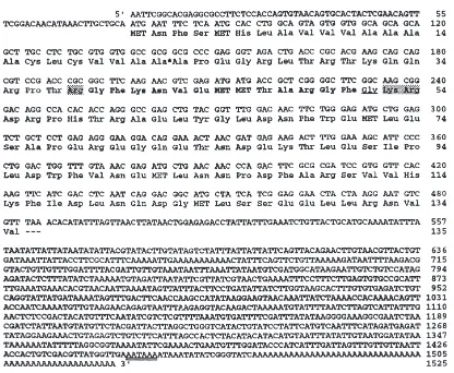
powder using a mortar and pestle. The powder was sus-pended in 10 mM Tris-HCl pH 8.0, 100 mM EDTA, 0.5% SDS and then processed as described (Ding et al., 1995). Aliquots containing 20µg of genomic DNA were digested with selected restriction enzymes, then separ-ated by electrophoresis on a 1% (w/v) agarose gel. The DNA was depurinated, denatured and neutralized prior to vacuum-assisted transfer to Hybond-N+ nylon mem-branes (Amersham). A 545 bp PCR fragment, amplifed from the P. unipuncta AT cDNA clone using primers TCGGAGGAACTACTAAGG (59primer; position 457–
475, Fig. 1) and CTTCCTTATATGGCTTGG (39
primer; position 984–1002, Fig. 1) was used as probe for hybridization after 32P-oligolabelling (Pharmacia).
Developmental stages from early sixth instar to 5-day-old adults were chosen from mRNA extraction as the
rates of JH biosynthesis are known in these stages (Cusson et al., 1993). Poly A+ enriched RNA was iso-lated from 100–500 brains from each developmental stage using the Quickprep micro mRNA purification kit (Pharmacia). Approximately 5 µg RNA was denatured and resolved by glyoxal-DMSO electrophoresis in a 1.2% agarose gel (Sambrook et al., 1989) using RNA as markers (RNA ladder, BRL). RNA was transferred to
Hybond N+ (Amersham) nylon membranes and
hybridized with the 545 bp AT PCR fragment described above for 1 h at 68°C in QuikHyb solution (Stratagene). RNA blots were washed twice for 15 mins with 2×SSC, 0.1% SDS and once for 30 min with 0.1% SSC, 0.1% SDS at 60°C and then exposed to Kodak XAR film. After exposure, the probe was removed from the mem-brane and hybridized with a labeled Pst/HindIII fragment containing the coding region of the Drosophila mel-anogaster a-tubulin gene (Mischke and Pardue, 1982). Densitometry was performed using an ImageQuant com-puting densitometer (Molecular Dynamics).
2.4. Wholemount immunocytochemistry
immunoreactiv-694 P.F. Truesdell et al. / Insect Biochemistry and Molecular Biology 30 (2000) 691–702
Fig. 1. Nucleotide sequence of the AT precursor cDNA inP. unipuncta.The translated portion of the sequence is organized into codons with the deduced amino acid sequence shown directly below. The allatotropin peptide sequence is shown in bold. Proteolytic cleavage sites are shaded and the glycine residue required for amidation is single underlined. A signal peptide cleavage site is indicated by a bold star and a potential polyadenylation signal is double underlined. This sequence has been deposited in the Genbank database (accession no. AF212926).
ity was visualized when either the primary or the second-ary antibody incubation was omitted from the procedure. Images of Manse-AT-like immunoreactivity were obtained with a Bio-Rad MRC600 laser scanning con-focal microscope employing an argon-krypton laser and BHS filter block.
2.5. Wholemount in situ hybridization analysis of AT mRNA expression
Allatotropin cDNA in pBluescript was linearized with Eco RI then treated with 50 µg/ml proteinase K in 20 mM EDTA and 0.4% (w/v) SDS for 1 h at 50°C. The template was purified by two phenol/chloroform/isoamyl alcohol extractions and then ethanol precipitated. The transcription reaction contained 1µg of template DNA, 1X transcription buffer (Promega), 50 mM DTT, 1.2 U/µl T7 RNA polymerase, 1.5 U/µl RNAGuard (Pharmacia) and 1X digoxigenin (DIG) RNA labeling mix (Boehringer Mannheim). This solution was incu-bated for 2 h at 37°C and then treated with DNAse I (20µg/ml) for 15 min. EDTA was added to a final con-centration of 20 mM and the RNA precipitated with 0.5
M sodium acetate and 4 volumes of 100% ethanol. The recovered DIG-labeled RNA probe was resuspended in water. Tissue preparation, hybridization and post-hybridization washes were as previously described (Fuse´ et al., 1998). Cells expressing AT mRNA were detected by incubating the tissues with 1% goat serum, PBS con-taining 0.3% Triton X-100 and 0.75 U/ml anti-digoxig-enin peroxidase conjugated antibody for 12 h at 4°C. Tissues were then incubated in PBS containing 3,39 diaminobenzidine tetrahydrochloride, 2 mg/ml b -d(+)glucose and 0.4 mg/ml NH4Cl. Glucose oxidase (2.5
3. Results
3.1. P. unipuncta cDNA characterization
The complete nucleotide sequence of the 1525 nucleo-tide P. unipuncta AT cDNA is shown in Fig. 1. The cDNA contains 78 nucleotides of untranslated sequence upstream of a single open reading frame beginning at position 79 and ending at position 484 with the trans-lation stop codon TAA. The untranslated sequence that follows is extremely A/T-rich (73%) and extends for 1042 nucleotides. There are two overlapping consensus polyadenylation signals (AATAAA) beginning at pos-itions 1450 and 1455, which are 19 and 24 nucleotides upstream of the poly A tail, respectively. The matureP. unipuncta AT peptide located between residues 39 and 52 (Fig. 1), is identical to the sequence of purified Manse-AT peptide (Kataoka et al., 1989) and to that pre-dicted from M. sexta AT cDNA (Taylor et al., 1996). The Pseun-AT peptide within the precursor is flanked by potential Arg and LysArg endoproteolytic cleavage sites, and ends with a glycine residue, the signal for carboxy-terminal amidation by peptidyl-glycine-a-amidating monooxygenase (Eipper et al., 1992). Recognition of the proteolytic cleavage sites would result in the production of two additional peptides of 15 and 81 amino acids. The basic organization of the Pseun-AT precursor is similar to that of A. convolvuli (Kataoka, unpublished) and the 131 amino acid precursor of M. sexta (Taylor et al., 1996) with 83% and 84% amino acid identity, respectively (Fig. 2). Comparison of the AT cDNA sequences between species exhibited an overall simi-larity of 78%, with the greatest sequence variation out-side the coding region.
Southern blot analysis was performed to determine the number of AT genes present in theP. unipunctagenome. For digestion of genomic DNA, restriction
endonucle-Fig. 2. Comparison of AT precursor peptides from P. unipuncta
(Pseun-AT),M. sexta(Manse-AT) (Taylor et al., 1996) andA. convol-vuli(Agrco-AT) (Kataoka, unpublished). The sequences of the AT pre-cursors were aligned using the PCGENE program, PALIGN. The shortest AT precursor fromM. sextawas used in this comparison. Star symbols represent amino acids identical to each of the precursors. The mature AT peptide is shown in bold and the processing sites are italic-ized.
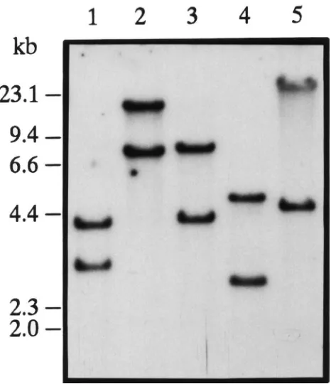
Fig. 3. Southern blot hybridization analysis ofP. unipunctagenomic DNA. Samples of genomic DNA (20µg/lane) from individual moths were digested with restriction endonucleasesEco RI,HindIII,SalI,
SmaI andXbaI (lanes 1-5) and separated on a 1% agarose gel. A
32P-labeled 545 bp PCR product, amplified from the cDNA clone, was
used as a probe.λ-Hind III DNA size markers (kbp) are indicated on the left.
Fig. 4. Graphical representation of the amounts of AT mRNA from brains at different developmental stages. Poly A+RNA was isolated
from two independent sets of 100 brains at each developmental stages of the armyworm,P. unipunctaand hybridized with a32P-labeled AT
696 P.F. Truesdell et al. / Insect Biochemistry and Molecular Biology 30 (2000) 691–702
ases were chosen based on their inability to digest the cDNA sequence. A 545 bp PCR fragment spanning resi-dues 457 to 1002 (Fig. 1) shown by PCR analysis with genomic DNA to be uninterrupted within the genome was used as a hybridization probe. Hybridization with this probe to genomic DNA from individual pupae resulted in two bands of hybridization of different size but similar intensity (Fig. 3). This indicates that the P. unipuncta AT gene is present as a single copy gene in the haploid genome but contains restriction fragment polymorphisms between the two alleles.
The pattern of mRNA expression of theP. unipuncta
AT gene during development was examined by Northern blots (Fig. 4). The AT transcript is not expressed in sixth instar larvae, pre-pupae or early pupae. A single tran-script of approximately 1.5 kb was detected first in late pupae. The level of expression in day 1 female and male moths was respectively 23% and 30% lower than that in late pupae. The level of expression increased slightly in day 3 moths and reached higher levels by day 5 for both sexes (Fig. 4).
3.2. Cellular localization of AT expression
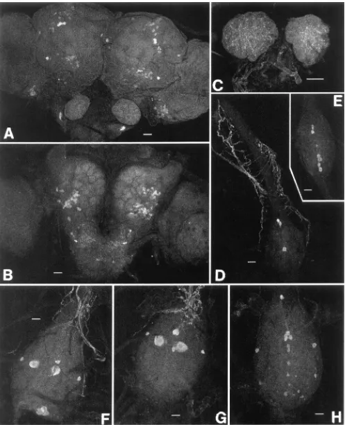
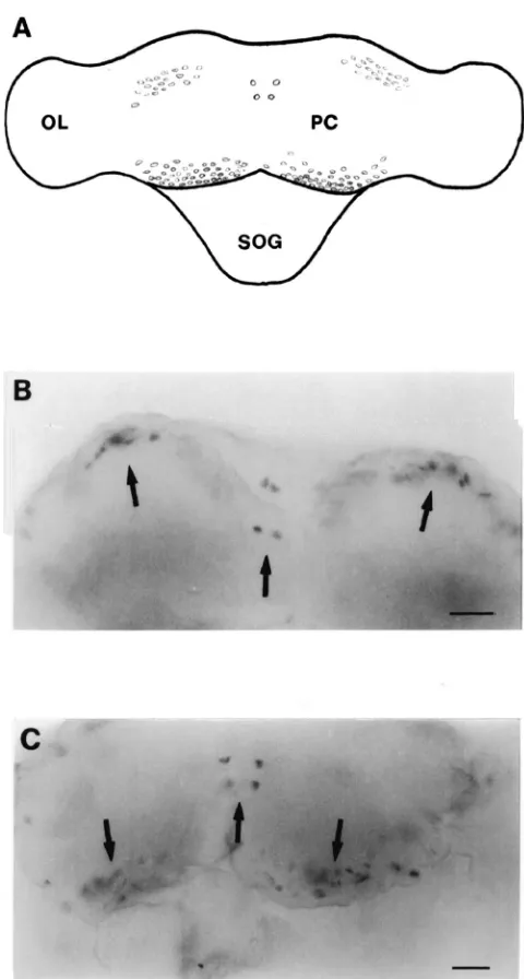
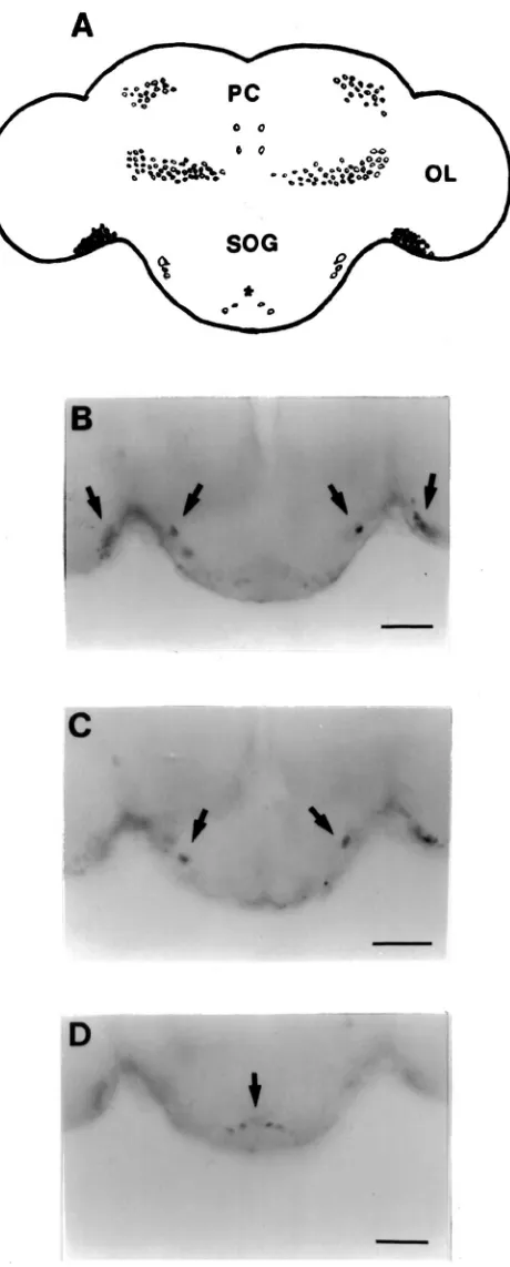
Several groups of cells were detected in the brain both by staining with a specific AT monoclonal antibody (Fig. 5A and B) and in situ hybridization with a gene-specific probe (Figs. 6–8). Both techniques detect two symmetri-cal pairs of strongly immunoreactive/hybridizing cells located along the midline of the brain in the PI (Figs. 5A and 6B and C). In the posterio-dorsal region, there was a population of approximately 10 to 15 cells which extended laterally from the medial region toward the optic lobes. In the anterior and lateral position from these cells, a group of 20–30 cells were also highly immunoreactive/hybridizing. A cluster of approximately 30–40 cells was found in the posterio-ventral region in each optic lobe adjacent to the protocerebrum (Figs. 5A and 6B and C). Both immunoreactivity and hybridization analysis revealed 65 to 70 cells in the anterio-ventral regions of each deuterocerebrum (Figs. 5B and 7). The SOG of adult P. unipuncta lies immediately below and appears fused to the ventral side of the brain. There were two relatively large cells in the anterio-lateral region of the SOG which displayed strong AT expression (Fig. 8B). Below each of these cells were a pair of cells of similar intensity (Fig. 8C). In the posterior region of the SOG at least two pairs of cells showed moderate hybridization to the probe (Fig. 8D).
No AT-hybridization was found in the corpora allata (CA). AT-specific antibodies detected faint immunoreac-tivity in axons and release points in the spaces surround-ing the CA cells (not the CA cells themselves) (Fig. 5C). The axons which contribute to this immunoreactivity have not been identified.
The abdominal nervous system in adultP. unipuncta
Fig. 6. Dorsal view of whole mount in situ hybridization analysis of the adult brain ofP. unipuncta. A digoxigenin-labeled antisense probe transcribed from Pseun-AT cDNA was used to localize the expression of AT mRNA. (A) Schematic diagram of the brain showing the cells that expressed AT mRNA. (OL) optic lobe, (PC) protocerebrum, (SOG) suboesophageal ganglion. (B), (C) Photographs of cells in the protocerebrum that contained AT mRNA. Lateral, medial and posterior cells are shown. Scale bar=125µm.
hybridiz-698 P.F. Truesdell et al. / Insect Biochemistry and Molecular Biology 30 (2000) 691–702
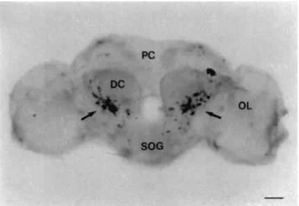
Fig. 7. Frontal view of whole mount in situ hybridization analysis of the adult brain ofP. unipuncta. Cells in the deutocerebrum (DC) that displayed a positive reaction are indicated by an arrow. (PC) protocerebrum, (OL) optic lobe, (SOG) suboesophageal ganglion. Scale bar=100µm.
ation was detected in three pairs of medial neurosecre-tory cells (Figs. 5D and E and 9D) for all adults (day 1, 3 and 5) of both sexes. The middle pair is located slightly dorsally, whereas the other two pairs are more ventrally located. Immunoreactive axonal projections form a network from each abdominal ganglia (Fig. 5D). The sixth or terminal ganglion is a fused ganglion which innervates segments six to nine. Both AT-spe-cific immunoreactivity and hybridization analysis revealed that the total number and pattern of cells was dependent on sex but not the age of the adults. For both males and females, there were three pairs of strongly hybridizing medial cells which were positioned proxi-mally within the ganglion. One pair was present in the extreme anterior region whereas the other two pairs of cells occupied a more central position in the ganglion. Laterally are one or two smaller cells showing moderate hybridization. The most distal region of the terminal ganglion in the male contained a concentrated group of eight to twelve strongly hybridizing medial cells (Fig. 9E). In contrast, many smaller and fainter cells were present in this region of the female (Fig. 9F). Identical cells were detected with AT-specific antibody (Figs. 5F, G and H).
4. Discussion
Fig. 8. Posterior view of whole mount in situ hybridization analysis of the adult brain and suboesophageal ganglion ofP. unipuncta. (A) Schematic representation showing cells that contain AT mRNA. (B– D) Photographs of the optic lobes (OL) and suboesophageal ganglion (SOG). Cells which expressed AT transcripts are indicated by arrows. PC, protocerebrum. Scale bar=70µm
to the regulation of CA activity in some species. In P. unipuncta, expression was detected in more than ten cells of the SOG that could potentially innervate the CA and regulate JH biosynthesis. Although the precise route of AT delivery to CA is unknown, AT-specific immuno-reactivity within the CA suggest that this peptide is released directly and specifically onto CA cells. In con-trast, the relatively high expression of AT during late pupal stages is difficult to explain in terms of JH biosynthesis. Kataoka et al. (1989) tested the effects of Manse-AT on CA of several insect species of different developmental stages, and suggested that the ability of Manse-AT to stimulate JH biosynthesis was restricted to adult Lepidoptera. Because JH is not believed to be synthesized by pupal CA, expression of AT at this stage may indicate that it is produced in a stable form and stored for later translation, or that the peptide has alter-native functions in the pupal brain. Taylor et al. (1996) found that, in addition to the adult, AT is expressed in both larval and pupal brains of M. sexta. The lack of effect of Manse-AT on larval and pupal CA (Kataoka et al., 1989) and the absence of JH in pupae suggest that inM. sexta,AT is not involved in regulating the activity of the CA in these stages. It is currently unknown if AT possesses any brain-specific functions during these or other stages of development.
AT mRNA and peptide have been localized to a num-ber of cells in the adult brain, SOG and ventral abdomi-nal nerve cord. Each deutocerebrum contained between 65 and 75 AT-expressing cells. A similarly large number of cells was detected in the dorso-posterior region of the protocerebrum. This region is not known to be involved in the regulation of JH biosynthesis. Although the distri-bution, arborization and termination of the axons of these cells is unknown, nerves extending from the deuto-cerebrum do innervate the antennae and antennal muscles. Possibly AT is involved in regulating the movement of the antennae and/or sensory perception. Alternatively, if the cells arborize extensively within the brain, then AT may function in a neuromodulatory or neurotransmitter capacity.
Strong AT expression was detected in three pairs of cells in each of the third, fourth and fifth abdominal gan-glia of adultP. unipuncta. Immunocytochemical analysis revealed that cells of similar position in M. sexta con-tained AT peptides (Veenstra et al., 1994). Several dif-ferent cardioactive peptides are known to be produced by cells of the abdominal ganglia and released into the hemolymph through the transverse nerve, a neurohemal organ in Lepidoptera. Application of AT purified from these cells, to the heart of adult M. sexta resulted in a significant increase in heart rate (Veenstra et al., 1994). Based on the similar pattern of expression between the two species, AT may be a cardioaccelerator in P. unipuncta.
700 P.F. Truesdell et al. / Insect Biochemistry and Molecular Biology 30 (2000) 691–702
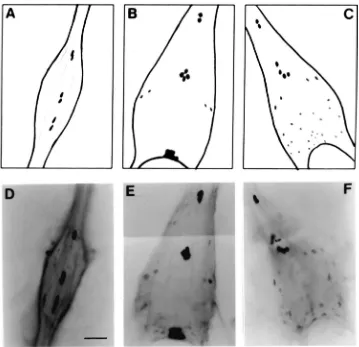
Fig. 9. Whole mount in situ hybridization analysis of the adultP. unipunctaabdominal nervous system. (A–C) Schematic representations of the fourth abdominal ganglion, and male and female terminal ganglia, respectively, showing cells which express AT mRNA. (D) Photograph of the fourth abdominal ganglion. Three pairs of cells containing AT mRNA are shown. Similar cells were identified in each of the third, fourth, and fifth abdominal ganglia of males and females. (E), (F) Photographs of male and female terminal ganglia, respectively. The anterior region of each ganglion contains three pairs of positively hybridized cells. Ten additional cells were detected in the posterior region of the male ganglion. Several smaller, weakly hybridized cells are present in the female ganglion. Scale bar=60µm.
female TAG contained AT mRNA and peptide. These cells have been identified inM. sextaand shown to con-tain AT peptides (Veenstra et al., 1994). InM. sexta, the most prominent AT expression in ventral nerve cord was in the larval TAG, and expression appeared to persist in the same TAG cells in pupae and pharate adults. Sex-specific TAG expression was not noted in M. sexta
(Bhatt and Horodyski, 1999). In contrast, strong expression of AT was also detected in a concentrated mass of ten cells in the posterior region of the terminal ganglion in P. unipuncta males. The pattern of expression suggests that AT has a role specifically in males, for example, in the contraction of muscles, such as the phallus protractor muscle required for copulation and sperm transfer. Several peptides from male insects (usually from the accessory glands) have been shown to influence female physiology and sexual behaviour when delivered through copulation (Paemen et al., 1992; Lange and Loughton, 1985; Moshitzky et al., 1996). Pae-man et al. (1991) have characterized a myotropic
pep-tide, Lom-AG-MT, that shares sequence identity with the AT peptide in ten of the thirteen amino acids. This pep-tide is found in the accessory reproductive glands of male L. migratoria and is transferred to females during copulation, to stimulate oviduct contractions. The simi-larity between the two peptides suggests that AT pos-sesses myotropic functions. However, it is possible that the peptides have evolved separately and assumed differ-ent roles. Using immunolocalization, sexual dimorphism has been previously reported in the TAG of several spec-ies:L. migratoria(Pfluger and Watson, 1988; Stevenson et al., 1994), G. bimaculatus(Yamaguchi et al., 1985),
to male sex organs; they correspond closely to a simi-larly dimorphic population of imaginal midline neurons inM. sextathat have been shown to innervate sex struc-tures (Thorn and Truman, 1994a,b).Whether the AT-immunoreactive TAG cells are DUM neurons in P. unipuncta is unclear.
In general, the nature of AT function inP. unipuncta
remains to be determined, but the data imply that it is involved in several different physiological processes. Although the AT peptides in P. unipunctaandM. sexta
are identical, differences in activity between species are not uncommon. For example, the AST peptide com-pletely inhibited JH biosynthesis in M. sexta(Kramer et al., 1991) but was only 60% effective in P. unipuncta
at ten times the concentration (Jansons et al., 1996). This may reflect differences between non-migratory and migratory species, especially as low JH is necessary for migratory flight initiated by sexually immature individ-uals. Cusson et al. (1993) have shown that environmental conditions affect the rate of JH biosynthesis in P. unipuncta, and one would predict similar influences on the level of AT expression. The developmentally regu-lated expression during summer-like conditions has been established but nothing is known of the effects of fall-like conditions. By determining the level of AT expressed under these conditions, it may be possible to better understand the regulation of JH biosynthesis under different environmental conditions. McNeil et al. (1995) have proposed that low levels of JH are responsible for the delay in sexual maturation and are associated with migratory flight. If this hypothesis is correct and the expression of AT is shown to correlate with JH levels, then AT may play an indirect role in the regulation of each of these events.
Acknowledgements
This work has been funded by Natural Sciences and Engineering Research Council (NSERC) grants OGP0036481 (WGB), A9407 (SST) and 008279 (JNM) and Fonds F.C.A.R. (JNM).
References
Barth, R.H., 1965. Insect mating behaviour: endocrine control of a chemical communication system. Science 149, 882–883. Bendena, W.G., Garside, C.S., Yu, C.G., Tobe, S.S., 1997.
Allatostat-ins: diversity in structure and function of an insect neuropeptide family. Annals. N.Y. Acad. Sci. 814, 53–66.
Bhatt, T.R., Horodyski, F.M., 1999. Expression of theManduca sexta
allatotropin gene in cells of the central and enteric nervous systems. J. Comp. Neurol. 403, 407–420.
Bogus, M., Scheller, K., 1994. Identification of allatotropin-secreting cells in the brain of an insect larva. Naturwissenschaften 81, 87–89. Carrow, G.M., Calabrese, R.L., Williams, C.M., 1984. Architecture
and physiology of insect cerebral neurosecretory cells. J. Neurosci. 4, 1034–1044.
Cusson, M., McNeil, J.N., 1989a. Involvement of juvenile hormone in the regulation of pheromone release activities in a moth. Science 243, 210–212.
Cusson, M., McNeil, J., 1989b. Ovarian development in female army-worm moths,Pseudaletia unipuncta: its relationship with phero-mone release activities. Can. J. Zool. 67, 1380–1385.
Cusson, M., McNeil, J.N., Tobe, S.S., 1990.In vitrobiosynthesis of juvenile hormone by corpora allata ofPseudaletia unipunctavirgin females as a function of age, environmental conditions, calling behaviour and ovarian development. J. Insect Physiol. 36, 139–146. Cusson, M., Yagi, K.J., Tobe, S.S., McNeil, J.N., 1993. Identification of release products of corpora allata of male and female armyworm moths,Pseudaletia unipuncta. J. Insect Physiol. 39, 775–783. Cusson, M., Yu, C.G., Carruthers, K., Wyatt, G., Tobe, S., McNeil, J.,
1994a. Regulation of vitellogenin production in armyworm moths,
Pseudaletia unipuncta. J. Insect Phys. 40, 129–136.
Cusson, M., Tobe, S.S., McNeil, J., 1994b. Juvenile hormones: their role in the regulation of the pheromonal communication system of the armyworm moth,Pseudaletia unipuncta. Arch. Biochem. Phy-siol. 25, 329–345.
Ding, Q., Donly, B.C., Tobe, S.S., Bendena, W.G., 1995. Molecular cloning of the gene for the allatostatin family of neuropeptides from the cockroach, Periplaneta americana. Eur. J. Biochem. 234, 737–746.
Eipper, B.A., Stoffers, D.A., Mains, R.E., 1992. The biosynthesis of neuropeptides: peptide-amidation. Annu. Rev. Neurosci. 15, 57–85. Ferenz, H., Diehl, I., 1983. Stimulation of juvenile hormone biosynth-esisin vitroby Locust allatotropin. Z. Naturforsch. 38, 856–858. Fuse´, M., Bendena, W.G., Donly, B.C., Tobe, S.S., Orchard, I., 1998.
In situ hybridization analysis of leucomyosuppressin mRNA expression in the cockroach,Diploptera punctata. J. Comp. Neurol. 395, 328–341.
Gadot, M., Rafaeli, A., Applebaum, S.W., 1987. Partial purification and characterization of Locust allatotropin I. Arch. Insect Biochem. Physiol. 4, 213–223.
Homberg, U., Davis, N.T., Hildebrand, J.G., 1991. Peptide-immunocy-tochemistry of neurosecretory cells in the brain and retrocerebral complex of the sphinx mothManduca sexta. J. Comp. Neurol. 303, 35–52.
Jansons, I.S., Cusson, M., McNeil, J., Tobe, S.S., Bendena, W.G., 1996. Molecular characterization of a cDNA from Pseudaletia unipunctaencoding theManduca sextaallatostatin peptide. Insect Biochem. Mol. Biol. 26, 767–773.
Kataoka, H., Toschi, A., Li, J.P., Carney, R.L., Schooley, D.A., Kramer, S.J., 1989. Identification of an allatotropin from adult
Manduca sexta. Science. 243, 1481–1483.
Kramer, S.J., Toschi, A., Miller, C.A., Kataoka, H., Quistad, G.B., Li, J.P., Carney, R.L., Schooley, D.A., 1991. Identification of an allatostatin from the tobacco hornwormManduca sexta. Proc. Natl. Acad. Sci. USA 88, 9458–9462.
Lange, A.B., Orchard, I., 1984. Dorsal unpaired median neurons, and ventral bilaterally paired neurons, project to a visceral muscle in an insect. J. Neurobiol. 15, 441–453.
Lange, A.B., Loughton, B., 1985. An oviposition-stimulating factor in the male accessory reproductive gland of the locust, Locusta migratoria. Gen. Comp. Endocrinol. 57, 208–215.
Lee, K.Y., Horodyski, F.M., Chamberlin, M.E., 1998. Inhibition of midgut ion transport by allatotropin (Mas-AT) and Manduca FLRFamides in the tobacco hornwormManduca sexta. J. Exp Biol. 201, 3067–3074.
Lorenz, M.W., Hoffmann, K.H., 1995. Allatotropic activity in the suboesophageal ganglia of crickets,Gryllus bimaculatusand Ach-eta domesticusA (Ensifera:Gryllidae). J. Insect Physiol. 41, 191– 196.
702 P.F. Truesdell et al. / Insect Biochemistry and Molecular Biology 30 (2000) 691–702
Physiological integration of migration. In: Drake, V.A., Gatehouse, A.G. (Eds.), Lepidoptera. Cambridge University Press, Cambridge, pp. 279–302.
Mischke, D., Pardue, M.L., 1982. Organization and expression ofα -tubulin genes in Drosophila melanogaster. J. Mol. Biol. 156, 446–449.
Moshitzky, P., Fleischmann, I., Chaimov, N., Saudan, P., Klauser, S., Kubli, E., Applebaum, S., 1996. Sex-peptide activates juvenile hor-mone biosynthesis in theDrosophila melanogastercorpus allatum. Arch. Insect Biochem. Physiol. 32, 363–374.
Muszynska-Pytel, M., 1987. Allatotropic activity of the medial cells of Galleria mellonella(Lepidoptera) larval brain. Experientia 43, 908–909.
Orchard, I., Lange, A.B., 1985. Evidence for octopaminergic modu-lation of an insect visceral muscle. J. Neurobiol. 16, 171–181. Orchard, I., Loughton, B., 1985. Neurosecretion. In: Kerkut, G.A.,
Gil-bert, L.I. (Eds.), Comprehensive Insect Physiology, Biochemistry and Pharmacology, vol. 7. Pergamon Press Ltd, pp. 61–108. Paeman, L., Tips, A., Shoofs, L., Prost, P., Vandamme, J., Deloof, A.,
1991. Lom-AG-myotropin: a novel myotropic peptide from the male accessory gland ofLocusta migratoria. Peptides 12, 7–10. Paemen, L., Schoofs, L., DeLoof, A., 1992. Localization of
Lom-AG-myotropin I-like substances in the male reproductive and nervous tissue of the locust, Locusta migratoria. Cell Tissue Res. 286, 91–97.
Pfluger, H.-J., Watson, A.H.D., 1988. Structure and distribution of dor-sal unpaired median (DUM) neurones in the abdominal nerve cord of male and female locusts. J. Comp. Neurol. 268, 329–345. Pratt, G.E., Pener, M.P., 1983. Precocene sensitivity in the adult female
Locusta migratoria after electrocoagulation of the pars intercer-ebralis neurosecretory cells. J. Insect Physiol. 29, 33–39. Rankin, M.A., Riddiford, L.M., 1978. Significance of hemolymph
juv-enile hormone titer changes in timing of migration and reproduction in adult Oncopeltus fasciatus. J. Insect Physiol. 24, 31–38. Rankin, M., McAnelly, M.L., Bodenhamer, J.E., 1986. The
oogenesis-flight syndrome revisited. In: Danthanarayana, W. (Ed.), Insect Flight, Dispersal and Migration. Springer Verlag, Berlin, pp. 27– 48.
Rembold, H., Schlagintweit, B., Ulrich, M., 1986. Activation of
juven-ile hormone biosynthesis in vitro by a corpus cardiacum factor from
Locusta migratoria. J. Insect Physiol. 32, 91–94.
Sambrook, J., Fritsch, E.F., Maniatis, T., 1989. Molecular Cloning: A Laboratory Manual, 2nd ed. Cold Spring Harbour Laboratory Press, Cold Spring Harbour.
Stevenson, P.A., Pfluger, H.-J., Eckert, M., Rapus, J., 1994. Octopam-ine-like immunoreactive neurones in locust genital abdominal gan-glia. Cell Tissue Res. 275, 299–308.
Taylor, P.A., Bhatt, T.R., Horodyski, F.M., 1996. Molecular charac-terization and expression analysis ofManduca sextaallatotropin. Eur. J. Biochem. 239, 588–596.
Thorn, R.S., Truman, J.W., 1994a. Sexual differentiation in the CNS of the moth,Manduca sexta. I. Sex and segment-specificity in pro-duction, differentiation and survival of the imaginal midline neu-rons. J. Neurobiol. 25, 1039–1053.
Thorn, R.S., Truman, J.W., 1994b. Sexual differentiation in the CNS of the moth,Manduca sexta. II. Target dependence for the survival of the imaginal midline neurons. J. Neurobiol. 25, 1054–1066. Tobe, S.S., Stay, B., 1977. Corpus allatum activity invitroduring the
reproductive cycle of the viviparous cockroach Diploptera punctata. Gen. Comp Endocinol. 31, 138–147.
Tobe, S.S., Stay, B., 1985. Structure and function of the corpus alla-tum. Adv. Insect Physiol. 18, 305–432.
Veenstra, J.A., Hagedorn, H.H., 1993. A sensitive enzyme immunoas-say forManducaallatotropin and the existence of an allatotropin-immunoreactive peptide in Periplaneta americana. Arch. Insect Biochem. Physiol. 23, 99–109.
Veenstra, J.A., Lehman, H.K., Davis, N.T., 1994. Allatotropin is a cardioacceleratory peptide in Manduca sexta. J. Exp. Biol. 188, 347–354.
Yamaguchi, T., Kushiro, N., Waki, T., 1985. Sexual dimorphism of the terminal abdominal ganglion of the cricket. Naturwissensch-aften 72, 153–154.
Zitnan, D., Sehnal, F., Bryant, P.J., 1993. Neurons producing specific neuropeptides in the central nervous system of normal and pupari-ation-delayedDrosophila. Dev. Biol. 156, 117–135.