Journal of Life Sciences
Volume 8, Number 5, May 2014 (Serial Number 73)
David Publishing Company www.davidpublishing.com
Publication Information
Journal of Life Sciences is published monthly in hard copy (ISSN 1934-7391) and online (ISSN 1934-7405) by David Publishing Company located at 240 Nagle Avenue #15C, New York, NY 10034, USA.
Aims and Scope
Journal of Life Sciences, a monthly professional academic journal, covers all sorts of researches on molecular biology, microbiology, botany, zoology, genetics, bioengineering, ecology, cytology, biochemistry, and biophysics, as well as other issues related to life sciences.
Editorial Board Members
Dr. Stefan Hershberger (USA), Dr. Suiyun Chen (China), Prof. Dr. Fadel Djamel (Algeria), Dr. Francisco Torrens (Spain), Dr. Filipa João (Portugal), Dr. Masahiro Yoshida (Japan), Dr. Reyhan Erdogan (Turkey), Dr. Grzegorz Żurek (Poland), Dr. Ali Izadpanah (Canada), Dr. Barbara Wiewióra (Poland), Dr. Amanda de Moraes Narcizo (Brasil), Dr. Marinus Frederik Willem te Pas (The Netherlands), Dr. Anthony Luke Byrne (Australia), Dr. Xingjun Li (China), Dr. Stefania Staibano (Italy), Prof. Dr. Ismail Salih Kakey (Iraq), Hamed Khalilvandi-Behroozyar (Iran).
Manuscripts and correspondence are invited for publication. You can submit your papers via Web Submission, or E-mail to [email protected] or [email protected]. Submission guidelines and Web Submission system are available online at http://www.davidpublishing.com.
Editorial Office
240 Nagle Avenue #15C, New York, NY 10034, USA
Tel: 1-323-9847526, 1-302-5977046; Fax: 1-323-9847374, 1-323-9080457 E-mail:[email protected], [email protected]
Copyright©2014 by David Publishing Company and individual contributors. All rights reserved. David Publishing Company holds the exclusive copyright of all the contents of this journal. In accordance with the international convention, no part of this journal may be reproduced or transmitted by any media or publishing organs (including various websites) without the written permission of the copyright holder. Otherwise, any conduct would be considered as the violation of the copyright. The contents of this journal are available for any citation. However, all the citations should be clearly indicated with the title of this journal, serial number and the name of the author.
Abstracted / Indexed in
Database of EBSCO, Massachusetts, USA Chemical Abstracts Service (CAS), USA
Database of Cambridge Science Abstracts (CSA), USA Database of Hein Online, New York, USA
Ulrich’s Periodicals Directory, USA Universe Digital Library S/B, Proquest
Chinese Database of CEPS, American Federal Computer Library center (OCLC), USA China National Knowledge Infrastructure, CNKI, China
Chinese Scientific Journals Database, VIP Corporation, Chongqing, China Index Copernicus, Index Copernicus International S.A., Poland
Google Scholar (scholar.google.com)
Subscription Information
Price (per year): Print $420, Online $300, Print and Online $560.
David Publishing Company
240 Nagle Avenue #15C, New York, NY 10034, USA
Tel: 1-323-9847526, 1-302-5977046; Fax: 1-323-9847374, 1-323-9080457 E-mail: [email protected]
David Publishing Company www.davidpublishing.com
DAV ID P UBL ISH IN G
J LS
Journal of Life Sciences
Volume 8, Number 5, May 2014 (Serial Number 73)
Contents
Physiology and Biochemistry
385 Intravascular Access in Chelonia mydas and Dermochelys coriacea Using the Seldinger Technique Ultrasound-Guided
Gustavo Henrique Pereira Dutra, Fábio Futema, Flávio Augusto Marques dos Santos and Cristiane
Lassalvia Nascimento
394 Arg-X Activity in Trypsin-like Complexes of the Nuclear Rroteins in the Suprastructures of Interphase Chromatin During Induction of Growth Morphogenesis Mature Germs of Wheat
Ivanova Evilina, Vafina Gulnara, Ivanov Ruslan and Tereshchenko Lidia
399 The Effect of Mixture of Alpinia Galanga, Eurycoma Longifolia Jack and Syzygium Aromaticum Crude Extract on the Growth of Saccharomyces Cerevisiae and Escherichia Coli
Nur Jasrina Jaafar, Kaswandi Md. Ambia, Hing Hiang Lian and Rahim Md. Noah
404 Association Between Interleukin-6 (IL-6) and Iron Status in Rheumatoid Arthritis Patients
Abbas Sabbar Dkhil and Musa Nima Mezher
Botany and Zoology
410 Genetic Diversity of Apple Cactus, Cereus peruvianus Mill. Clones (Cactaceae) and Its F1 Hybrids Using Random Amplified Polymorphic DNA in Indonesia
Sukaya, Nandariyah and Bambang Pujiasmanto
418 Modeling of Date Palm (Phoenix dactylifera L.) Vegetative Aerial Architecture, Example of Two Tunisian Cultivars
Sana Gammoudi, René Lecoustre and Mohamed Ben Salah
425 Architecter Study of the Young Date Palm (Phoenix dactylifera L.) Root System
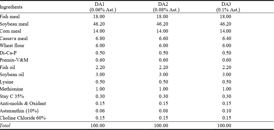
433 Study on Development of Formulated Feed for Improving Growth and Pigmentation of Koi Carp
(Cyprinus carpio L., 1758) Juveniles
Nguyen Van Nguyen, Tran Van Khanh and Pham Duy Hai
442 Study of Caspian Goby Neogobius sp. Karyotype Flexibility from Several Biotops
Aithazha Bigaliev, Saidina Kobegenova, Viktor Vasil’ev, Elena Vasil’eva, Aiman Imentai and Ashan Shametov
Interdisciplinary Researches
447 Diel Vertical Distribution of Zooplankton in Lake Baringo, Kenya
Reuben Omondi, Andrew W. Yasindi and Adiel M. Magana
461 Heavy Metals in Dust Deposition in the Vicinity of Coal Ash Disposal Site Divkovici II
Abdel Dozic, Vahida Selimbasic, Amira Cipurkovic, Aida Crnkic, Zorica Hodzic and Ilvana Trumic
473 A Potential Weed Control—Using Robotic Implement
May 2014, Vol. 8, No. 5, pp. 385-393
Journal of Life Sciences, ISSN 1934-7391, USA
Intravascular Access in
Chelonia mydas
and
Dermochelys coriacea
Using the Seldinger Technique
Ultrasound-Guided
Gustavo Henrique Pereira Dutra1, Fábio Futema2, Flávio Augusto Marques dos Santos3 and Cristiane Lassalvia
Nascimento1
1. Aquário Municipal de Santos, Av. Bartolomeu de Gusmão sn. Ponta da Praia. Santos-SP 11030-500, Brazil 2. Universidade de Guarulhos, Av. Anton Philips, 01, Vila Hermínia, Guarulhos 07030-010, Brazil
3. Universidade Anhembi Morumbi, Rua Casa do Ator, 294. Vila Olímpia 04546-000-Sao Paulo, SP, Brasil
Received: March 12, 2014/ Accepted: May 12, 2014 / Published: May 30, 2014.
Abstract: Strand-ed turtles need fluid parenterally. The jugular access is best for the maintenance and patency of the catheter. The Seldinger technique guided by ultrasound seems to be the safest for catheter insertion. Five juveniles Chelonia mydas with fibropapillomatosis and an adult male leatherback turtle (Dermochelys coriacea) with altered buoyancy were sent to Santos Municipal Aquarium for rehabilitation. Turtles underwent catheterization of the jugular vein using the Seldinger technique with central venous polyurethane catheter monolumen 14 G to Dermochelys and 18 G for Chelonia, guided by ultrasound with 10 MHz transducer catheters were sutured to the skin and animals were subjected to fluid. In all turtles, the catheters were patency and were well established. There were no granulomatous reactions or related infections. The Dermochelys improved clinically after fluid resuscitation, and the catheter was removed one week after. In one Chelonia excision was 21% of its weight in tumors, and the animals received colloid catheter by enabling the mitigation of bleeding due to removal. The use of ultrasonography has enabled monitoring of all access as the patency of the catheter and fluid diffusivity.
Key words: Sea turtles, Chelonia, Dermochelys, ultrassonography, Seldinger technique.
1. Introduction
Stranded sea turtles usually require hydration care; they have varying degrees of gastrointestinal obstruction, cold stunning paralysis, trauma, infectious diseases, parasitism and fibropapillomatosis. Hypoglycemic turtles need to receive fluid resuscitation with glucose. Most stranded animals have hydroelectrolytic imbalances such as hyperkalemia and hyponatremia should receive fluids parenterally [1, 2]. In severely compromised turtles, routes of administration of IV (intravenous fluids) or IO (intraosseous) allow rapid rehydration and
Corresponding author: Gustavo Henrique Pereira Dutra, research fields: experimental and comparative pathology, pathology of testudines. E-mail: [email protected].
emergency therapy. However, the placement and maintenance of catheters in these locations can be technically challenging, especially in aquatic species, and should be reserved for patients who are unconscious or minimally sensitive [3]. A constant infusion of fluids is preferable over intermittent bolus, but any effort should be made to place an intravenous catheter, or alternatively intraosseous. Intraosseous catheters can be placed in the distal humerus, femur or bone bridges. The main disadvantages of intraosseous catheters are limited flow rate of fluid due to the short bone marrow, administration of drugs possibly painful and the needle may undergo metal fatigue and rupture [4, 5]. The catheter should not remain in place for more than 72 h. The catheter site should be prepared aseptically [6]. Intraosseous fluids are best provided to
D
Intravascular Access in Chelonia mydas and Dermochelys coriacea Using the Seldinger Technique Ultrasound-Guided
386
patients with poorly calcified shells. It is indicated for adult species of tortoises, drilling the bone bridge before than needle is inserted. The usual site is the bone bridge and limb bones [7]. Young et al. [8] evaluated the effectiveness of intraosseous catheterization in Gopherus agassizii, comparing four possible locations of maintenance intraosseous catheters (humerus, femur, bridge and plastron gular region) to the jugular vein. Compared to the jugular vein, the intraosseous sites of the humerus and femur were the best access, near 84.4 and 61.8% respectively of the distribution by jugular vascular access. Insertion sites in the bridge and gular shell were less effective with only 41.9 and 40.8% of the systemic activity of flow, respectively. The subcarapacial vein and the cervical sinus are most commonly used in marine turtles for venipuncture and bolus of fluids drugs. Benefits for the IV bolus method include easy accessibility of the vessel and minimal stress for the patient [5]. In clinical practice with sea turtles, it is often necessary to maintain a permanent venous access for rapid rehydration and emergency therapy in critically ill turtles. In addition, venous catheterization can be used to administer drugs and fluids during and after surgery [9]. The supravertebral sinus is commonly used to collect blood in sea turtles. Blood is drawn from adults and juveniles in such access. If necessary, an ultrasound can be used to identify the location of the sinus, especially useful in large patients. Long spinal needles may be necessary to achieve supravertebral sinus and sedation may be necessary to prevent injury to the medulla of the animal. In addition, incidental aspiration of lymph within the sample is not uncommon and essentially invalidates the sample parameters. This site is important for obtaining samples, but for the maintenance and patency of the vessel to fluid therapy, the best access is from the jugular vein [3, 5, 7, 10]. The skin of the access site should be cleaned aseptically. In some patients, anesthesia must be made, even if local anesthesia. The risk versus benefit should be assessed
Seldinger Technique Ultrasound-Guided
ultrasound to catheterization of the jugular vein and
cephalic in Caretta caretta. The authors worked with
22 loggerhead turtles referred for surgery of the gastrointestinal tract. The ultrasound examinations were performed with a device connected to a transducer of ultrasound of linear multifrequency with frequency set at 11 MHz. Once locating the vein during the longitudinal ultrasound examination, a catheter was inserted into the skin below the transducer, 15-25 degrees relative to the skin surface. For the cephalic vein, a catheter 51 mm 20 gauge or 22 is inserted and proximally of this dorso-ventrally, to the jugular vein caliber of 51 mm 16 or 18 g inserted into the craniocaudal direction.
2. Materials and Methods
Five juvenile turtles of undetermined sex of the
species Green turtle (Chelonia mydas) and an adult
male individual of the species leatherback turtle (Dermochelys coriacea) with 360 kg and 2.2 m long were used. The animals arrived at the shore of stranded beach, and were sent to Santos Municipal Aquarium for rehabilitation. Green turtles were admitted to Santos Municipal Aquarium in different degrees and clinical stages of fibropapillomatosis for surgical excision of tumors in different dates.
The leatherback turtle was found in the channel of the Port of Santos, with changes in buoyancy on March 17, 2011. Blood was collected from the hind flippers and the animal was subjected to subcutaneous fluid therapy. The animal was transferred via crane to a room with a pool of 5.000 L, and underwent digital radiography for the evaluation of its postural change. On March 19, the animal underwent jugular vein catheterization using the Seldinger technique, guided
by an ultrasound machine SONOSITE NanoMaxx
® L38n connected to transducer 10-5 MHz with depth scan of 9.0 cm (Fig. 1). After viewing the jugular vein in a sagittal section by transducer, a 14 G catheter it was inserted under visual ultrasound monitoring of the catheter within the vessel (Figs. 2 and 3). The needle
Fig. 1 Transducer on skin of dorso-lateral region of neck.
inside the vessel was slipped the metallic flexible guide wire with tip in J inside the lumen of the needle, until the end of the guide wire, with no resistance to
the passage of the guide wire (Fig. 4). This
procedure allowed the maintenance of the position
of the jugular vein. The needle was withdrawn,
leaving the guide wire (Fig. 5) and an vascular
expander (dilator) immediately inserted into the
jugular vein, slipped around the guide wire by expander lumen to dilate the path of the catheter (Fig. 6). After using the expander, it was withdrawn. Later
a central venous catheter polyurethane monolumen
14 L of 20 cm it was fully inserted around the guide wire toward the jugular vein (Fig. 7). Withdrawing the guide wire and the catheter was fixed to the skin of the animal. After catheterization, the catheter was tapped with luer device (PRN Luer Lock ® BD device), sutured to the skin with nylon monofilament and the
animal was subjected to intravenous fluid with 1
Intravascular Access in Chelonia mydas and Dermochelys coriacea Using the Seldinger Technique Ultrasound-Guided
388
Fig. 2 Inserting a catheter 14 G in jugular vein.
Fig. 3 US image of needle within the vessel.
Fig. 4 Wire guide slipped inside the lumen needle.
Fig. 5 Withdraw of needle, leaving patent the wire guide.
Fig. 6 Insertion of vascular expander (dilator).
Fig. 7 Catether inserted and intravenous fluid therapy in
Seldinger Technique Ultrasound-Guided
cells and total leukocyte counts made in a Neubauer chamber with diluted aliquots in Natt and Herrick solution at 1:100 dilutions. Hematocrit was obtained by rotating closed microcapillary filled with heparinized whole blood at 5000 rpm for 5 min, and the hemoglobin concentration was obtained from an photocolorimetric method (CELM HB ®) at 530 nm.
For the green sea turtles, the animals were found at different dates and shores and were taken to rehab at Santos Municipal Aquarium, all turtles suffering from different grades and clinical stages of fibropapillomatosis. The animals were taken to the University of Guarulhos to remove the tumors and the use of cryosurgery on June 30, 2011. The animals were initially induced with intravenous bolus of propofol at a dose of 5.0 mg/kg to allow intubation with an endotracheal tube. All animals had the jugular vein catheterized using the Seldinger technique with a central venous catheter polyurethane monolumen 18 L 20 cm peripherally inserted, with wire guide, ultrasound guided (MyLab30VetGold ®) frequency often set at 10 Mhz, in the same way (Figs. 8 to 13) as
described above for Dermochelys coriacea. The tube
was tapped with luer device (PRN Luer Lock ® BD device) and sutured to the skin with nylon monofilament. All animals were anesthetized with isoflurane in veterinary vaporizer (HB Conquest Shape ®) with isoflurane minimum alveolar concentration at 2.5%. A single 25 kg turtle was anesthetized with propofol 1% (10 mg/mL) with a syringe infusion pump (Baxter ® Model AS40A), in the ratio of 30 mL/h, at a dose of 200 mg/kg/min, a three-way stopcock device connected to a central venous catheter and peripherally inserted. Associated with the three-way stopcock device was an infusion set providing 25 mL/kg of sodium chloride solution at 0.9%. In this way, the animal received hetastarch at dosage of 4.0 mL/kg. All animals had their tumors excised with an electronic multiprocessor bipolar scalpel and underwent cryosurgery with Cryogun ® device inserted into 500 mL liquid nitrogen with metal
tips. The animals were kept with their jugular vein accessed after the procedure.
3. Results
The results of digital radiography of leatherback turtle showed only radiopaque and distended bowel loops, for this exam was possible to observe the lungs with dense parenchyma, typically with a fibrous appearance. During the rehabilitation period, the animal may be hydrated and receive medication quickly and efficiently. The animal defecated five times in the enclosure, and improved survivability. The animal was kept in rehab in the enclosure until it started to hurt itself in the edges of the pool, as the animal already had better buoyancy, to prevent bacterial contamination lacerations to the patent. The animal received the metal tags on the hind limbs (BR 75672 and BR 75673), and the catheter was removed and the chelonian was released in the vicinity of Parque Estadual Marinho da Lage de Santos (24°20'503" S46°07'317" W) at 03/25/2011.
For green turtles, the catheters maintained adequate
patency and it was well established. There was no
granulomatous or infectious changes related to
catheters reaction. Just one green turtle died after the procedure. At necropsy of this turtle showed grayish
tumor of firm consistency of approximately two
Intravascular Access in Chelonia mydas and Dermochelys coriacea Using the Seldinger Technique Ultrasound-Guided
390
Fig. 8 Insertion of wire guide within catheter lumen.
Fig. 9 After withdraw of the needle, maintaining the wire guide patent.
Fig. 10 Insertion of vascular dilator in C. mydas.
Fig. 11 Insertion of polyurethane monolumen catheter.
Fig. 12 Proving of patency of catheter, with blood drainage.
Seldinger Technique Ultrasound-Guided
Fig. 14 Neoplasia in ventricular cavum pulmonale.
Fig. 15 Propofol anesthesia and fluid therapy under infusion pump in Chelonia mydas.
green turtle of 25 kg, the excision was 5.4 kg of skin fibropapillomas and after the procedure, the animal received hydroxyethyl starch at dosage of 4.0 mL/kg IV (Fig. 15). Despite extensive areas of neoplastic tissue excised, preventing a wound healing by first intention, there was excellent healing and repair of surgical wounds and the use of cryosurgery was responsible for fibropapillomas.
4. Discussion
According to the Red List of Threatened Animals [16], there are 129 species of turtles (62.3% of 207 listed species, 39.3% of all species) officially considered globally threatened, especially marine species [16]. The low-density population decline associated with loss of alleles leads to a loss of genetic variability and consequent fixing of deleterious alleles
in the population; this reduces the potential adaptive, and increases the likelihood of extinction [17]. Therefore, any conservation effort, including clinical care, to keep individuals of hazardous situations, whether or not anthropogenic nature, it is important for the turtle population as a whole [18].
Similar to the findings of Di Bello et al. [9], progress on the catheter until insertion were successful in veins and were monitored by ultrasound. A bolus of sterile saline was injected to test the correct positioning of the catheter. In all animals, the catheters were maintained successfully. For the leatherback turtle, the authors believed to be the most effective and most appropriate (perhaps the only) for volume replacement in this species of large chelonian, unable to stay in the dorsal recumbency to receive intracelomic fluid. As this species critically endangered, all efforts should be made in the conservation of this adult male. The results corroborate what has been postulated by Pittiruti et al. (2000) [12], for being the Seldinger technique the easiest and safest technique of percutaneous percussion. Despite reporting Yildizeli et al. (2004) [13], there was not contamination of the site of insertion into the jugular vein in this specie, with over a week of catheterization, confirming the findings of Pittiruti et al (2000) [12], in a marine animal of absolutely pelagic habits. The use of ultrasonography has enabled the monitoring of safe and adequate access from the jugular vein, confirming anterior studies [14, 15]. The animal improved buoyancy during treatment and in this way was released in pelagic environment.
Intravascular Access in Chelonia mydas and Dermochelys coriacea Using the Seldinger Technique Ultrasound-Guided
392
which receives blood from the right atrium (this in turn receives blood from the vena pre cava and jugular catheter was inserted where). At the time of fluid administration during the surgical procedure, there was overhead in large-circulation return, the systolic mechanical impossibility, due to the presence of heart disease at cavum pulmonale, generating an increase in preload, an insufficiency in cardiac pump, an increase the hydrostatic pressure in wide circulation, generating the events of hepatic and splenic congestion visualized at autopsy. Visceral fibromas are reported in some turtles that have fibropapillomas skin and major organs are lung (which was verified in this case), kidneys, liver and heart. For the green turtle that underwent excision of 5.4 kg of tumors, we believe that the loss of blood suffered due to excision was quite significant and this animal was only kept alive today because of the patency of central venous catheter peripherally inserted, which allowed administering to restore colloidal plasma volume suitable for infusion.
5. Conclusions
The authors concludes that the Seldinger technique, guided by ultrasound is the most appropriate, safe and effective vascular access for fluid replacement in marine turtles needing veterinary care and intensive care.
References
[1] M. Walsh, Rehabilitation of Sea Turtles, in: K.L. Eckert, K.A. Bjorndal, F.A. Abreu-Grobois, M. Donnelly (Eds.), Research and Management Techniques for the Conservation of Sea Turtles, IUCN/SSC Marine Turtle Specialist Group Publication, Blanchard, 1999, No. 4, pp. 202-207.
[2] J. Wyneken, D.R. Mader, E.S. Weber III, C. Merigo, Medicine Care of Sea Turtle, in: D.R. Mader (Ed.), Reptile Medicine and Surgery, Saunders Elsevier, St. Louis, 2006, pp. 972-1007.
[3] D.R. Mader, E. Rudloff, Emergency and Critical Care, in: D.R. Mader (Ed.), Reptile Medicine and Surgery, Saunders Elsevier, St. Louis, 2006, pp. 533-548.
[4] H. Krum, Intraosseous Fluid Administration in Sea Turtles, in: Proceedings of the 4th Annual Association of
Reptilian and Amphibian Veterinarians Conference, Houston, ARAV, 1997, p. 125.
[5] T.M. Norton, Chelonian emergency and critical care, Seminars in Avian and Exotic Pet Medicine 14 (2) (2005) 106-130.
[6] D. Martinez-Jimenez, S.J. Hernandez-Divers, Emergency care of reptiles, Veterinary Clinics of North America- Exotic Animal Practice 10 (2007) 557-585.
[7] S. McArthur, Problem-solving Approach to Conditions of Marine Turtles, in: S. McArthur, R. Wilkinson, J. Meyer (Eds.), Medicine and Surgery of Turtles and Tortoises, Blackwell Publishing, Oxford, 2004, pp. 301-307. [8] B.D. Young, N. Stegeman, B. Norby, J.J. Heatley,
Comparison of intraosseous and peripheral venous fluid dynamics in the desert tortoise (Gopherus agassizii), Journal of Zoo and Wildlife Medicine 43 (1) (2012) 59-66.
[9] A. Di Bello, C. Valastro, D. Freggi, V. Saponaro, D. Grimaldi, Ultrasound-guided vascular catheterization in loggerhead sea turtles (Caretta caretta), Journal of Zoo and Wildlife Medicine 41 (3) (2010) 516-518.
[10] S.J. Divers, Emergency care of the critically ill reptiles, in: Proceedings of Association of Reptilian and Amphibian Veterinarians, 1997, pp. 153-161.
[11] J.A. Briscoe, R. Syring, Techniques for emergency airway and vascular access in special species, Seminars in Avian and Exotic Pet Medicine 13 (3) (2004) 118-131. [12] M. Pittiruti, M. Buononato, M. Malerba, C. Carriero, L.
Tazza, D. Gui, Which is the easiest and safest technique for central venous access? A retrospective survey of more than 5,400 cases, Journal of Vascular Access 1 (3) (2000) 100-107.
[13] B. Yildizeli, T. Lacin, H.F. Batirel, M. Yüksel, Complications and management of long-term central venous access catheters and ports, Journal of Vascular Access 5 (4) (2004) 174-178.
[14] A.J.H. Van Boxtel, M.C. Fliedner, D.M. Borst, S.C.C.M. Teunissen, Peripherally inserted central venous catheters: First results after the introduction in a Dutch University Medical Center, Journal of the Association for Vascular Access 13 (3) (2008) 128-133.
[15] W.G. Warrington Jr., D.A. Penoyer, T.A. Kamps, V. Hoeck, Outcomes of using a modified Seldinger technique for long term intravenous therapy in hospitalized patients with difficult venous access, Journal of the Association for Vascular Access 17 (1) (2012) 24-30.
Seldinger Technique Ultrasound-Guided
[17] A.T. Hahn, J.C. Castilho, L. Soares, S.L. Bonatto, There was a significant loss of genetic variability of olive turtle (Lepidochelys olivacea) on the Brazilian coast due to human action?, in: XVI Encontro de Geneticistas do Rio Grande do Sul, Porto Alegre, de 27-29 Julho, 2008.
May 2014, Vol. 8, No. 5, pp. 394-398
Journal of Life Sciences, ISSN 1934-7391, USA
Arg-X Activity in Trypsin-like Complexes of the Nuclear
Rroteins in the Suprastructures of Interphase Chromatin
During Induction of Growth Morphogenesis Mature
Germs of Wheat
Ivanova Evilina, Vafina Gulnara, Ivanov Ruslan and Tereshchenko Lidia
Institute of Biology Ufa Science Centre, Russian Academy of Science, Ufa, Bashkortostan 450054, Russia
Received: March 05, 2014/ Accepted: April 11, 2014 / Published: May 30, 2014.
Abstract: The purpose of this study was to analyze spatio-temporal dynamics of localization of protease-sensitive sites Arg-X in non-histone and histone blocks of heteropolymer suprastructures (nucleoplasm, chromatin, nuclear matrix) as possible zones affecting the conformational rearrangements of the total interphase chromatin at the induction of increasing morphogenesis of mature embryos-germs of spring and transformed from its winter wheat. Germinated embryos-germs were detached from endosperm after 24 hours from the start of soaking. Cell nuclei have been allocated from embryos-germs and cleared, and then from their heteropolymer suprastructures (nucleoplasm, chromatin loosely bound with nuclear matrix and chromatin tightly bound with nuclear matrix, and nuclear matrix) were extracted by increasing ionic strength of solution. From isolated nuclear suprastructures, non-histone proteins were separated from histones using ion exchange chromatography. Trypsin-like complexes from non-histone proteins and histone blocks were isolated using the affinity chromatography. The Arg-X (tryptase) activity was assessed by cleavage of Arg-X bonds in the arginine-enriched protein protamine. Hypersensitivity to the Arg-X proteolysis in trypsin-like complexes detected at the level suprastructures of chromatin tightly bound with the nuclear matrix was shown. The most active changes of the nuclear proteome have occurred at the level of the non-histone proteins and the core histones (H2A + H2B) (H3 + H4) of induced to growth embryos-seedlings of winter wheat (compared to the initial spring form of wheat). Perhaps hypersensitivity to the Arg-X activity of the trypsin-like complexes in the non-histone proteins and the core blocks of chromatin tightly bound with nuclear matrix have been entrenched during the transforming of the winter wheat from the initial spring wheat.
Key words: Arg-X protease-sensitive, trypsin-like complexes, non-histones, histones, supramolecular structures, cell nuclei, spring and winter wheat.
1. Introduction
In recent years, interests have increased to the morphogenesis significantly. Evidence of this re-edit the book Rene Thom "Structural Stability and Morphogenesis" in 2002 [1]. Retrospective, active interests to the problem of morphogenesis arise in connection with well-known successes in the fields of molecular biology and genetics, morphogenesis
Corresponding author: Evilina Ivanova, Ph.D., chief research officer, research field: biochemistry. E-mail: [email protected].
includes numerous processes. In the literature, there is no report on the classification of morphological processes. The modern biology has established membership in the number of morphological processes: molecular and supramolecular morphogenesis. Processing programmed molecular (or covalent) information assume the transition to self-organization of supramolecular architecture from individual compound components based on non-covalent intermolecular interactions [2]. Currently, some progress has been made in the study of chromatin
D
Chromatin During Induction of Growth Morphogenesis Mature Germs of Wheat
structure on the level of the whole nucleus [3]. In plants, there is a highly organized epigenetic system. They are capable to modify the histone proteins and DNA almost the same way as well as do animals. Plants use the same epigenetic enzymes like that in animals and even humans. Many of plants responsing to the environmental conditions are directly related with the change program of cells. However, still now the molecular mechanisms of adaptive evolution of features have been studied less considerably [4]. At present, a certain interest appears to elucidate the role of proteolysis at the level of histones [5], and even modified proteolytic of histones by tryptase [6], as well as epigenetic regulation of the expression of genes vernalization [7]. Morphological limits and epigenetic influences on the nuclear architecture, on the evolutionary stability of chromosome territory mechanisms, and on the changes in their architecture during the transcription and reparation were discussed [8]. Thus, the dynamics of chromosomes in the interphase nuclei is still not well studied to build more than adequate mathematical logic circuits of the theory of biological specificity of development. The cell nuclei of mature embryos initial spring and transformed from its winter wheat used as a model system, in which there was an adaptive formation of the functional group of coordinately expressed genes. Problems of self-organization of biological systems are in the focus of specialists in macromolecular compounds, in particular biopolymers [2]. Chromatin structure is a supra-hetero-polymer "coherent and extremely viable organism" where effected intermolecular interactions of different supramolecular structures: DNA, RNA, proteins and polysaccharides. They have spatial (geometrical, topological) characteristics, which depend on the surrounding environmental conditions exhibiting their ability to adapt. The purpose of this work was the analysis of spatial-temporal dynamics of localization of protease-sensitive sites Arg-X in non-histone and histone blocks of heteropolymer
suprastructures (nucleoplasm, chromatin, nuclear matrix) as possible zones affecting the conformational rearrangements of the total interphase chromatin at the induction of growth morphogenesis of mature embryos-germs of spring and transformed from its winter wheat.
2. Materials and Methods
Highest-quality seeds of wheat (Triticum aestivum
L.) cv.: Artemovka (spring), and transformed from it
Arg-X Activity in Trypsin-like Complexes of the Nuclear Proteins in the Suprastructures of Interphase Chromatin During Induction of Growth Morphogenesis Mature Germs of Wheat
396
evaluated by cleavage Arg-X bonds in the arginine—enriched protein-protamine Salmine-A-1 («Merk»)—the molecule of which consists of 33 amino acids (22 molecules of Arg, 4-Ser, 3-Pro, 2-Glu and Val) in all fractions of the nuclei [11]. Activity of Arg-X proteolysis was calculated in nanomoles of arginine per second per microgram of protein (n mol/s·mkg protein).
3. Results and Discussion
3.1. Physiology-morphological state of embryos-germs
The growth of morphogenesis begins with the initiation of physiological processes of growth and development of the germs. This period of differentiated growth at the expense of stretching cells is characterized the root emergence. In the 24 h embryos-germs, the distal part of the root is pushed on account of stretching the cells of the embryonic axis of coleoptile and the root. The apex of coleoptile and the root tip are the sensory organs, which are specifically responding to one or another inducible influence. The metabolic system of regulation at the level of signaling "language" of the apical meristem of shoot and root are formed in the period preceding the root emergence. It provides the plasticity, coordinating and simultaneous growth of different tissues of the germ in the close interrelation between them. During the formation of the root meristem, the final "destination of the cells" was fixed only after germination. In growing continuous cellular-tissue masses there will always be mechanical tension due to passive-smoothly deformable cell layers [14]. Moreover, cell-tissue layers may lead to the formation of geometrically striking complex forms. In plants this is observed during the morphogenesis of growth shoot cones. Interrelation of growth with mechanical tensions, naturally distributed in the wide areas of growing tissues, suggests that growth is undoubtedly obeying certain forms of holistic control of the
organism [15].
3.2. Arg-X activity of trypsin-like complexes of the nuclear proteins
Earlier, at the level of suprastructures of the cell nuclei of embryos induced to growth (21 h) [16], and on identifying of sites of hypersensitive to proteolysis at the level of the linker and core histones were shown the dynamics of functioning of Arg-X proteolysis [17]. In the next step of this experiment, Arg-X proteolysis activity was decided to isolate the TLC (trypsin-like complexes) from the proteome of all these suprastructures cell nuclei, and identify in these complexes the sites of sensitivity to the Arg-X protease. Sites of hypersensitivity to the Arg-X proteolysis in TLC are at the level suprastructures of chromatin tightly bound with the nuclear matrix (Chr II) which was shown in Fig. 1.
Chromatin During Induction of Growth Morphogenesis Mature Germs of Wheat
Fig. 1 Arg-X protease sensitivity in TLC of proteins from the suprastructures cell nuclei of embryos-germs wheat (Triticum aestivum L.) cv.: Artemovka (spring) (1) and Mironovsky 808 (winter) (2) after 24 h from the start of soaking. TLC: Trypsin-Like Complexes isolated using the affinity chromatography. Blocks of proteins of the cell nucleus: NHP: non-histone proteins; HI: lysine-rich histone, H2A + H2B: moderately lysine-rich histones, H3 + H4: arginine-rich histones; Suprastructures of cell nucleus: Np: nucleoplasm, Chr I: chromatin loosely bound with the NM, Chr II: chromatin tightly bound with NM, NM: nuclear matrix.
4. Conclusions
In this paper, features of Arg-X proteolysis were considered as a possible mechanism for the architecture reorganization of chromatin matrix in connection with the physiology of growth and development of the embryos-germs. Perhaps hypersensitivity to the Arg-X activity of the TLC in the NHP and the core blocks of chromatin tightly bound with NM has been entrenched during the transforming of the winter wheat from the initial spring wheat. Since the nuclear matrix is considered as an active dynamic structure, which participates in the formation of large enzymatic and regulatory complexes that control the topology and function of DNA. And actively responds to external and internal stimuli, thereby regulating the fundamental biological processes.
Acknowledgments
The reported study was supported by RFBR, research project No. 14-04-3124314.
References
[1] R.F. Tom, Structural Stability and Morphogenesis, Logos, Moscow, 2002, pp. 253. (in Russian)
[2] J.M. Lehn, Supramolecular Chemistry, Nauka Publishers, Novosibirsk, 1998. (in Russian)
[3] T. Mistelli, D.L. Spector, The Nucleus, Cold Spring Harbor Laboratory Press, New York, 2011.
[4] K.V. Gubin, V.V. Suslov, N.A. Kolchanov, Molecular-genetic systems of development: The dynamics of functioning and molecular evolution, Russian Journal of Biochemistry 73 (2) (2008) 270-282. (in Russian)
Arg-X Activity in Trypsin-like Complexes of the Nuclear Proteins in the Suprastructures of Interphase Chromatin During Induction of Growth Morphogenesis Mature Germs of Wheat
398
[6] F.R. Melo, F. Vita, B. Berent-Maoz, F. Levi-Schaffer, G. Zabucchi, G. Pejler, Proteolytic histone modification by mast cell tryptase, a serglycin proteoglycan-dependent secretory granule protease, The Journal of Biological Chemistry (2014). (in press)
[7] A.B. Scherban, E.A. Salina, Epigenetic regulation expression of genes vernalization, Russian Journal of Cytology 55 (4) (2013) 234-237. (in Russian)
[8] I. Schubert, P. Shaw, Organization and dynamics of plant interphase chromosomes, Trends in Plant Science 16 (2011) 273-281.
[9] V.N. Remeslo, Winter Wheat Mironovskaya 264 and 808, Kolos, Moscow, 1964. (in Russian)
[10] E.A. Ivanova, G.H. Vafina, Method of isolation of plant cell nucleus, RF Patent, 1701747 B48 (1991). (in Russian)
[11] E.A. Ivanova, G.H. Vafina, Method of obtaining of nuclear fractions possessing proteinase and inhibition activity, RF Patent, 1733471 B18 (1992). (in Russian) [12] E.A. Ivanova, G.H. Vafina, Method of preparative
isolation of basic proteins from suprastructures of cell nuclei of plants, RF Patent, 2408602 B1 (2011). (in Russian)
[13] E.A. Ivanova, Fractionation of plant histones on columns with amberlite IRC-50, Materials of 3rd scientific conference of young scientists, Publishing House of Bashkir Department Academy of Science USSR, Ufa
(1972) 54-55. (in Russian)
[14] L.V. Belousov, Morpho-mechanical aspect of epigenetic, Russian Journal of Genetics 42 (9) (2006) 1165-1169. (in Russian)
[15] L.V. Belousov, Growth Morphogenesis and Morphological Integrity of the Adult Organism, in: Biological Morphogenesis, Publishing House of Moscow University, Moscow, 1987, pp. 186-204. (in Russian)
[16] R.S. Ivanov, G.H. Vafina, E.A. Ivanova, Arg-X protease-sensitive in supramolecular structures of interphase cell nucleus during growth morphogenesis mature germs of wheat, Journal of Life Sciences 6 (12) (2012) 1351-1355.
[17] E.A. Ivanova, G.H. Vafina, R.S. Ivanov, Analysis of localization protease sensitive sites of Arg-X in interphase chromatin in the dynamics of superstructures during induction the growth morphogenesis of mature embryos spring and winter wheat, Plant Physiology and Genetics (Kiev) 46 (2014). (in press) (in Russian)
[18] E.A. Ivanova, G.H. Vafina, Physiology-biochemical analysis of interphase chromosomes during germination of seeds of wheat, Physiology and Biochemistry of Cultivated Plants (Kiev) 24 (1992) 577-584. (in Russian)
May 2014, Vol. 8, No. 5, pp. 399-403
Journal of Life Sciences, ISSN 1934-7391, USA
The Effect of Mixture of Alpinia Galanga, Eurycoma
Longifolia Jack and Syzygium Aromaticum Crude
Extract on the Growth of Saccharomyces Cerevisiae and
Escherichia Coli
Nur Jasrina Jaafar1, Kaswandi Md. Ambia1, Hing Hiang Lian2 and Rahim Md. Noah1
1. University Kuala Lumpur, Institute of Medical Science, Blok A1-1, Jalan TKS 1, Taman Kajang Sentral, 43000 Kajang, Selangor Darul Ehsan, MALAYSIA
2. School of Diagnostic & Applied Health Sciences, Faculty of Health Science, Universiti Kebangsaan Malaysia, 50300 Jalan Raja Muda, Kuala Lumpur, MALAYSIA
Received: March 05, 2014/ Accepted: April 25, 2014 / Published: May 30, 2014.
Abstract: Alpinia galanga, Eurycoma longifolia Jack and Syzygium aromaticum have been widely used in traditional medicine for decades. Antimicrobial activities for individual crude extract were well established. Crude methanolic extracts of Alpinia galanga, Eurycoma longifolia Jack and Syzygium aromaticum and the combination for all extracts were tested using well diffusion techniques against Saccharomyces cerevisiae (ATCC 9763) and Escherichia coli (ATCC 25922). The mixed extracts were prepared based on the concentration ratio of 50 µg/µL which are 1:1, 1:1:1 and 1:2:2. Single Syzygium aromaticum extract showed higher inhibition zone on Saccharomyces cerevisiae compared to Escherichia coli. There is reduction in diameter of inhibition zone for single extract and mixture extracts either in combination of two or three extracts tested on Saccharomyces cerevisiae and Escherichia coli growth but they are not significant. In conclusion, Syzygium aromaticum showed highest activity against Saccharomyces cerevisiae and Escherichia coli. Reduction in diameter of inhibition zone indicated that Alpinia galanga and Eurycoma longifolia Jack extracts had antagonistic effect with Syzygium aromaticum.
Keywords: Antifungal, antibacterial, extract, Alpinia galanga, Eurycoma longifolia, Syzygium aromaticum, Saccharomyces cerevisiae and Esherichia coli.
1. Introduction
The medicinal plants that are frequently used locally such as langkuas galanga (Alpinia galanga) [1], Tongkat Ali (Eurycoma longifolia Jack) [2] and clove (Syzygium aromaticum) [3] exhibited many antimicrobial effects.
Alpinia galanga (L.) Willd or Lengkuas a plant in the ginger (Zingiberaceae) family was commonly used in the Arabian and in Unani systems of medicine for the treatment of dyspepsia, gastralgia, chronic enteritis
Corresponding author: Kaswandi Md. Ambia, Ph.D., associate professor, research field: natural Product. E-mail: [email protected].
[4] and sea sickness [5]. Different parts of A. galanga are traditionally claimed to be used for the treatment including anti-fungal, anti-tumor, anti-used as a carminative, to increase hydrochloric acid in the stomach and to improve peristalsis [6]. It was also used in dentistry where the essential oil of clove was used as anodyne for dental emergencies [7].
Eurycoma longifolia Jack also known as Tongkat Ali, a flowering plant in the Simaroubaceae family claimed to have aphrodisiac properties was mainly used as aphrodisiac, anti-cancer, anti-pyretic and anti-malarial remedy [8].
D
The effect of mixture of Alpinia galanga, Eurycoma longifolia Jack and Syzygium aromaticum crude extract on the growth of Saccharomyces cerevisiae and Escherichia coli
400
A wide range of medical plants parts was used for extract as raw drugs and they possess varied medical properties [9]. Many of bacteria species are resistant to multiple agents, making choosing an antibiotic to treat such infections difficult [10]. The increase effects were associated with conventional treatments [11].
This paper described the mixture of Alpinia galanga, Eurycoma longifolia Jack and Syzygium aromaticum extract and their effect on Escherichia coli and Saccharomyces cerevisiae.
2. Materials and Methods
2.1 Preparation of extractsMethanolic plant extraction for this experiment was prepared by Faculty of Health Science, Universiti Kebangsaan Malaysia using modified technique of Shimada [12]. A. galanga, E. longifolia and S. aromaticum extracts weighted respectively (0.5 g) and dissolved into 0.5 m/L of 5% DMSO and 9.5 m/L of distilled water to obtain the stock concentration of 500 µg/µL for all single extracts. Mixture extracts were prepared by mixing the stock solution of the three single extracts that are A. galanga, E. longifolia and S. aromaticum extracts by volume of the ratio 1:1:1 and 1:2:2. The ratio of 1:2:2 mixtures of the extracts is the concentration proportional to the concentration 500 µg: 1000 µg: 1000 µg. The mixture extracts 50 µL are filled into the wells of the agar plate for the survival test. The ratio of the concentration for the mixed extract of A. galanga is equivalent to 50 µg of the concentration of E. longifolia and S. aromaticum extracts.
2.2 Growth of bacteria and fungi
E. coli (ATCC 25922) was grown on the nutrient
agar and incubated at 37 C about 24 h. While for S.
cerevisiae (ATCC 9763), it was incubated at 30 C
about 2 days. Bacteria suspension of E. coli was
prepared by transferring the isolated colonies to 2.0 mL of normal saline (0.08OD) using spectrophotometer at wavelength of 625 nm. S.
cerevisiae was prepared using the same procedure but at different temperature incubation i.e. 30 C for 48 h.
2.3 The survival test
The survival test was conducted using well diffusion method to detect the antibacterial and antifungal activity towards the extract solution [13]. A sterile cotton swap was dipped in a bacterial
suspension of E. coli and streaked on nutrient agar.
The inoculums were allowed to dry for 3 to 5 min to allow diffusion of bacteria into the agar. A sterilized Durham tube was used to punch wells on the agar plates with maximum 4 wells per plate. A total of 50
L of the single extracts, mixture of extracts, positive controls and negative control respectively were filled into the labeled wells. The test was repeated five times and the mean value was recorded. The same procedure was applied for S. cerevisiae. All the plates used, containing E. coli are incubated at 37 C for 24 h,
while S. cerevisiae was incubated at 30 C for 48 h.
The inhibition zone was measured after incubation. Statistical analysis for each test was done using SPSS version 18.
3. Results and Discussion
A. galanga and E. longifolia extracts does not showed any positive effect on S. cerevisiae (Table 1). S. aromaticum extract showed the highest inhibitory effect against S. cerevisiae with mean diameter of inhibition zone at 22.4 mm. Previous studies have reported antifungal activity for clove oil and eugenol against yeasts and filamentous fungi [14], and human pathogenic fungi [15]. S. aromaticum extract showed inhibition at concentration of 500 µg/µL on S. cerevisiae. This showed that S. aromaticum extract was suitable to be utilized to obtain the active components as compared to A. galanga and E. longifolia for antifungal activities.
A. galanga produce negative result when tested on
extract on the growth of Saccharomyces cerevisiae and Escherichia coli
coli with mean diameter inhibition zone of 5.0 mm
and 17.2 mm respectively. However, single extract of S. aromaticum still showed higher inhibition effect as
compared to E. longifolia extract when tested on E.
coli. The results of this study are in line with those reported by Burt and Reinders [16] that clove oil was found to be effective against non-toxigenic strains of
E. coli O157:H7 [17]. When S. aromaticum extract was compared to gentamicin which acts as a positive
control toward E. coli, it showed significant
differences between the extract and the positive control that produce the higher inhibition zone at 25.0 mm.
The extract mixture ratio of L:TA (1:1) did not provide a positive effect on S. cerevisiae, while, extracts mixture ratios for L:C (1:1), L:TA:C (1:1:1) and L:TA:C (1:2:2) have positive effect on S. cerevisiae with inhibition zone of 24.4 mm, 22.4 mm and 22.6 mm respectively. The results showed that L:C (1:1) recorded the highest inhibition zone diameter means among all the extracts at 24.4 mm. When compared the various mixture extract ratio to fungal inhibition effect, showing similar inhibition
zone to single extract of S. aromaticum, A. galanga mixed with S. aromaticum (L:C) in the ratio 1:1, the inhibition zone increase as compared with single extract of S. aromaticum. This shows that A. galanga demonstrate the suppression of growth when tested on S. cerevisiae. When A. galanga was mixed with E. longifolia and S. aromaticum (L:TA:C) in the ratio of 1:1:1 and 1:2:2, the inhibition effect obtained was almost similar to that of single extract of S. aromaticum at 22.4mm. Based on the above result, E. longifolia does not have the suppression effect against S. cerevisiae as shown by a decrease in inhibition zone diameter.
The extract mixture ratio L:TA (1:1) does not have any effect on E. coli but extracts mixture ratio of L:C (1:1), however, L:TA:C (1:1:1) and L:TA:C (1:2:2)
have positive effect on E. coli. The results also
revealed that L:C (1:1) recorded the highest mean of inhibition zone diameter among all the extracts used, which is at 15.4 mm. By comparing the various mixture extract ratio with bacteria inhibition effect which produce almost the same inhibition with the single extract of S. aromaticum. A. galanga mixed
Table 1 The effect of a single extract and mixture of extracts ratio between A. galanga, E. longifolia and S. aromaticum extract on growth of S. cerevisiae and E. coli.
Extracts Inhibition zone (mm) standard deviation
Saccharomyces cerevisiae Escherichia coli
Alpinia galanga (L) - -
Eurycoma longifolia (TA) - 5.0 2.07
Syzygium aromaticum (C) 22.4 7.16 17.2 2.28
L:TA (1:1) - -
L:C (1:1) 24.4 5.46 15.4 2.07
L:TA:C (1:1:1) 22.4 3.43 12.4 1.82
L:TA:C (1:2:2) 22.6 3.65 13.8 1.79
Positive control 15.0 0.00 25.0 0.00
Negative control - -
Indicators: (-) = no inhibition zone; L = Alpinia galanga extract; TA = Eurycoma longifolia extract; C = Syzygium aromaticum extract Positive control:
Amphotericin B: act as the positive control for Saccharomyces cerevisiae Gentamicin: act as the positive control for Escherichia coli
The effect of mixture of Alpinia galanga, Eurycoma longifolia Jack and Syzygium aromaticum crude extract on the growth of Saccharomyces cerevisiae and Escherichia coli
402
with S. aromaticum (L:C) in the ratio 1:1, the inhibition zone decreases to 15.4 mm compared with single extract of S. aromaticum [3]. This further proved that A. galanga do not suppressed the growth of E. coli. If A. galanga was mixed with E. longifolia and S. aromaticum (L:TA:C) in the ratio of 1:1:1, the inhibition effect on the bacteria decreases to 12.4 mm. While for the ratio of 1:2:2, it increased slightly to 13.8 mm. Based on this result, both extracts E. longifolia and A. galanga did not produce inhibition effect on E. coli [18].
Statistical analysis showed that there is no significant difference between diameters of single or mixture of crude extract compared with control for both fungus and bacteria.
4. Conclusions
Clove extract produce positive inhibition on S. cerevisiae and E. coli. S. aromaticum extract could be a strong candidate as an antimicrobial agent to substitute/compliment the antibiotics in the market. A. galanga and E. longifolia also showed antimicrobial activity when the mixture was added together but both extracts do not showed as strong an inhibition as Syzygium aromaticum extracts. It remains that single extracts were more effective than combination of mixture extracts on the inhibition of S. cerevisiae and
E. coli.
Acknowledgments
The authors would like to thank Faculty of Health Science UKM for supplying the extracts, Mr. Mohd Rohani Jais, Mr. Shazwan Shazdee Wahab and Miss Faridah Parid for their assistance in laboratory works.
References
[1] A.K. Chudiwal, D.P. Jain, R.S. Somani, Alpinia galanga Willd: An Overview on Phyto-Pharmacological Properties, Indian Journal of Natural Products and Resources 1 (2) (2010) 143-149.
[2] R. Bhat, A.A. Karim, (2010), "Tongkat Ali (Eurycoma
longifolia Jack): A review on its ethnobotany and pharmacological importance", Fitoterapia 81 (7) (2010) 669-679.
[3] E. Pinto, L. Vale-Silva, C. Cavaleiro, L. Salgueiro, Antifungal activity of the clove essential oil from syzygium aromaticum on Candida, aspergillus and dermatophytes species, Journal of Medical Microbiology 58 (2009) 1454-1462.
[4] A.K. Ghosh, M. Banerjee, N.K. Bhattacharyya, Anti-inflammatory activity of root of Alpinia galanga willd, Chronicles of Young Scientists 2 (2011) 139-143. [5] B. Hemabarathy, B. S-Balkis, V. Feizal, Paracetamol
hepatotoxicity in rats treated with crude extract of Alpinia galangal, Journal of Biological Sciences 9 (2009) 57-62. [6] S. Dhinahar, T. Lakshmi, Role of botanicals as
antimicrobial agents in management of dental infections: A review, International Journal of Pharma and Bio Sciences 2 (4) (2009) 690-697.
[7] A.N. Daniel, S.M. Sartoretto, G. Schmidt, Caparroz S.M. Assef, C.A. Bersani-Amado, R.K.N. Cuman, Anti-inflammatory and antinociceptive activities of eugenol essential oil in experimental animal models, Brazilian Journal of Pharmacognosy 19 (1B) (2009) 212-217.
[8] M. Rashid, S. Kumar, S.B. Ahmad, Medicinal uses of eurycoma longifolia: A review, The Pharma Research (2) (2009) 70-78.
[9] V. Ambikapathy, V.S. Gomathi, A. Panneerselvam., Effect of antifungal activity of some medicinal plants against pythium debaryanum (Hesse), Asian Journal of Plant Science and Research 1 (3) (2011) 131-134. [10] A. Pallett, K. Hand, Complicated urinary tract infections:
Practical solutions for the treatment of multiresistant gram-negative bacteria, Journal of Antimicrobial Chemotheraphy 65 (3) III (2010) 25-33.
[11] J. Davies, D. Davies, Origins and evolution of antibiotic resistance, Microbiology and Molecular Biology Reviews 74 (3) (2010) 417-433.
[12] S. Shimada, Antifungal steroid glycoside from sea cucumber, Science 163 (1969) 1462.
[13] C.M. Mattana, S.E. Satorres, A. Sosa, M. Fusco, L. E. Alcaraz, Antibacterial activity of extracts of acacia aroma against methicillin-resistant and methicillin-sensitive staphylococcus, Brazilian Journal of Microbiology 41 (2010) 581-587.
[14] P. Thobunluepop, Implementation of bio-fungicides and seed treatment in organic rive Cv. KDML 105 farming, Pakistan Journal of Biological Sciences 12 (16) (2009) 1119-1126.
extract on the growth of Saccharomyces cerevisiae and Escherichia coli
calcium stress and inhibition of The TOR pathway. Antimicrobial Agents and Chemotheraphy 54 (12) (2010) 5062-5069.
[16] S.A. Burt, R.D. Reinders, Antibacterial activity of selected plant essential oils against Escherichia coli O157:H7, Lett. Appl. Microbiol. 36 (2003) 162-167. [17] A. Pandey, P. Singh, Antibacterial activity of syzygium
aromaticum (clove) with metal ion effect against food borne pathogens, Asian Journal of Plant Science and Research 1 (2) (2011) 69-80.
May 2014, Vol. 8, No. 5, pp. 404-409
Journal of Life Sciences, ISSN 1934-7391, USA
Association Between IL-6 (Interleukin-6) and Iron Status
in Rheumatoid Arthritis Patients
Abbas Sabbar Dkhil1, and Musa Nima Mezher2
1. Physiology Department, Faculty of Medicine, University of Qadisiyah, Qadisiyah 58002, Iraq 2. Biology Department, Faculty of Science, University of Kufa, Najaf, Qadisiyah 58002, Iraq
Received: March 08, 2014/ Accepted: April 21, 2014 / Published: May 30, 2014.
Abstract: The study was performed to determine whether the srum concentrations of IL (interleukin)-6 are elevated in patients with RA (rheumatoid arthritis) and to investigate the relationship between IL-6 levels and iron status in RA patients. 95 serum samples were obtained, 70 of them from patients with RA who had visited the department of Rheumatology at Al-Sadder medical city in Najaf governorate (Iraq) and 25 age and sex-matched healthy controls. The authors assessed the clinical parameters of the disease, including ESR (erythrocyte sedimentation rate), CRP (C-reactive protein), and RF (rheumatoid factor). Serum levels of iron and TIBC (total iron binding capacity) were measured spectrophotometrically, while TS% (transferrin saturation percentage) and transferrin concentration were calculated mathematically. Serum concentrations of IL-6 (interleukin-6) and ferritin were measured using an ELISA (enzyme-linked immunosorbent assay). The results of serum concentration of IL-6 (interleukin-6) and ferritin were significantly elevated (P < 0.0001) in patients with RA compared to those of healthy controls. On the other hand, serum concentrations of iron, TIBC (total iron binding capacity), TS% (transferrin saturation percentage) and transferrin concentration were significantly decreased in patients with RA compared with those of healthy controls. These findings suggest that anemia is the most frequent observations in patients with RA and mostly associative with increasing level of interleukin-6.
Key words: Rheumatoid arthritis, interleukin-6, Iron status, ACD (anemia of chronic diseases), proinflammatory cytokines.
1. Introduction
RA (rheumatoid arthritis) is a chronic disease that causes inflammation mainly in the synovium and produces destruction and deformity of the joints. The etiology of RA remains unclear, but it is known to be associated with genetic and environmental factors [1].
Various proinflammatory cytokines, such as IL-6, TNF (tumor necrotic factor)-α, IL (interleukin)-1β, and IFN (interferon)-γ, are increased in the synovial tissue or synovial fluid of patients with RA [2, 3]. Increased levels of proinflammatory cytokines lead to the proliferation of synovial tissue, and thereby cause damage in the articular cartilage and bone destruction in the adjacent area [4, 5]. In particular, IL-6 is a cytokine with various functions.
When IL-6 is activated, acute inflammatory
Corresponding author: Dr. Abbas Sabbar Dkhil, lecturer, research field: physiology. E-mail: [email protected].
responses such as fever or anemia are induced. IL-6 promotes the proliferation of B cells and thus is involved in the production of the rheumatoid factor [6]. Recently, RA has been observed to be associated with high levels of IL-6 in the synovial membrane and serum [6, 7]. This led to the speculation that IL-6 plays a pathogenic role in RA.
Recently, an IL-6 receptor antagonist, tociliumab, was developed and showed clinical efficacy in the treatment of RA [8-10].
IL-6 is a pleiotropic cytokine with a wide range of biological activities on various target cells. It regulates immune responses, hematopoiesis, acute phase responses and bone metabolism [11]. Despite its important physiological roles, dysregulated overproduction of IL-6 is responsible for systemic inflammatory manifestations and abnormal laboratory findings in patients with RA. In fact, elevated IL-6
D
levels have been observed in both serum and synovial fluid in patients with RA, and there have been correlations between serum IL-6 levels and clinical and laboratory indices of RA [12]. These findings have led to the concept that interference with the actions of IL-6 could represent a therapeutic approach to RA.
In RA, it is estimated that 30-60% of patients are anemic. One of the most frequent causes of anemia in RA patients is IDA (iron deficiency anemia). ACD (anemia of chronic disease) which does not usually respond to iron supplementation is another major cause of anemia in patients with RA [13]. However, Bloxham and his coworkers found that the majority of anemic patients were ACD, with rather fewer patients demonstrating iron deficient [14].
In the current study, the authors examined whether serum levels of IL-6 are increased in patients with RA and whether the increased levels are significantly correlated with iron status. The authors compared the serum concentrations of IL-6 in patients with RA and those in normal controls and then investigated the correlation between serum levels of IL-6 and the iron status alterations of RA.
2. Materials and Methods
2.1 Subjects and clinical assessmentThe study was conducted in 50 patients who had visited Al-Sadder Medical Cityat Najaf Governorate (Iraq) between February and May 2013, and who fulfilled the ACR (American College of Rheumatology) 2010 revised criteria for the diagnosis of RA [15]. Twenty age and sex-matched healthy adults without any evidence of chronic inflammatory disease served as the controls. The patients underwent thorough clinical and laboratory evaluation, including complete medical history, seropositivity test for RA (Rheumatoid Arthritis), CRP (C-reactive protein), and estimation of ESR (erythrocyte sedimentation rate). At the time of clinical assessment for disease, 6 mL of blood samples were collected intravenously from each
patient, 1 mL for evaluation of ESR, whereas serum samples were collected in glass tubes without anticoagulant, stored for one hour at room temperature, centrifuged (2,500 r.p.m. for 10 min at 4 °C) and then aliquoted in plastic tubes before being stored at -20 °C until analysis.
2.2 Study Design
The patients were classified according to the duration of RA disease [15]. As follows:
GI (Group I): the group of patients with disease length of (less than one year) was considered as the group with very early disease duration.
GII (Group II): the group of patients with disease length of (1-5) years was considered as the group with early disease duration.
GIII (Group III): the group of patients with disease length of (6-15) years was considered as the group with median disease duration.
Group IV (GIV): the group of patients with disease length of (16-25) years was considered as the group with long disease duration.
GV (Group V): the group of patients with disease length of (more than 25) years was considered as the group with very long disease duration.
2.3 Methods
2.3.1 Rapid test
RF (rheumatoid factor) and C-reactive protein were performed by a rapid latex agglutination test kit for the presumptive detection of the RF and C-reactive protein in serum.
2.3.2 ESR (Erythrocyte sedimentation rate)
The ICSH (International Committee on Standardization in Hematology) recommends the use of the Westergren method [16].
2.3.3 Serum iron and TIBC (total iron binding capacity)
Association Between IL-6 (Interleukin-6) and Iron Status in Rheumatoid Arthritis Patients
406
Hospital. The Colorimetric test method was used via RANDOX reagents (RANDOX kit, U.K.) [17].
2.3.4 Transferrin saturation percentage (%)
TS% (transferrin saturation percentage) was calculated mathematically [18].
2.3.5 Transferrin concentration
Transferrin concentration was calculated mathematically [19].
2.3.6 Measurement of serum IL-6and ferritin The serum concentration of IL-6 was measured using an AssayMax ELISA (enzyme-linked immunosorbent assay) kit (Assaypro, USA) at Virology Laboratory of AL-Sadder Medical City in Najaf Governorate. Fifty µL each of serum sample and assay diluent were placed in each well of a 96-well plate coated with a monoclonal mouse IgG against IL-6. This mixture was incubated for two h at room temperature, and each well was aspirated and washed five times with wash buffer. Subsequently, 50
μL of Biotinylated IL-6 Antibody was added to each
well and incubated for two h. Again, each well was washed five times with wash buffer. Following this, 50
μL of Streptavidin-Peroxidase Conjugate was added per
well and incubated for 30 min and each well was aspirated and washed five times with wash buffer.
Subsequently, 50 μL of substrate solution, which was
prepared with equal amounts of stabilized hydrogen peroxide (H2O2) and tetramethylbenzidine, was added
for a 20 min reaction under dark conditions. The
reaction was quenched by the addition of 50 μL stop
solution (0.5 N of HCl). Within 30 min, the optical density was measured at a wavelength of 450 nm using the bioelisa reader ELX 800 (Molecular Device Co., biokit, CA, USA). The serum concentration of IL-6 was determined based on a standard concentration curve. The correlation coefficient (r) of the standard concentration curve was 0.990.
The serum ferritin was also purchased from Assaypro, USA, and determined using similar methods. The serum concentration of ferritin was determined based on a standard concentration curve.
The correlation coefficients (r) were 0.993.
2.4 Statistical analysis
Data analyses were performed with SAS version 9.1 (SAS Institute Inc., Cary, NC, USA). All of the descriptive variables were expressed as the mean ±SE (standard error). The correlations between the concentrations of IL-6 and iron status were tested using Pearson’s correlation test. The group analyses were performed using one-way ANOVA and Tukey’s post-hoc analyses. For all tests, a p value less than 0.05 was considered statistically significant.
3. Results
3.1. Serum concentrations of iron
TIBC (total iron binding capacity), TS% (transferrin saturation percentage) and transferrin
concentration were significantly decreased (P <
0.0001) in patients with RA compared to those of healthy controls, while serum concentration of ferritin was significantly elevated (P < 0.0001) in patients with RA compared to those of healthy controls (Table 1).
3.2. Serum concentration of IL-6
Serum level of IL-6 of RA patients showed significant increase (P < 0.0001) in the average values comparatively to the healthy group. In the very early duration of the disease was much higher (Fig. 1).
3.3. Relationship of IL-6 levels to iron status
Serum concentration of IL-6 showed inverse correlations with serum iron, and serum TIBC, respectively (Table 2).
3.4. Relationship of IL-6 levels to serum ferritin and ESR
Table 1 Iron status in healthy control group and in the groups of patients suffering from RA.
Iron status Healthy Control (n = 20)
Ferritin (ng/mL) 131.2±5.96 220.4±23.73a ns
Data are expressed as means ±SE (standard error).
The asterisks indicate significant difference based on Tukey’s multiple comparison tests.
The same letters indicate no significant difference between groups based on Tukey’s multiple comparison tests. *** indicate extremely significant (P < 0.001).
* indicate significant (P value 0.01 to 0.05). Ns: not significant. groups of patients suffering from rheumatoid arthritis.
Data are expressed as means ±SE (standard error).
*** indicate significant difference based on Tukey’s multiple comparison tests.
P < 0.0001
Table 2 Correlation between IL-6 and iron status in RA patients.
Table 3 Correlation between IL-6 and serum ferritin and ESR in RA patients. *Pearson’s correlation analysis was performed.
4. Discussion
The current study showed that serum concentrations of IL-6, were significantly elevated in patients with RA compared to those in healthy controls (Fig. 1). As seen in previous reports, this finding supports the hypothesis that IL-6 is involved in the pathogenesis of RA [20-23]. In addition to these previous observations, the significant increase in the IL-6 cytokine observed in the current study indicates that this cytokines might play a role in inducing inflammatory responses or mediating anti-inflammatory responses in the pathogenesis of RA [24].
The present study also indicates significant decrease in serum iron, serum transferrin in all groups of patients with RA (Table 1). As seen in previous reports, this finding supports the hypothesis that the anemia is the most frequent extra-articular manifestation of the disease [25, 26].
In RA, it is estimated that 30-60% of patients are anemic. One of the most frequent causes of anemia in RA patients is IDA (iron deficiency anemia). ACD (anemia of chronic disease) which does not usually respond to iron supplementation is another major cause of anemia in patients with RA [27].